

Embryo toxic effects of depleted uranium on the morphology of the mouse fetus
- PMID: 24734072
- PMCID: PMC3985252
Embryo toxic effects of depleted uranium on the morphology of the mouse fetus
Abstract
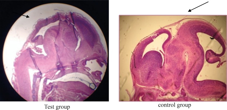
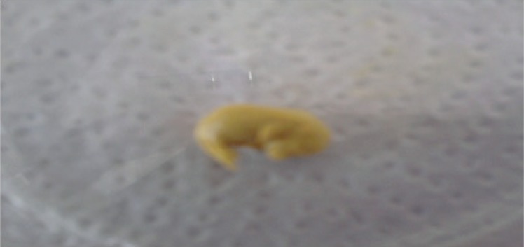
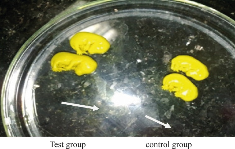
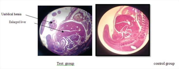
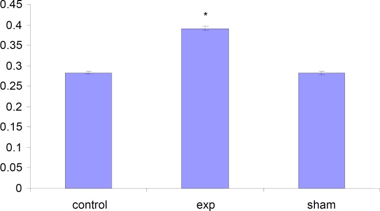
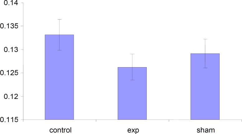
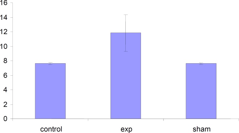
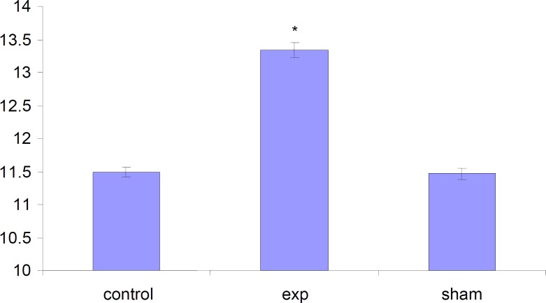
Although the biokinetics, metabolism, and chemical toxicity of uranium are well known, until recently little attention was paid to the potential toxic effects of uranium on reproduction and development in mammals. In recent years, it has been shown that uranium is a developmental toxicant when given orally or subcutaneously (SC) to mice. Decreased fertility, embryo/fetal toxicity including teratogenicity, and reduced growth of the offspring have been observed following uranium exposure at different gestation periods. For investigating the effects of DU on pregnant animals, three groups (control, sham and test) of NMRI mice were chosen. In test group 4 mg/Kg of DU were administered intraperitonealy at 11 day of gestation, in sham group only normal saline injected to interior peritoneum as indicated in the test group and in Control group which was considered as the comparison base line of our research, no injection was made. Caesarean sections were performed at 15 day of the gestation; and their placentas were examined externally. Base on our results DU caused significant external anomalies, and caused a significant decrease (p < 0.05) in the weight and diameter of placentas, the number of the embryos, their body weight and crown-rump length of fetuses.
Keywords: Depleted uranium; Embryotoxicity; Morphology; Mouse fetus.
Figures
References
-
- Jiri P, Jiri K, Rudolf S, Gustav S, Josef H. Toxicological Aspects of Depleted Uranium. Appl Biomed. . 2004;2:37–42.
-
- Cothern CR, Lappenbusch WL. Occurrence of Uranium in Drinking Water in the US. Health phys. . 1983;45:89–99. - PubMed
-
- Cothern CR, Lappenbusch WL, Cotruvo JA. Health Effects Guidance for Uranium in Drinking Water. Health phys. 1983;44:377–384. - PubMed
-
- Pourahmad J, Shaki F, Hosseini MJ, Ghazi-Khansari M. Depleted uranium induces disruption of energy homeostasis and oxidative stress in isolated rat brain mitochondria. Metallomics . 5:736–744. - PubMed
-
- Jalal Pourahmad, Monireh Ghashang, Hossein Ali Ettehadi, Ruhollah Ghalandari. A search for cellular and molecular mechanisms involved in depleted uranium (DU) toxicity. Environ Toxicol. . 2006;21:349–354. - PubMed
LinkOut - more resources
Full Text Sources