Review
. 2014 Aug;72(2):236-58.
doi: 10.1111/aji.12252. Epub 2014 Apr 16.

Regulation of mucosal immunity in the female reproductive tract: the role of sex hormones in immune protection against sexually transmitted pathogens

Affiliations
Review

Regulation of mucosal immunity in the female reproductive tract: the role of sex hormones in immune protection against sexually transmitted pathogens

Charles R Wira et al. Am J Reprod Immunol. 2014 Aug.

Abstract

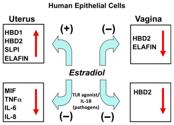
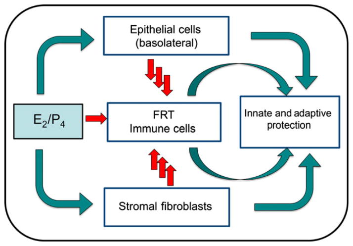
The immune system in the female reproductive tract (FRT) does not mount an attack against human immunodeficiency virus (HIV) or other sexually transmitted infections (STI) with a single endogenously produced microbicide or with a single arm of the immune system. Instead, the body deploys dozens of innate antimicrobials to the secretions of the FRT. Working together, these antimicrobials along with mucosal antibodies attack viral, bacterial, and fungal targets. Within the FRT, the unique challenges of protection against sexually transmitted pathogens coupled with the need to sustain the development of an allogeneic fetus, has evolved in such a way that sex hormones precisely regulate immune function to accomplish both tasks. The studies presented in this review demonstrate that estradiol (E2 ) and progesterone secreted during the menstrual cycle act both directly and indirectly on epithelial cells, fibroblasts and immune cells in the reproductive tract to modify immune function in a way that is unique to specific sites throughout the FRT. As presented in this review, studies from our laboratory and others demonstrate that the innate and adaptive immune systems are under hormonal control, that protection varies with the stage of the menstrual cycle and as such, is dampened during the secretory stage of the cycle to optimize conditions for fertilization and pregnancy. In doing so, a window of STI vulnerability is created during which potential pathogens including HIV enter the reproductive tract to infect host targets.

Keywords: Epithelial cells; female reproductive tract; fibroblasts; immune cells; mucosal immunity; sex hormones.


Figures

Fig. 1
Fig. 1
Schematic of the mucosal immune system throughout the human female reproductive tract (FRT). As seen in the drawing on the left side, the vagina and ectocervix are lined with squamous epithelial cells Columnar cells are present throughout the upper FRT including the endocervix, uterine endometrium (EM), and Fallopian tubes. Panel a–d: are confocal photomicrographs showing the distribution of immune cells throughout out the uterus. Frozen sections were directly stained with three fluorescently tagged monoclonal antibodies: an epithelial cell specific antibody (Cy3 labeled clone BerEP4, Red color panels a–d), anti-CCR5 (FITC labeled clone 2D7, Green color panels a–d) and anti-CXCR4 (Cy5 labeled 12G5, blue color panels a–d). Panel (a) Epithelial gland extending from the myometrial interface (far left) to the luminal epithelium (right-hand side). Panels (b) through d show higher magnification fields from the same region. The luminal epithelium, in contrast with the adjacent glands, retains a relatively intense BerEP4 expression (red color panels a and d). CCR5 expression is prominent on the lymphoid aggregates located in the stratum basalis immediately adjacent to the myometrium (‘LA’ in panel b). Epithelial expression of CCR5 is low in the stratum basalis and increases in the proximal third of the stratum functionalis. Panel f: This photo plate consists of a lymphoid aggregate in the uterine EM at the late proliferative stage of the menstrual cycle. The three fluorochromes are T cells (Cy3-anti CD3, red), B cells (FITC anti CD19, green) and macrophages (Cy5-anti CD14, blue). Panel e: Vaginal squamous cells expressing GalCer (green) is expressed on parabasal epithelial cells (p) and in the surface regions of the cornified layer. Panel g: CD8+ T cells present in both the submucosa and the squamous epithelium. Panel h: CD14 expression is found on both stroma and squamous epithelium macrophages. Panel i: CD1a positive dendritic cells (DC) are present in the squamous epithelium (blue). Panel a–d; Panel f; Panel e, g, h, i.
Fig. 2
Fig. 2
Estradiol inhibits transepithelial resistance (TER) in the human uterine epithelial cell line ECC-1. ECC-1 cells were grown in media supplemented with stripped FBS until maximal TER of about 2000 ohms/well in cell inserts was obtained. Estradiol (5 × 10⁻⁸ M) and/or progesterone (1 × 10⁻⁷ M) were added to the basolateral compartment of some inserts and TER was measured after 48 hr. Four wells per group; **Significantly different (P < 0.01) from Control. From reference (145).
Fig. 3
Fig. 3
Estradiol stimulates secretion of secretory leukocyte protease inhibitor (SLPI) by ECC-1 cells. ECC-1 cells were grown to confluence and high transepithelial resistance in cell inserts. The media was changed and some inserts were cultured with 5 × 10⁻⁸ M E2 and/or progesterone (1 × 10⁻⁷ M) for 48 hr. Apical conditioned media were recovered, centrifuged to remove cell debris and SLPI was measured by ELISA. Four wells per group; **Significantly different (P < 0.01) from control or progesterone. From reference (12).
Fig. 4
Fig. 4
Estradiol inhibits lipopolysaccharide (LPS)-induced IL-6 secretion by uterine epithelial cells. Monolayer cultures of uterine epithelial cells at high transepithelial resistance were treated with 5 × 10⁻⁸ M E2 for 48 hr in the basolateral compartment of cell inserts with some wells receiving an apical treatment of ultra pure LPS (100 mg/mL) after 24 hr. Apical conditioned media were recovered, centrifuged to remove cell debris and IL-6 was measured by ELISA. LPS stimulated a significant increase in IL-6 secretion, which was mostly abrogated by pre-treatment with E2. Four wells per group; **Significantly different (P < 0.01) from Control, E2 and E2 + LPS. Adapted from reference (12).
Fig. 5
Fig. 5
Estradiol inhibits IL-1β mediated secretion of HBD2 and IL-8 and the inhibition occurs though involvement of the estrogen receptor. ECC-1 cells were preincubated with E2, the pure ER antagonist ICI182780, or a combination of steroid and antagonist for 72 hr before stimulation with IL-1β (5 ng/mL). Apical and basolateral conditioned medium were collected following 24 hr stimulation and analyzed for HBD2 and IL-8 protein secretion by ELISA. The results are shown as the mean ± S.E.M. **Significantly different (P < 0.01) from control. Red bars, apical conditioned medium; Blue bars, basolateral conditioned medium. Adapted from reference (57).
Fig. 6
Fig. 6
Estradiol decreases the production of HBD2 and elafin in primary vaginal squamous epithelial cells. Vaginal secretions and cells were recovered from volunteers using an Instead Menstrual Cup following insertion for 1 hr. Freshly isolated vaginal epithelial cells were incubated overnight in triplicate wells prior to treatment with P4, E2 or a combination of both for 48 hr. Conditioned media were recovered and analyzed by ELISA for the presence of HBD2 (a) and elafin (b). Data shown are from one volunteer that is representative of two experiments from two different volunteers. ***P < 0.001 with respect to control. From reference (98).
Fig. 7
Fig. 7
Schematic of estradiol regulation of innate immune function by human epithelial cells in the upper and lower female reproductive tract. In the uterus, E2 enhances the secretion of antimicrobial factors and reduces the secretion of induced pro-inflammatory mediators. In contrast with the uterus, E2 inhibits both the constitutive and induced secretion of antimicrobials in the vagina.
Fig. 8
Fig. 8
Estradiol stimulates secretion of hepatocyte growth factor (HGF) by human uterine stromal fibroblasts. Uterine fibroblasts were cultured to confluence in 24 well plates and 1 × 10⁻⁸ M E2 or control media was added on Day 0 and every 2 days thereafter with media change. The conditioned media was recovered every 2 days, centrifuged and supernatants analyzed for HGF by ELISA. HGF secretion increased with E2 treatment and with on-going culture. Four wells per group; **significantly different from Control on the day shown (P < 0.01).
Fig. 9
Fig. 9
Fibroblasts were isolated from hysterectomy tissues of the Fallopian tubes (FT), endometrium (EM), cervix (Cx), ectocervix (ECx), and vagina (VG), and grown in culture for 48 hr. Conditioned medium from four patients per tissue was assayed for CCL20/MIP3α by ELISA. Fibroblasts from the upper female reproductive tract (FRT; FT, EM, Cx), but not the lower tract (ECx, VG), secrete CCL20/MIP3α.
Fig. 10
Fig. 10
Conditioned medium (CM) from EM fibroblasts in culture for 48 hr from two patients were diluted 1:4 in culture media and tested for anti-human immunodeficiency virus (HIV) activity in a TZM-bl assay. Fibroblast CM from each patient had potent anti-HIV activity against an R5 (BaL) reference virus. The mean ± S.E.M. relative light units (RLU) for media, virus alone (BaL) and the conditioned media from each patient are shown from quadruplicate treatments. ***P < 0.001.
Fig. 11
Fig. 11
Estradiol augments lipopolysaccharide (LPS)-induced IL-1β levels in monocytes (a). Peripheral blood monocytes were incubated with indicated concentrations of E2 for 24 hr and then stimulated with 10 ng/mL LPS for an additional 12 hr. Supernatants were collected from these cultures. IL-1β production was measured by ELISA. (b) Estradiol inhibits interleukin-1 receptor type I (IL-1RtI) protein expression. Whole cell lysates were generated from ECC-1 cells incubated with various concentrations of E2 for 72 hr. Proteins were resolved by 10% SDS-PAGE and detected with an anti-IL-1RtI antibody. Individual bands were scanned, and the intensity was quantified by computer analysis. The amount of IL-1RtI was normalized to GAPDH levels and plotted as a percentage of the control, **significantly different from Control (P < 0.01) (representative of two experiments).,
Fig. 12
Fig. 12
Effect of estradiol on Susceptibility of Blood CD4+ T cells and Macrophages to human immunodeficiency virus (HIV) Infection (BaL). (a) Released p24 levels in the culture media after 7 days of infection when CD4+ T cells where pre-treated with E2 (pre E2), treated with E2 before and after infection (prepost E2) or only after infection (post E2). (b) p24 levels released in to culture media after 7 days of infection when macrophages where pre-treated with E2 (pre E2), treated with E2 before and after infection (prepost E2) or only after infection (post E2). Bars represent mean ± S.E.M. from 8 (a) and 4 (b) independent experiments with different donors. *P < 0.05; **P < 0.01; ***P < 0.001. Adapted from reference (116).
Fig. 13
Fig. 13
Inhibition of dendritic cells (DC)-SIGN on iDC by Uterine Epithelial Cell conditioned media (CM) Decreases Trans-infection of human immunodeficiency virus (HIV) to TZM-bl cells. Panel a. Averaged mean fluorescence intensity (MFI) values for DC-SIGN expression by DC from individual donors (n = 5) are presented as% MFI of Control DC ± standard error of mean (S.E.M.). *P = 0.035 (S.E.M.). Panel b and c. Reduced trans infection of HIV-1 by DC cultured with primary UEC CM. Shown is the effect of primary UEC CM on trans-infection by DC of HIV-1 reference HIV-1 (BaL or YU-2) (a) or transmitted/founder variants (b) (n = 5 blood donors). HIV-1 trans-infection assay was performed using TZM-bl reporter cells as targets as described in materials and methods. Unshaded histograms are Control DC and the black histograms are CM DC. The data are presented as% transmission 6 S.E.M. (Calculated from 4 or 5 separate experiments: see Table I) of HIV-1 by CM DC relative to Control DC. *P = 0.05, **P = 0.001. From reference (131).
Fig. 14
Fig. 14
Effect of estradiol and TGFβ on natural killer (NK) cell immune function. To examine the effect of E2 on chemokine expression, endometrial tissue sections were incubated with different concentrations of E2 as indicated for 48 hr, then snap frozen and stored at −80°C. Chemokines CXCL11 (a) and CXCL10 (b) were measured following the isolation of total RNA using TRIzol. Quantitative Real-Time PCR was used to determine the relative fold expression of each gene compared with medium only. In other studies, endogenous TGFβ suppression of poly (I:C)-induced interferon-γ (IFN-γ) production by uterine NK cells was measured (c and d). Uterine cells were isolated from a hysterectomy patient and cultured with media, IL-12 and IL-15, or poly (I:C) as indicated for 18 hr in the presence of blocking anti-TGFβ monoclonal antibodies (αTGFβ), control IgG (IgG) or media only (media). Uterine NK cells were then analyzed for intracellular IFN-γ production by flow cytometry (c). IL-12 and IL-15 in combination and poly (I:C) increase the percent of IFN-γ+ producing NK cells and antibody to TGFβ enhances the number of uterine NK cells induced by poly (I:C) that produce IFN-γ. In (d), data are expressed in terms of fold increase in IFN-γ+ NK cells. Adapted from (bottom) Adapted from reference (139). *P < 0.05.
Fig. 15
Fig. 15
Comparison of antimicrobial activity in secretions from uterine epithelial cells against human immunodeficiency virus (HIV)-1, Neisseria gonorrhoeae, Candida albicans, and Lactobacillus crispatus. Results shown are the percent inhibition obtained with uterine apical conditioned media (CM) derived from four cell inserts of one patient. For each pathogen, the colony-forming unit (CFU) obtained with CM incubation was compared with four controls of the microorganism incubated with media. **P < 0.01 compared with control. For HIV-1 analysis, CM (48 hr) from polarized uterine epithelial cells was diluted 1:10 with fresh media and incubated with BaL HIV-1 for 1 hr before addition to TZM-bl cells to assess infection. Results are compared with infection data obtained with each virus incubated with media alone. Data presented for HIV-1 are for 4 inserts per patient). **P < 0.01 compared with control. From reference (144).
Fig. 16
Fig. 16
Effect of estradiol on anti-bacterial activity by ECC-1 epithelial cells in culture. Monolayer cultures were treated with or without two concentrations of E2 for 48 hr. The apical conditioned media was recovered after 2 days, centrifuged and an aliquot of supernatants was then incubated with Staphylococcus aureus for 1 hr and the bacteria were cultured overnight. Control colony forming units (CFU) refers to the colonies grown in media in the absence of uterine epithelial cells. The inhibition seen with no E2 represents the bacteria that grew out in the presence of antimicrobials constitutively produced by the cells. Estradiol by itself had no effect on bacterial CFU (not shown). Four wells per group; **Significantly different (P < 0.01) from Control. From reference (12).
Fig. 17
Fig. 17
Schematic indicating the role of sex hormones in epithelial cell, fibroblast, and immune cell protection in the female reproductive tract (FRT) tissues from the upper and lower FRT. Estradiol and progesterone act on epithelial cells, fibroblasts, and immune cells both directly and indirectly through cytokines, chemokines, and antimicrobials to modulate innate and adaptive immune protection. Immune regulation varies with the site in the reproductive tract and may be either enhanced or suppressed by sex hormones to meet the combined challenges of procreation and pathogenic challenge.

References

    1. UNAIDS. AIDS Epidemic Update, Joint United Nations Programme on HIV/AIDS (UNAIDS) and World Health Organization (WHO) Geneva, Switzerland: 2008. UNAIDS 2008.
    1. UNAIDS. AIDS Epidemic Update JUNPoHAUaWHOW. Geneva, Switzerland: 2007.
    1. Ogra P, Yamanaka T, Losonsky GA. Reproductive Immunology. In: Fleicher N, editor. Local immunologic defenses in the genital tract. New York: Alan R. Liss, Inc; 1981. pp. 381–394. - PubMed
    1. Wira CR, Fahey JV, White HD, Yeaman GR, Given AL, Howell AL. The mucosal immune system in the human female reproductive tract: influence of stage of the menstrual cycle and menopause on mucosal immunity in the uterus. In: Glasser S, Aplin J, Guidice L, Tabibzadeh S, editors. The Endometrium. New York: Taylor and Francis; 2002. pp. 371–404.
    1. Wira CR, Fahey JV. A new strategy to understand how HIV infects women: identification of a window of vulnerability during the menstrual cycle. AIDS. 2008;22:1909–1917. - PMC - PubMed

Publication types

MeSH terms