Review
. 2014 Jun;37(6):315-24.
doi: 10.1016/j.tins.2014.03.004. Epub 2014 Apr 13.

Parkin and PINK1: much more than mitophagy

Affiliations
Review

Parkin and PINK1: much more than mitophagy

Leslie A Scarffe et al. Trends Neurosci. 2014 Jun.

Abstract

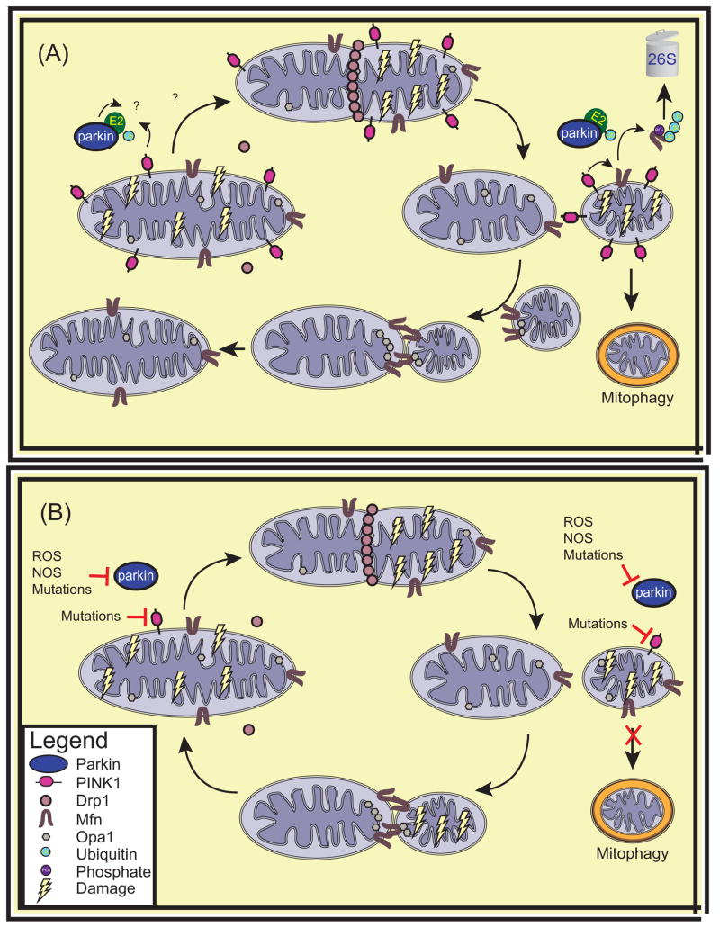
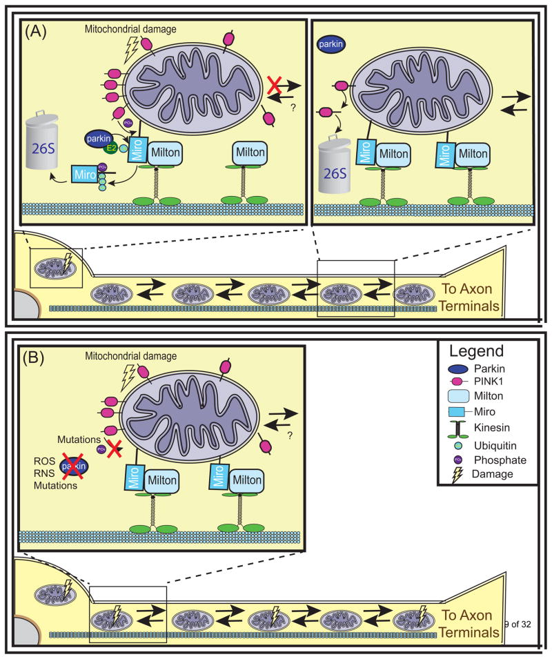
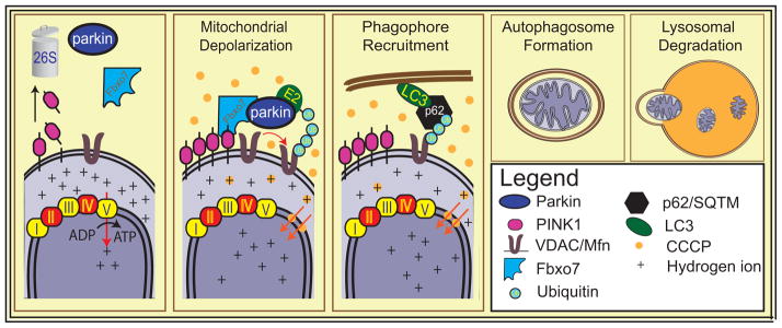
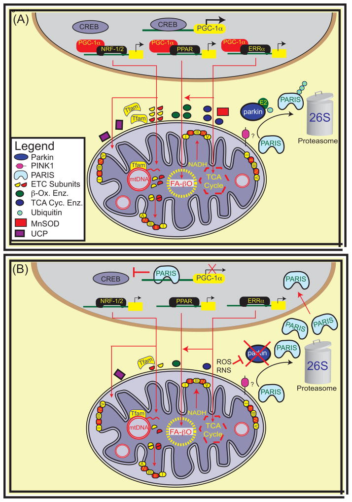
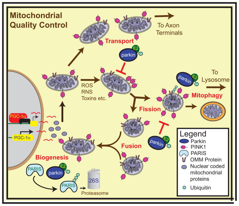
Parkinson's disease (PD) is a progressive neurodegenerative disease that causes a debilitating movement disorder. Although most cases of PD appear to be sporadic, rare Mendelian forms have provided tremendous insight into disease pathogenesis. Accumulating evidence suggests that impaired mitochondria underpin PD pathology. In support of this theory, data from multiple PD models have linked Phosphatase and tensin homolog (PTEN)-induced putative kinase 1 (PINK1) and parkin, two recessive PD genes, in a common pathway impacting mitochondrial health, prompting a flurry of research to identify their mitochondrial targets. Recent work has focused on the role of PINK1 and parkin in mediating mitochondrial autophagy (mitophagy); however, emerging evidence casts parkin and PINK1 as key players in multiple domains of mitochondrial health and quality control.


Figures

Figure 1
Figure 1. Mitochondrial Fission and Fusion
PINK1 and Parkins’ role in regulating the balance of mitochondrial fission to fusion. (a) PINK1 localized to healthy mitochondria is cleaved and exported from mitochondria to be rapidly degraded by the proteasome. Drp1 accumulates on mitochondria targeted for fission and allows mitochondria to divide. Conversely, in mitochondria destined to fuse, Mfn molecules from separate mitochondria tether mitochondria together and allow fusion of the OMM to occur. Subsequently, fusion of the IMM of the two mitochondria ensues via Opa1. PINK1 localized on damaged mitochondria is stabilized on the OMM. PINK1 phosphorylates Mfn, priming it for degradation by parkin. This results in a decreased rate of mitochondrial fusion. It is unknown whether parkin or PINK1 directly affect any of the proteins involved in mitochondrial fission. (b) In PD, the loss of PINK1 or parkin prevents cells from responding to mitochondrial damage by altering the balance of fission to fusion. Abbreviations: Ub, ubiquitin; Drp1, Dynamin related protein 1, Mfn, mitofusin; Opa1, optic atrophy one; E2, ubiquitin conjugating enzyme
Figure 2
Figure 2. Mitochondrial Transport
PINK1 and Parkin halt the transport of damaged mitochondria. (a) Mitochondria are transported along microtubules throughout the cell by interacting with kinesin motor proteins. The OMM protein Miro tethers mitochondria to kinesins via its interaction with Milton. PINK1 localized to healthy mitochondria is cleaved and exported from mitochondria to be rapidly degraded by the proteasome, allowing mitochondria to be transported throughout the cell. Conversely, PINK1 localized to severely damaged mitochondria is stabilized on the OMM where it can phosphorylate Miro. This phosphorylation primes Miro for ubiquitination by Parkin and degradation by the 26S proteasome. As a result, mitochondria dissociate from kinesins and their transport is halted. (b) In PD, the loss of PINK1 or parkin prevents cells from halting transport as a response to mitochondrial damage. Abbreviations: Ub, ubiquitin; ROS, reactive oxygen species; RNS, reactive nitric oxide species.
Figure 3
Figure 3. Mitochondrial Autophagy
Parkin mediates removal of damaged mitochondria. After widespread mitochondrial depolarization by CCCP, PINK1 is stabilized on the OMM, enabling parkin to translocate to mitochondria and ubiquitinate OMM proteins including mitofusins and VDAC. Some of these ubiquitin moieties may serve as signals to recruit autophagic machinery including HDAC6, or p62. Mitochondria are engulfed by the autophagosome, which eventually fuses with the lysosome, leading to the degradation of the dysfunctional organelles. Abbreviations: Ub, ubiquitin; Mfn, mitofusin; VDAC, valtage dependent anion channel; Fbxo7, F-box protein 7; p62/SQTM, p62 sequestome; CCCP, Carbonyl cyanide m-chlorophenyl hydrazine; LC3, light chain 3
Figure 4
Figure 4. Mitochondrial Biogenesis
Mitochondrial biogenesis involves the transcription, translation and assembly of new mitochondrial proteins and the replication of mitochondrial DNA. A common link in the coordination of cellular responses to metabolic demands and oxidative stress is the Peroxisome proliferator-activator receptor gamma co-activator (PGC) family of proteins and in particular PGC-1α. PGC-1α is often called the master regulator of mitochondrial biogenesis. The gene for PGC-1α is inducible and responsive to many cellular events leading to AMPK activation and CREB dependent transcription. PGC-1α increases expression of nuclear respiratory factor 1 (NRF-1) and estrogen response receptor (ERR) α and also catalyzes the transcriptional activity of NRF-1/2, ERR and Peroxisome proliferator-activated receptor (PPAR) families of transcription factors. Converging evidence from sporadic PD and adult-onset parkin KO mice implicate transcriptional repression of PGC-1α by PARIS as an important pathogenic mechanism in dopamine neuron degeneration downstream of parkin loss [66]. Abbreviations: Tfam, Mitochondrial transcription factor A; CREB, cAMP responsive element binding protein, mtDNA, mitochondrial DNA; Enz, enzyme; ETC, electron transport subunits; β-Ox, β-oxidation; TCA Cyc, tricarboxylic acid cycle; MnSOD, manganese superoxide dismutase; UCP, mitochondrial uncoupling proteins; NADH, Nicotinamide adenine dinucleotide; FA-βO; fatty acid β-oxidation.
Figure 5
Figure 5. Mitochondrial Quality Control
Parkin and PINK1 regulate mitochondrial quality control. PINK1 and parkin modulate mitochondrial dynamics both by tipping the fission-fusion balance towards fission and by stalling the transport of damaged mitochondria. PINK1 and parkin may regulate the turnover of mitochondria by recruiting the autophagic machinery to damaged mitochondria and allowing mitophagy to occur, while also playing an important role in the production of novel mitochondrial components by biogenesis. Abbreviations: PGC-1α, Peroxisome proliferator-activator receptor gamma co-activator 1α; ROS, reactive oxygen species; RNS, reactive nitric oxide species; E2, ubiquitin conjugating enzyme; Ub, ubiquitin; OMM, outer mitochondrial membrane.

References

    1. Dawson TM, Dawson VL. Molecular pathways of neurodegeneration in Parkinson’s disease. Science. 2003;302:819–822. - PubMed
    1. Exner N, et al. Mitochondrial dysfunction in Parkinson’s disease: molecular mechanisms and pathophysiological consequences. The EMBO journal. 2012;31:3038–3062. - PMC - PubMed
    1. Schon EA, Przedborski S. Mitochondria: the next (neurode)generation. Neuron. 2011;70:1033–1053. - PMC - PubMed
    1. Corti O, et al. What genetics tells us about the causes and mechanisms of Parkinson’s disease. Physiol Rev. 2011;91:1161–1218. - PubMed
    1. Martin I, et al. Recent advances in the genetics of Parkinson’s disease. Annu Rev Genomics Hum Genet. 2011;12:301–325. - PMC - PubMed

Publication types

MeSH terms