. 2014 Apr 14;29(1):88-101.
doi: 10.1016/j.devcel.2014.02.005.

An enhancer composed of interlocking submodules controls transcriptional autoregulation of suppressor of hairless

Affiliations

An enhancer composed of interlocking submodules controls transcriptional autoregulation of suppressor of hairless

Feng Liu et al. Dev Cell. .

Abstract

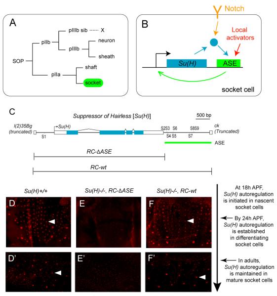
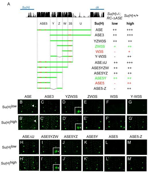
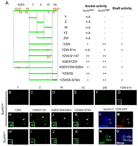
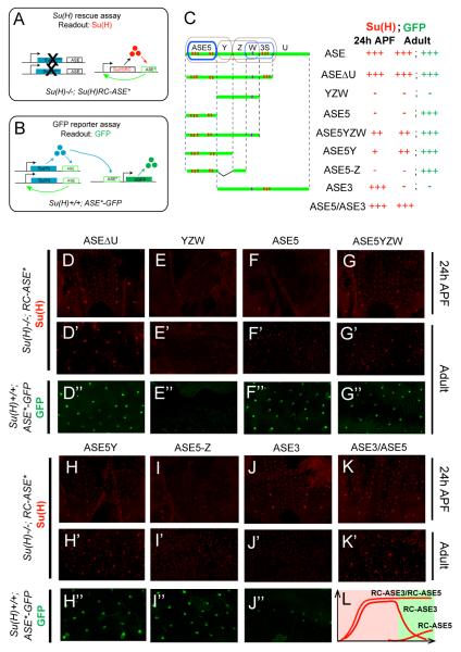
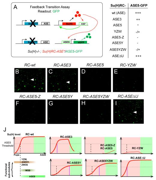
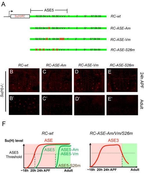
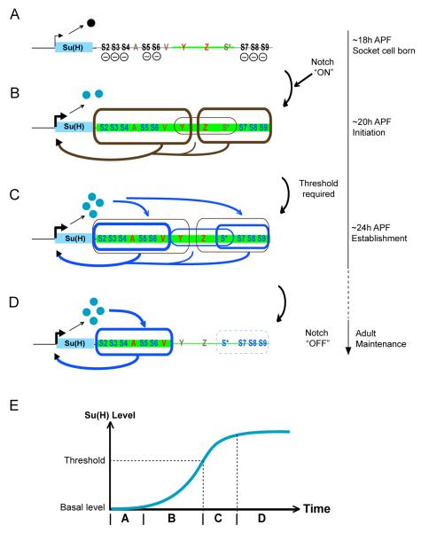
Positive autoregulation is an effective mechanism for the long-term maintenance of a transcription factor's expression. This strategy is widely deployed in cell lineages, where the autoregulatory factor controls the activity of a battery of genes that constitute the differentiation program of a postmitotic cell type. In Drosophila, the Notch pathway transcription factor Suppressor of Hairless activates its own expression, specifically in the socket cell of external sensory organs, via an autoregulatory enhancer called the ASE. Here, we show that the ASE is composed of several enhancer submodules, each of which can independently initiate weak Su(H) autoregulation. Cross-activation by these submodules is critical to ensure that Su(H) rises above a threshold level necessary to activate a maintenance submodule, which then sustains long-term Su(H) autoregulation. Our study reveals the use of interlinked positive-feedback loops to control autoregulation dynamically and provides mechanistic insight into initiation, establishment, and maintenance of the autoregulatory state.


Figures

Figure 1
Figure 1. The ASE Controls Transcriptional Autoactivation of Su(H) in the Socket Cell
(A) Lineage of Drosophila adult external mechanosensory organs. The socket cell is highlighted in green. SOP, sensory organ precursor cell. (B) Su(H) autoactivates its expression specifically in the socket cell via a dedicated cis-regulatory module, the autoregulatory socket enhancer (ASE) (Barolo et al., 2000). The socket cell-specific activation of the ASE is dependent on synergies between Notch signaling, via Su(H), and inputs from other activators in the sensory organ lineage (Barolo et al., 2000). (C) Diagram of the Su(H) gene. The ASE is included within a 1.9-kb genomic segment located downstream of Su(H), and contains eight high-affinity Su(H) binding sites (S2-S9) (Barolo et al., 2000). The RC-wt genomic DNA fragment fully rescues all known functions of Su(H) when placed in a Su(H) null mutant background; RC-ΔASE lacks the ASE, and hence the autoregulatory activity of the gene, but rescues all other functions, including the broad basal level of Su(H) expression (see D-F and D’-F’) (Barolo et al., 2000). (D-F’) Anti-Su(H) antibody (red) marks the high level of the protein in socket cells in the pupal notum at 24 hours after puparium formation (APF) (D-F) and in the abdominal epithelium of adult flies (D’-F’). Note the lack of strong staining in the Su(H)−/−; RC-ΔASE genotype (E-E’). Individual socket cells are indicated by arrowheads. Su(H)−/− refers to the null genotype Su(H)AR9/Su(H)SF8 (Barolo et al., 2000; Schweisguth and Posakony, 1992). The relationship between the imaging timepoints and the development of microchaete socket cells is described to the right of these panels.
Figure 2
Figure 2. Identification of Functional Sequence Elements of the ASE in the Nascent Socket Cell
(A) The pattern of conservation of ASE sequences in Drosophila species, as displayed by the UCSC genome browser (genome.ucsc.edu), is shown at the top. Diagrams of ASE fragments (green lines) tested in GFP reporter transgene constructs are shown below. Previously identified Su(H) binding sites are marked in red. Observed levels of GFP expression in nascent socket cells are summarized at right. Reporter gene activities were assayed in two genetic backgrounds: Wild type [Su(H)+/+ or Su(H)high] and Su(H)−/−; RC-ΔASE [only the basal level of Su(H), or Su(H)low]. GFP levels were scored using the following semi-quantitative system: strong +++, moderate ++, weak +, weak stochastic −/ +, and negative −. Two comparisons (ZW3S/ W3S and ASE5Y/ ASE5) are highlighted in green/ red type. (B-M) GFP expression in the pupal notum at 24 hours APF; Su(H)low background, lacking the autoregulatory activity of Su(H). (B’-M’) GFP expression in the pupal notum at 24 hours APF; wild-type [Su(H)high] background. Arrowheads in B and B’ mark the positions of single mechanosensory organs. Insets in D, D’, I, and I’ show socket cell (<) specificity of GFP expression.
Figure 3
Figure 3. The ASE is Activated by the Synergistic Function of a Low Level of Su(H) and Weak Local Activators
(A) Diagrams of ASE fragments (green lines) tested in GFP reporter transgene constructs in the pupal notum at 24 hours APF. Previously identified Su(H) binding sites (S2-S9) are marked in red; a Su(H) binding site, S*, identified here is marked in purple. Mutated Su(H) binding sites are indicated by “x”. Observed levels of GFP expression are summarized at right, using the same semi-quantitative scoring system as in Figure 2. “n.d.” means not determined. (B-K) GFP expression in the pupal notum at 24 hours APF in the wild-type [Su(H)high] background. Arrowheads in H-K mark the positions of single mechanosensory organs. (H’-K’) GFP expression in the pupal notum at 24 hours APF in the mutant [Su(H)low] background [Su(H)−/−; RC-ΔASE] lacking the autoregulatory activity of Su(H). Insets in G-K and H’-K’ show higher-magnification views of GFP expression at single mechanosensory organ positions. Cells displaying GFP signal are marked (<). (L-O) The YZW fragment of the ASE drives weak expression in both the shaft (Sh) and socket (So) cells (denoted by <). Shown is a single developing mechanosensory organ in the pupal notum at 20 hours APF in the wild-type background. (L) The cells of the mechanosensory organ lineage (see Figure 1A) are marked by anti-D-Pax2 antibody (blue); the strongest signal is in the shaft cell (Kavaler et al., 1999). (M) The cells of the pIIb branch of the lineage are marked by anti-Prospero (Pros) antibody (red). (N) Anti-GFP antibody (green) marks the activity of the YZW-GFP transgene. (O) Merged three-channel image. The socket and shaft cells are distinguished by their enlarged nuclei (due to endoreplication) and lack of anti-Prospero reactivity. See also Figures S1, S2, and S3.
Figure 4
Figure 4. The ASE’s Sub-modules Dynamically Control Su(H) Autoregulation
(A) The Su(H) transgene rescue assay. Left: The endogenous Su(H) autoregulatory loop is disrupted by Su(H) null mutations (X’s). Right: The Su(H) gene [Su(H)RC] and ASE* (wild-type ASE or fragment thereof) carried in the rescue transgene construct form a positive feedback loop. Anti-Su(H) antibody staining is used as the readout, indicating whether ASE* is sufficient to establish Su(H) autoregulation in the socket cell. (B) GFP reporter transgene assay in the wild-type background. Once the endogenous ASE-Su(H) feedback loop is established (left), its output [Su(H)] acts on the ASE fragment (ASE*) carried in the reporter transgene construct to drive GFP expression (right). GFP fluorescence in adult flies is used as the readout, indicating whether ASE* is able to respond to the high maintenance levels of Su(H). (C) Diagrams of ASE fragments (green lines) tested in both the Su(H) transgene rescue and GFP reporter transgene assays. Sub-modules of the ASE are encircled: Brown indicates a sub-module active in the Su(H)low background in pupal stages; blue indicates a sub-module active only in the Su(H)high background in pupal stages. Observed levels of Su(H) and GFP expression are summarized at right, using the same semi-quantitative scoring system as in Figure 2. (D-K) Su(H) rescue assay: Anti-Su(H) antibody staining in the pupal notum at 24 hours APF. (D’-K’) Su(H) rescue assay: Anti-Su(H) antibody staining in the adult abdominal epithelium. (D”-J”) GFP reporter assay: GFP expression in socket cells in the adult abdominal epithelium. (L) Cartoon illustration of Su(H) expression in the Su(H) rescue assays using RC-ASE3, RC-ASE5, or RC-ASE3/RC-ASE5. Pink panel indicates pupal stage; green panel indicates adult stage; red lines indicate levels of Su(H) expression (relative levels are arbitrary). See also Figure S4.
Figure 5
Figure 5. The Transition From Initiation to Maintenance of Su(H) Autoregulation Depends on Interlinked Functions of the ASE’s Sub-modules
(A) Diagram of the feedback transition assay. One copy each of a Su(H) rescue construct and the ASE5-GFP reporter construct, both on the third chromosome, are placed into the Su(H) null background. Observed levels of GFP expression are summarized at right, using the same semi-quantitative scoring system as in Figure 2. (B-I) Activity of the ASE5-GFP reporter in the pupal notum at 28 hours APF in the Su(H)−/−; RC-ASE* background. Arrowheads in B, C, and I indicate single microchaete socket cells that express high levels of GFP. (J) Cartoon illustrations of the time window s in which fragments of the ASE are active in development. Pink panels indicate pupal stage; green panels indicate adult stage; red lines represent levels of Su(H) expression. Su(H) autoregulation is established in the pupal notum between 18 and 24 hours APF, and persists into adulthood. The dashed vertical line marks the time (24 hours APF) when ASE5 is normally activated by an above-threshold level of Su(H); this threshold is indicated by the dashed horizontal line. In each illustration, the purple vertical line marks the suggested time (if ever) when the indicated Su(H) rescue transgene reaches the threshold required to activate ASE5. See also Figures S5 and S6.
Figure 6
Figure 6. ASE5 is Combinatorially Activated in Pupal Stages to Maintain Su(H) Autoregulation in Adults
(A) Diagrams of Su(H) rescue constructs carrying point mutations in the ASE5 sub-module of the ASE. Note that the ASE5 region has been expanded out of proportion to the rest of the ASE in order to illustrate the sequence motif composition of ASE5. Red X’s indicated mutated motifs. (B-E) Anti-Su(H) antibody staining in the pupal notum at 24 hours APF. (B’-E’) Anti-Su(H) antibody staining in the adult abdominal epithelium. (F) (Left) Cartoon summary of the effects of mutating the A motif or the Vvl sites on the function of an ASE5-GFP reporter (green lines) in a wild -type background (Liu and Posakony, 2012). In the presence of normal levels of Su(H) (red line), ASE5 normally becomes active in the socket cell of the microchaetes in the pupal notum between 20 and 24 hours APF; ASE5-Am and ASE5-Vm are not active until pre-pharate adult stages; and ASE5-S26m is silent throughout development. (Right) Based on these prior results, on those shown in Figures 4J-4K’, and on those shown in B-E’, we suggest that Su(H) expression directed by RC-ASE-Am, RC-ASE-Vm, and RC-ASE-S26m is due to ASE3 (red line). Like RC-ASE3 (see Figures 4J-4J’), these constructs all generate very substantial levels of Su(H) in pupal stages (C-E), but this early expression is not maintained into adults (C’-E’). We suggest that this is because by the time ASE3 becomes inactive, ASE5 has not been activated in time (RC-ASE-Am and RC-ASE-Vm) or has not been activated at all (RC-ASE-S26m).
Figure 7
Figure 7. Model of How the ASE Integrates Multiple Feedback Loops to Control Su(H) Autoregulation in the Differentiating Socket Cell
(A-D) Diagrams depicting the dynamic interaction between the ASE and its functional inputs in the socket cell. The timeline at right refers to the developmental stages of microchaete socket cells in the pupal notum. The body of the Su(H) gene is represented as a blue box; the accompanying arrow shows the direction of transcription, with its thickness denoting the intensity of transcriptional activity in the socket cell. The ASE is shown downstream of the gene, with functionally important sequence motifs and sub-regions labeled. Segments of the ASE receiving local activator inputs are highlighted in green, with the input level indicated by the thickness of the green line. The repressor form of Su(H) is shown as a black ball, the activator form as blue balls. (A) Shortly after the socket cell is born, the ASE receives local activator inputs via the sequences in YZW (thin green line, red Y and Z), but is silenced by the default repression function of Su(H), acting via binding sites S2-S9 (—). (B) Activation of the Notch pathway in the socket cell converts Su(H) into a transcriptional activator (Barolo et al., 2000). Su(H) and various local activator inputs selectively synergize to activate two sub-modules, ASE5Y and ZW3S (brown boxes, thick lines), which drive an increase in Su(H) transcription (thick brown arrows). YZW (brown box, thin line) may also contribute independently to this activity (thin brown arrow). (C) Once Su(H) expression rises above a threshold level, two Su(H) response elements, ASE5 and W3S (blue boxes, thick lines), are activated to fully establish the ASE-Su(H) feedback loop (blue arrows). Activation of ASE5 and W3S depend s in part on other local activator inputs; in particular, ASE5 receives two activator inputs via a single A motif (red A) and multiple binding sites for Vvl (red V), a POU-homeodomain transcription factor (Liu and Posakony, 2012). (D) As the socket cell differentiates, the local activator inputs on YZW disappear (brown Y and Z) and W3S is silenced (blue box, dashed line), but the activator inputs on ASE5 persist into adulthood, keeping ASE5 active to maintain Su(H) autoregulation (blue arrows). See also Figure S7. (E) The dynamic pattern of activation of the ASE’s sub-modules progressively establishes Su(H) autoregulation in the socket cell. Letters on the time axis refer to stages A-D above.

Similar articles

Cited by

References

    1. Bailey AM, Posakony JW. Suppressor of Hairless directly activates transcription of Enhancer of split Complex genes in response to Notch receptor activity. Genes Dev. 1995;9:2609–2622. - PubMed
    1. Barolo S, Walker RG, Polyanovsky AD, Freschi G, Keil T, Posakony JW. A Notch-independent activity of Suppressor of Hairless is required for normal mechanoreceptor physiology. Cell. 2000;103:957–969. - PubMed
    1. Ben-Tabou de-Leon S, Davidson EH. Information processing at the foxa node of the sea urchin endomesoderm specification network. Proc Natl Acad Sci USA. 2010;107:10103–10108. - PMC - PubMed
    1. Bischof J, Maeda RK, Hediger M, Karch F, Basler K. An optimized transgenesis system for Drosophila using germ-line-specific phiC31 integrases. Proc. Natl. Acad. Sci. USA. 2007;104:3312–3317. - PMC - PubMed
    1. Brandman O, Ferrell JEJ, Li R, Meyer T. Interlinked fast and slow positive feedback loops drive reliable cell decisions. Science. 2005;310:496–498. - PMC - PubMed

Publication types

LinkOut - more resources