. 2014 Jun 1;306(11):E1264-73.
doi: 10.1152/ajpendo.00438.2013. Epub 2014 Apr 15.

Hepatic mitochondrial and ER stress induced by defective PPARα signaling in the pathogenesis of hepatic steatosis

Affiliations

Hepatic mitochondrial and ER stress induced by defective PPARα signaling in the pathogenesis of hepatic steatosis

Qiaozhu Su et al. Am J Physiol Endocrinol Metab. .

Abstract

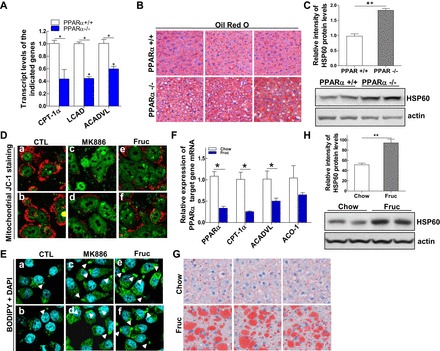
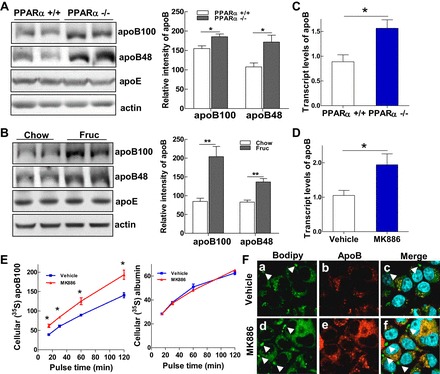
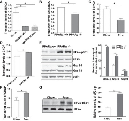
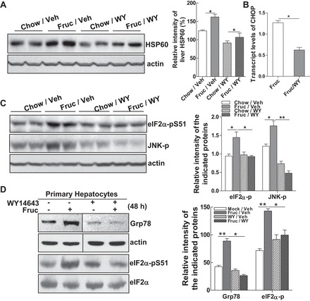
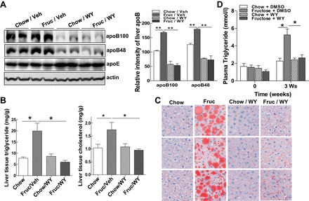
Emerging evidence demonstrates a close interplay between disturbances in mitochondrial function and ER homeostasis in the development of the metabolic syndrome. The present investigation sought to advance our understanding of the communication between mitochondrial dysfunction and ER stress in the onset of hepatic steatosis in male rodents with defective peroxisome proliferator-activated receptor-α (PPARα) signaling. Genetic depletion of PPARα or perturbation of PPARα signaling by high-fructose diet compromised the functional activity of metabolic enzymes involved in mitochondrial fatty acid β-oxidation and induced hepatic mitochondrial stress in rats and mice. Inhibition of PPARα activity further enhanced the expression of apolipoprotein B (apoB) mRNA and protein, which was associated with reduced mRNA expression of the sarco/endoplasmic reticulum calcium ATPase (SERCA), the induction of hepatic ER stress, and hepatic steatosis. Restoration of PPARα activity recovered the metabolic function of the mitochondria and ER, alleviated systemic hypertriglyceridemia, and improved hepatic steatosis. These findings unveil novel roles for PPARα in mediating stress signals between hepatic subcellular stress-responding machinery and in the onset of hepatic steatosis under conditions of metabolic stress.

Keywords: apolipoprotein B; endoplasmic reticulum; hepatic steatosis; mitochondrial and endoplasmic reticulum stress; peroxisome proliferator-activated receptor-α; very-low density lipoprotein.


Figures

Fig. 1.
Fig. 1.
Impairment of peroxisome proliferator-activated receptor-α (PPARα) signaling disrupts hepatic mitochondrial integrity and induces mitochondrial stress. A: livers from wild-type (WT) (+/+) and PPARα-knockout (−/−) male mice at the age of 12 wk were used to prepare total RNA to detect mRNA expression of PPARα target genes carnitine palmitoyltransferase-1α (CPT-1α), long-chain acyl-CoA dehydrogenase (LCAD), and acetyl-CoA dehydrogenase very-long chain (ACADVL) by quantitative (q)RT-PCR. B: representative photographs demonstrating the appearance of livers from 12-wk-old PPARα+/+ (top) or PPARα−/− (bottom) male mice. The livers were harvested and frozen liver sections subjected to staining with the lipid-specific Oil Red O dye to positively reveal lipid droplets. C: protein mass of heat shock protein 60 (HSP60) detected by immunoblot analysis in the livers of PPARα+/+ and PPARα−/− mice. D: primary mouse hepatocytes treated for 24 h with either DMSO (0.5%), MK-886 (20 μM), or fructose (12 μM) were stained with JC-1 staining reagent for 15 min at 37°C and subjected to confocal analysis. Healthy cells (mainly JC-1 aggregates) appear red; unhealthy/apoptotic cells (mainly JC-1 monomers) appear green. E: McA cells were untreated or treated with PPARα antagonist MK-886 (10 μM) or fructose (6 mM) for 48 h and then stained with BODIPY for confocal imaging to visualize lipid droplets (green) and 4,6-diamidino-2-phenylindole (DAPI) for nuclear staining (blue). F: livers from rats after 3 wk of feeding with chow or fructose diets were used to prepare total RNA to measure relative mRNA expression of PPARα and its target genes CPT-1α, ACADVL, and acyl-CoA oxidase-1 (ACO-1) by qRT-PCR. G: livers from rats fed chow (Chow) or fructose (Fruc) for 3 wk were harvested, and frozen liver sections were subjected to staining with the lipid-specific Oil Red O dye to positively reveal lipid droplets. H: protein mass of HSP60 detected by immunoblot analysis in the livers of rats fed Chow or Fruc for 3 wk. For animal studies, results are shown as means ± SE (n = 5–6/group). For in vitro studies, data represent 3 experiments. *P < 0.05; **P < 0.01 vs. controls (CTL).
Fig. 2.
Fig. 2.
Impairment of PPARα activity augments hepatic VLDL biogenesis. A and B: PPARα+/+ and PPARα−/− mice were fasted for 5 h, and blood collection was performed every hour after ip injection of poloxamer. Triglyceride (TG) and cholesterol (CHOL) levels were assessed at 0, 1, 2 and 3 h (A), and VLDL TG was assessed at 3 h (B). C: McA cells (1 × 10⁶) were untreated or treated with MK-866 (10 μM) for 48 h, deprived of methionine and cysteine for 1 h in the presence or absence of MK-866 (10 μM), and then pulsed with 100 μCi/ml [35S]methionine in the presence of oleate (400 μM) for 3 h. Media were collected and subjected to fractionation by ultracentrifugation, fractions were collected, and 35S-labeled apolipoprotein B (apoB)-100 and 35S-labeled apolipoprotein E (apoE) were immunoprecipitated with anti-apoB and anti-apoE antibodies, respectively. For animal studies, results are shown as means ± SE (n = 5–6/group). For in vitro studies, data represent 3 experiments. *P < 0.05 vs. controls.
Fig. 3.
Fig. 3.
PPARα signaling transcriptionally regulates hepatic apoB synthesis. A and B: immunoblot analysis of apoB-100, apoB-48, apoE, and actin in liver homogenates of PPARα+/+ and PPARα−/− mice (A) and rats fed chow or fructose for 3 wk (B). C and D: relative mRNA expression of apoB detected by qRT-PCR in livers of PPARα+/+ and PPARα−/− mice (C) and McA cells treated with MK-886 (10 μM) for 48 h (D). E: McA cells (1 × 10⁶) were untreated or treated with MK-866 (10 μM) for 48 h. Cells were then deprived of methionine and cysteine for 1 h in the presence or absence of MK-866 (10 μM) and pulsed with 100 μCi/ml [35S]methionine for 15, 30, 60 and 120 min. Cell lysates were prepared, and cellular 35S-labeled apoB-100 was immunoprecipitated with an anti-apoB antibody, followed by immunoprecipitation of 35S-labeled albumin, with an anti-albumin antibody as control. F: McA cells were untreated or treated with MK-886 (10 μM) for 48 h and then stained for confocal imaging with anti-apoB antibody (red), BODIPY to visualize lipid droplets (green), and DAPI for nuclear staining (blue). Colocalization of apoB with lipid droplets is shown in yellow. For animal studies, results are shown as means ± SE (n = 5–6/group). For in vitro studies, data represent 3 experiments. *P < 0.05 and **P < 0.01 vs. controls.
Fig. 4.
Fig. 4.
Accumulation of apoB in the hepatic endoplasmic reticulum (ER) induces ER stress. A: total mRNA was extracted from McA cells either containing a control vector or stably expressing a truncated form of apoB WT or mutant, and mRNA transcripts of the sarcoendoplasmic reticulum ATPase (SERCA) gene were measured. B and C: relative mRNA expression of SERCA detected by qRT-PCR in livers of PPARα+/+ and PPARα−/− mice (B) and rats fed Chow or Fruc diets for 3 wk (C). D: relative mRNA expression of C/EBP homologous protein (CHOP) detected by qRT-PCR in livers of PPARα+/+ and PPARα−/− mice. E: immunoblot analysis of the ER stress markers phospho-eukaryotic initiation factor-2α (eIF2α), Grp94, or Grp78 in livers of PPARα+/+ and PPARα−/− mice and quantification of their signal intensity. F: relative mRNA expression of CHOP detected by qRT-PCR in livers of rats fed Chow or Fruc diets for 3 wk. G: immunoblot analysis of phospho-eIF2α and its total protein in livers of rats fed Chow or Fruc diet for 3 wk. For animal studies, results are shown as means ± SE (n = 5–6/group). For in vitro studies, data represent 3 experiments. *P < 0.05; **P < 0.01 vs. controls.
Fig. 5.
Fig. 5.
Restoring PPARα signaling normalizes mitochondrial and ER homeostasis. A and B: livers from rats fed Chow or Fruc and concomitantly treated with vehicle (Veh) or PPARα agonist WY-14643 (WY; 6 mg/kg ip) once every 2 days for 3 wk were used to prepare liver homogenants. A: protein mass of HSP60 detected by immunoblot analysis. B: relative mRNA expression of CHOP detected by qRT-PCR in livers of Fruc-fed rats untreated or treated with WY for 3 wk. C: immunoblot analysis of phospho-eIF2α and phospho-JNK levels. D: primary rat hepatocytes were untreated or treated with fructose (6 mM) in the presence or absence of WY (40 μM) for 48 h. Whole cell lysates were subjected to immunoblot analysis for Grp78 and phospho-eIF2α. Lanes were run on the same gels but were noncontiguous. For animal studies, results are shown as means ± SE (n = 5–6/group). For in vitro studies, data represent 3 experiments. *P < 0.05; **P < 0.01 vs. controls.
Fig. 6.
Fig. 6.
Restoration of mitochondrial and ER homeostasis improves hepatic steatosis and alleviates systemic hyperlipidemia. A: immunoblot analysis of apoB-100, apoB-48, apoE, and actin from liver homogenants of rats with conditions as shown in Fig. 5A. B: liver TG and cholesterol contents from rats in Fig. 5A were determined by procedures detailed in materials and methods. C: liver sections from rats as shown in Fig. 5A were stained with Oil Red O dye to identify neutral lipid species. D: plasma TG from rats receiving the diets and treatments as indicated for 3 wk. For animal studies, results are shown as means ± SE (n = 5–6/group). For in vitro studies, data represent 3 experiments. *P < 0.05; **P < 0.01 vs. controls.

References

    1. Aoyama T, Peters JM, Iritani N, Nakajima T, Furihata K, Hashimoto T, Gonzalez FJ. Altered constitutive expression of fatty acid-metabolizing enzymes in mice lacking the peroxisome proliferator-activated receptor alpha (PPARalpha). J Biol Chem 273: 5678–5684, 1998. - PubMed
    1. Basciano H, Miller AE, Naples M, Baker C, Kohen R, Xu E, Su Q, Allister EM, Wheeler MB, Adeli K. Metabolic effects of dietary cholesterol in an animal model of insulin resistance and hepatic steatosis. Am J Physiol Endocrinol Metab 297: E462–E473, 2009. - PubMed
    1. Bensinger SJ, Tontonoz P. Integration of metabolism and inflammation by lipid-activated nuclear receptors. Nature 454: 470–477, 2008. - PubMed
    1. Bernal-Mizrachi C, Weng S, Feng C, Finck BN, Knutsen RH, Leone TC, Coleman T, Mecham RP, Kelly DP, Semenkovich CF. Dexamethasone induction of hypertension and diabetes is PPAR-alpha dependent in LDL receptor-null mice. Nat Med 9: 1069–1075, 2003. - PubMed
    1. Choi SH, Ginsberg HN. Increased very low density lipoprotein (VLDL) secretion, hepatic steatosis, and insulin resistance. Trends Endocrinol Metab 22: 353–363, 2011. - PMC - PubMed

Publication types

MeSH terms

LinkOut - more resources