

Early alteration of retinal neurons in Aipl1-/- animals
- PMID: 24736053
- PMCID: PMC4034756
- DOI: 10.1167/iovs.13-13728
Early alteration of retinal neurons in Aipl1-/- animals
Abstract
Purpose: Mutations in the photoreceptor cell-specific gene encoding aryl hydrocarbon receptor-interacting protein-like 1 (AIPL1) lead to Leber congenital amaurosis (LCA4), retinitis pigmentosa, and cone-rod dystrophy. Gene therapy appears to be promising in the treatment for AIPL1-mediated vision loss in humans. Prior to initiating these treatments, however, it is crucial to understand how the retinal neurons remodel themselves in response to photoreceptor cell degeneration. In this study, using an animal model for AIPL1-LCA, Aipl1(-/-) mice, we investigate the changes in postreceptoral retinal neurons during the course of photoreceptor cell loss.
Methods: Morphology of the Aipl1(-/-) retina from postnatal day 8 to 150 was compared to that of age-matched, wild-type C57Bl6/J retina (WT) by immunocytochemistry using cell-specific markers.
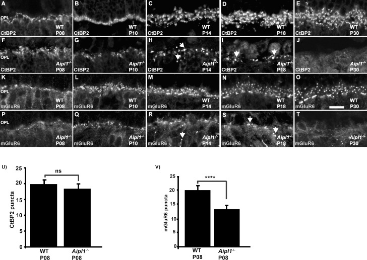
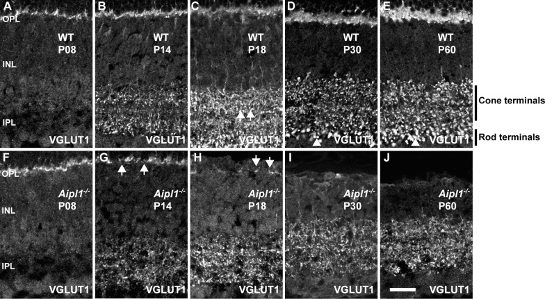
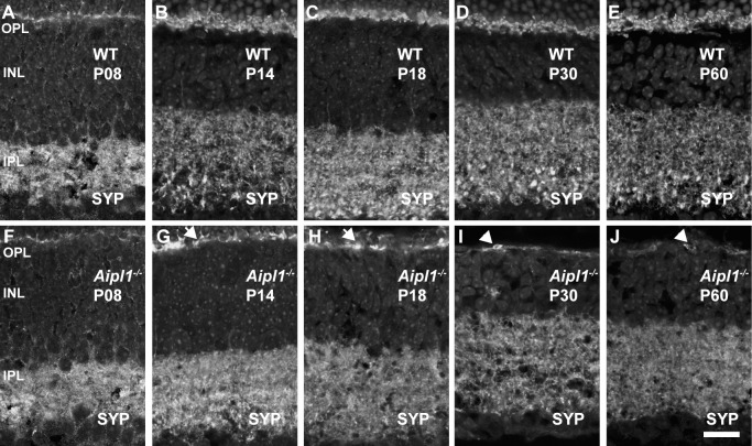
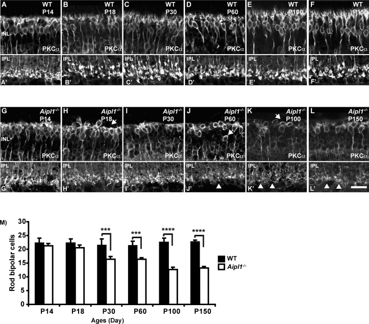
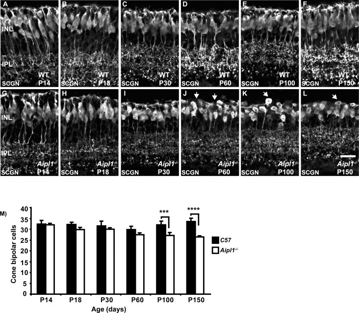
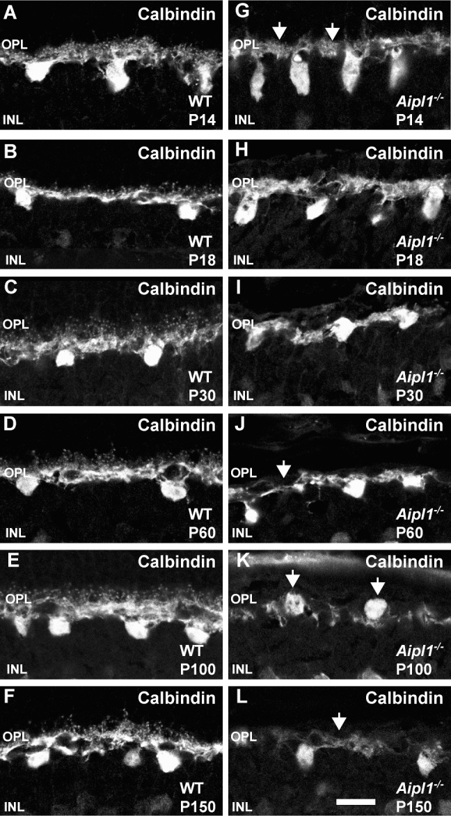
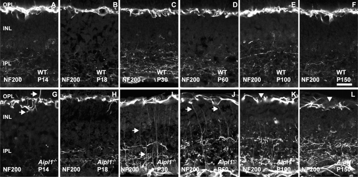
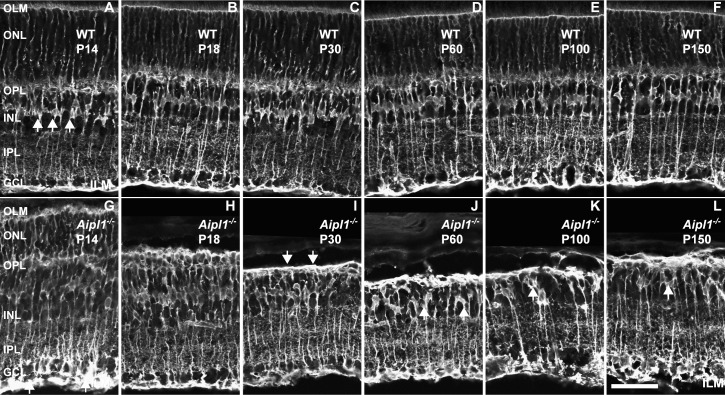
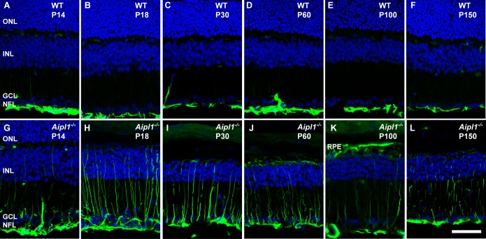
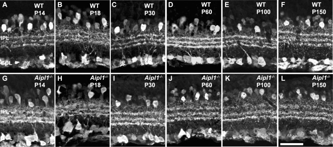
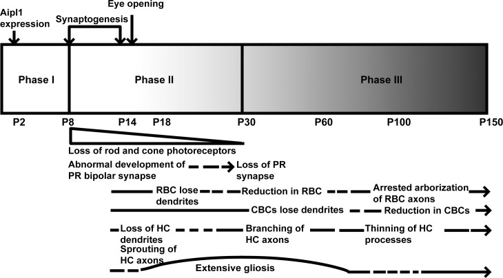
Results: Expression of postsynaptic proteins in bipolar cells is reduced prior to photoreceptor cell degeneration at postnatal day 8. Bipolar and horizontal cells retract their dendrites. Cell bodies and axons of bipolar and horizontal cells are disorganized during the course of degeneration. Müller cell processes become hypertrophic and form a dense fibrotic layer outside the inner nuclear layer.
Conclusions: An early defect in photoreceptor cells in the AIPL1-LCA mouse model affects the expression of postsynaptic markers, suggesting abnormal development of bipolar synapses. Once degeneration of photoreceptor cells is initiated, remodeling of retinal neurons in the Aipl1(-/-) animal is rapid.
Keywords: LCA; childhood blindness; photoreceptor degeneration; remodeling; retina.
Copyright 2014 The Association for Research in Vision and Ophthalmology, Inc.
Figures
Similar articles
-
Gene therapy for retinitis pigmentosa and Leber congenital amaurosis caused by defects in AIPL1: effective rescue of mouse models of partial and complete Aipl1 deficiency using AAV2/2 and AAV2/8 vectors.Hum Mol Genet. 2009 Jun 15;18(12):2099-114. doi: 10.1093/hmg/ddp133. Epub 2009 Mar 19. Hum Mol Genet. 2009. PMID: 19299492 Free PMC article.
-
The Leber congenital amaurosis protein, AIPL1, is needed for the viability and functioning of cone photoreceptor cells.Hum Mol Genet. 2010 Mar 15;19(6):1076-87. doi: 10.1093/hmg/ddp571. Epub 2009 Dec 30. Hum Mol Genet. 2010. PMID: 20042464 Free PMC article.
-
Predominant rod photoreceptor degeneration in Leber congenital amaurosis.Mol Vis. 2005 Jul 22;11:542-53. Mol Vis. 2005. PMID: 16052170
-
AIPL1 protein and its indispensable role in cone photoreceptor function and survival.Adv Exp Med Biol. 2014;801:43-8. doi: 10.1007/978-1-4614-3209-8_6. Adv Exp Med Biol. 2014. PMID: 24664679 Review.
-
Gene and Cell Therapy for AIPL1-Associated Leber Congenital Amaurosis: Challenges and Prospects.Adv Exp Med Biol. 2019;1185:97-101. doi: 10.1007/978-3-030-27378-1_16. Adv Exp Med Biol. 2019. PMID: 31884595 Review.
Cited by
-
Proteoglycan IMPG2 Shapes the Interphotoreceptor Matrix and Modulates Vision.J Neurosci. 2020 May 13;40(20):4059-4072. doi: 10.1523/JNEUROSCI.2994-19.2020. Epub 2020 Apr 7. J Neurosci. 2020. PMID: 32265257 Free PMC article.
-
Pluripotent Stem Cells for Retinal Tissue Engineering: Current Status and Future Prospects.Stem Cell Rev Rep. 2018 Aug;14(4):463-483. doi: 10.1007/s12015-018-9802-4. Stem Cell Rev Rep. 2018. PMID: 29675776 Free PMC article. Review.
-
Neuroplastin 65 deficiency leads to the impairment of visual function through affecting ribbon synapse in retina of mice.Front Cell Neurosci. 2025 May 8;19:1558334. doi: 10.3389/fncel.2025.1558334. eCollection 2025. Front Cell Neurosci. 2025. PMID: 40406567 Free PMC article.
-
Transplantation of Human Embryonic Stem Cell-Derived Retinal Tissue in the Subretinal Space of the Cat Eye.Stem Cells Dev. 2019 Sep 1;28(17):1151-1166. doi: 10.1089/scd.2019.0090. Epub 2019 Jul 22. Stem Cells Dev. 2019. PMID: 31210100 Free PMC article.
-
Comparative localization of cystathionine beta synthases and cystathionine gamma lyase in canine, non-human primate and human retina.Exp Eye Res. 2019 Apr;181:72-84. doi: 10.1016/j.exer.2019.01.007. Epub 2019 Jan 14. Exp Eye Res. 2019. PMID: 30653965 Free PMC article.
References
-
- Masland RH. The fundamental plan of the retina. Nat Neurosci. 2001; 4: 877–886 - PubMed
-
- Masland RH. Neuronal diversity in the retina. Curr Opin Neurobiol. 2001; 11: 431–436 - PubMed
-
- Jones BW, Watt CB, Frederick JM, et al. Retinal remodeling triggered by photoreceptor degenerations. J Comp Neurol. 2003; 464: 1–16 - PubMed
Publication types
MeSH terms
Substances
Grants and funding
LinkOut - more resources
Full Text Sources
Other Literature Sources
Medical
Molecular Biology Databases
Research Materials
