

Comparison of human septal nuclei MRI measurements using automated segmentation and a new manual protocol based on histology
- PMID: 24736183
- PMCID: PMC4180657
- DOI: 10.1016/j.neuroimage.2014.04.026
Comparison of human septal nuclei MRI measurements using automated segmentation and a new manual protocol based on histology
Abstract
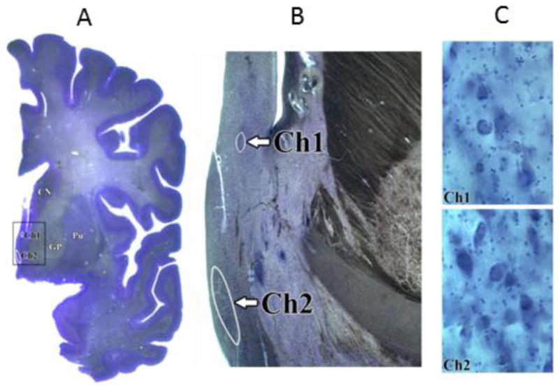
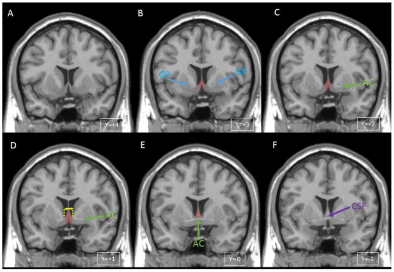
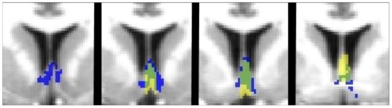
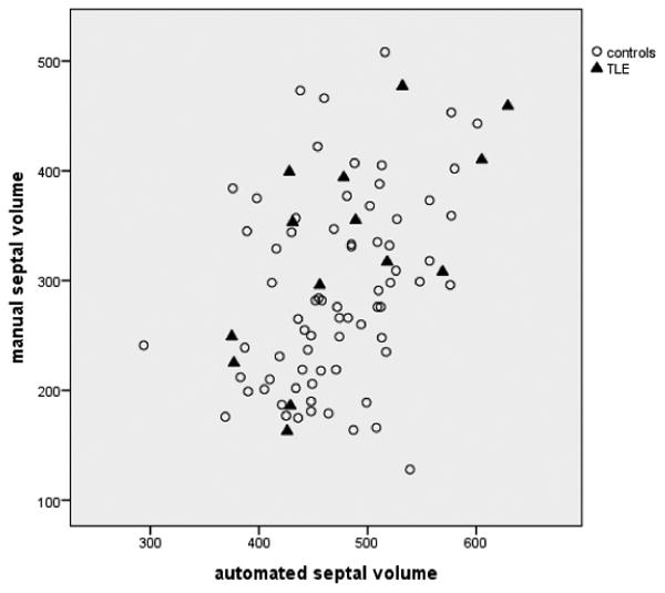
Septal nuclei, located in basal forebrain, are strongly connected with hippocampi and important in learning and memory, but have received limited research attention in human MRI studies. While probabilistic maps for estimating septal volume on MRI are now available, they have not been independently validated against manual tracing of MRI, typically considered the gold standard for delineating brain structures. We developed a protocol for manual tracing of the human septal region on MRI based on examination of neuroanatomical specimens. We applied this tracing protocol to T1 MRI scans (n=86) from subjects with temporal epilepsy and healthy controls to measure septal volume. To assess the inter-rater reliability of the protocol, a second tracer used the same protocol on 20 scans that were randomly selected from the 72 healthy controls. In addition to measuring septal volume, maximum septal thickness between the ventricles was measured and recorded. The same scans (n=86) were also analyzed using septal probabilistic maps and DARTEL toolbox in SPM. Results show that our manual tracing algorithm is reliable, and that septal volume measurements obtained via manual and automated methods correlate significantly with each other (p<.001). Both manual and automated methods detected significantly enlarged septal nuclei in patients with temporal lobe epilepsy in accord with a proposed compensatory neuroplastic process related to the strong connections between septal nuclei and hippocampi. Septal thickness, which was simple to measure with excellent inter-rater reliability, correlated well with both manual and automated septal volume, suggesting it could serve as an easy-to-measure surrogate for septal volume in future studies. Our results call attention to the important though understudied human septal region, confirm its enlargement in temporal lobe epilepsy, and provide a reliable new manual delineation protocol that will facilitate continued study of this critical region.
Copyright © 2014 Elsevier Inc. All rights reserved.
Figures
References
-
- Amaral DG, Cowan WM. Subcortical afferents to the hippocampal formation in the monkey. Journal of Comparative Neurology. 1980;189:573–591. - PubMed
-
- Andy OJ, Stephan H. The septum in the human brain. J Comp Neurol. 1968;133:383–410. - PubMed
-
- Ashburner J. A fast diffeomorphic image registration algorithm. NeuroImage. 2007;38:95–113. - PubMed
-
- Brisch R, Bernstein HG, Krell D, Stauch R, Trübner K, Dobrowolny H, Kropf S, Bielau H, Bogerts B. Volumetric analysis of septal region in schizophrenia and affective disorder. European archives of psychiatry and clinical neuroscience. 2007;257:140–148. - PubMed
-
- Butler T, Blackmon K, Zaborszky L, Wang X, DuBois J, Carlson C, Barr WB, French J, Devinsky O, Kuzniecky R, Halgren E, Thesen T. Volume of the Human Septal Forebrain Region Is a Predictor of Source Memory Accuracy. Journal of the International Neuropsychological Society. 2012;18:157–161. - PMC - PubMed
Publication types
MeSH terms
Grants and funding
- R01 AG022374/AG/NIA NIH HHS/United States
- M01 RR000096/RR/NCRR NIH HHS/United States
- K23 NS057579/NS/NINDS NIH HHS/United States
- NS023945/NS/NINDS NIH HHS/United States
- M01RR0096/RR/NCRR NIH HHS/United States
- RF1 NS023945/NS/NINDS NIH HHS/United States
- R01AG022374/AG/NIA NIH HHS/United States
- R01AG12101/AG/NIA NIH HHS/United States
- P30 AG008051/AG/NIA NIH HHS/United States
- UL1TR000038/TR/NCATS NIH HHS/United States
- R01 AG012101/AG/NIA NIH HHS/United States
- P30AG008051/AG/NIA NIH HHS/United States
- R01 NS023945/NS/NINDS NIH HHS/United States
- NS057579/NS/NINDS NIH HHS/United States
- R24-MH 068855/MH/NIMH NIH HHS/United States
- R24 MH068855/MH/NIMH NIH HHS/United States
LinkOut - more resources
Full Text Sources
Other Literature Sources
Medical
Miscellaneous
