. 2014 Apr 15;5(2):e00896.
doi: 10.1128/mBio.00896-14.

A new group of phage anti-CRISPR genes inhibits the type I-E CRISPR-Cas system of Pseudomonas aeruginosa

A new group of phage anti-CRISPR genes inhibits the type I-E CRISPR-Cas system of Pseudomonas aeruginosa

April Pawluk et al. mBio. .

Abstract

CRISPR-Cas systems are one of the most widespread phage resistance mechanisms in prokaryotes. Our lab recently identified the first examples of phage-borne anti-CRISPR genes that encode protein inhibitors of the type I-F CRISPR-Cas system of Pseudomonas aeruginosa. A key question arising from this work was whether there are other types of anti-CRISPR genes. In the current work, we address this question by demonstrating that some of the same phages carrying type I-F anti-CRISPR genes also possess genes that mediate inhibition of the type I-E CRISPR-Cas system of P. aeruginosa. We have discovered four distinct families of these type I-E anti-CRISPR genes. These genes do not inhibit the type I-F CRISPR-Cas system of P. aeruginosa or the type I-E system of Escherichia coli. Type I-E and I-F anti-CRISPR genes are located at the same position in the genomes of a large group of related P. aeruginosa phages, yet they are found in a variety of combinations and arrangements. We have also identified functional anti-CRISPR genes within nonprophage Pseudomonas genomic regions that are likely mobile genetic elements. This work emphasizes the potential importance of anti-CRISPR genes in phage evolution and lateral gene transfer and supports the hypothesis that more undiscovered families of anti-CRISPR genes exist. Finally, we provide the first demonstration that the type I-E CRISPR-Cas system of P. aeruginosa is naturally active without genetic manipulation, which contrasts with E. coli and other previously characterized I-E systems. IMPORTANCE The CRISPR-Cas system is an adaptive immune system possessed by the majority of prokaryotic organisms to combat potentially harmful foreign genetic elements. This study reports the discovery of bacteriophage-encoded anti-CRISPR genes that mediate inhibition of a well-studied subtype of CRISPR-Cas system. The four families of anti-CRISPR genes described here, which comprise only the second group of anti-CRISPR genes to be identified, encode small proteins that bear no sequence similarity to previously studied phage or bacterial proteins. Anti-CRISPR genes represent a newly discovered and intriguing facet of the ongoing evolutionary competition between phages and their bacterial hosts.


Figures

FIG 1
FIG 1
Anti-CRISPR phages encode type I-F anti-CRISPR genes and other uncharacterized open reading frames at a conserved genomic location. All of the phages shown are similar to E. coli phage Mu in terms of organization of this genomic region. The anti-CRISPR region is found between the gene homologous to gene G from Mu (black boxes) and the protease/scaffold gene (gray boxes). The genes included in the anti-CRISPR region are represented by colored boxes. The genes with the letter F in a white circle were previously shown to have anti-CRISPR activity against the type I-F CRISPR-Cas system of P. aeruginosa strain PA14. Genes sharing high sequence identity are indicated by boxes of the same color, and the percent identity of the encoded proteins is shown. The gene boxes and intergenic gaps are drawn proportionally according to the size marker shown. The 3′ conserved gene homologous to JBD30-36 is present in all anti-CRISPR-encoding phages, and all homologs shown are greater than 90% identical in amino acid sequence. The related P. aeruginosa phage JBD25 and E. coli phage Mu lacking the anti-CRISPR region are shown at the bottom of the figure for comparison.
FIG 2
FIG 2
The type I-E CRISPR-Cas systems of P. aeruginosa strains SMC4386 and SMC4389 are active. (A) The CRISPR loci from strains SMC4386 and SMC4389, sequenced by Cady et al. (10), are shown. CRISPR repeat sequences (black diamonds) and CRISPR spacers (white boxes) are indicated. CRISPR 1 and CRISPR 2 loci are separated by a line segment, representing the location of the cas genes between the two loci. The direction of transcription for both loci is from left to right. The following three protospacer sequences were constructed: PS1, corresponding to SMC4386 CRISPR 1 spacer 3 (CR1_sp3) and SMC4389 CR1_sp4; PS2, corresponding to SMC4386 CR2_sp7 and SMC4389 CR2_sp11; and PS3, corresponding to SMC4386 CR2_sp8 and containing no match within SMC4389 CRISPR loci. (B) Representative CRISPR RNA (crRNA), protospacer (gray box), and protospacer adjacent motif (PAM) (black box) are shown. (C) Transformation efficiencies of the PS1, PS2, and PS3 plasmids. Here, transformation efficiency is defined as the number of transformants per nanogram of the protospacer construct, represented as a percentage of the number of transformants per nanogram of empty vector in the same experiment. Error bars represent standard deviations of three biological replicates. (D) The PAM recognition specificity of the SMC4386 type I-E CRISPR-Cas system is assessed. The transformation efficiency of the PS2 protospacer flanked by the PAM sequence indicated on the x axis is shown. Error bars represent standard deviations of three biological replicates.
FIG 3
FIG 3
P. aeruginosa SMC4386 lysogenic strains display type I-E anti-CRISPR activity that is mediated by four unique genes. (A) Transformation efficiency assays using the PS2 construct with CTT PAM in wild-type (WT) SMC4386 or SMC4386 lysogens of the indicated anti-CRISPR phages are shown. The transformation efficiency of the protospacer/PAM construct is normalized to that of the empty vector and indicated as a percentage. Error bars represent standard deviations of biological replicates (n ≥ 2). (B) Tenfold serial dilutions of a lysate of phage JBD8 were spotted on lawns of wild-type strain SMC4386 or strain SMC4386 harboring a JBD5 prophage. (C) Tenfold serial dilutions of a lysate of a CRISPR-sensitive phage, JBD8, were spotted on lawns of SMC4386 expressing individual putative anti-CRISPR genes from a plasmid.
FIG 4
FIG 4
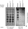
Anti-CRISPRs work during the process of infection to protect a targeted phage. Tenfold serial dilutions of lysates of three phages were spotted on either P. aeruginosa strain PA14 csy3::lacZ (a strain with no type I-E or type I-F CRISPR-Cas activity) or strain SMC4386. Phage DMS3m contains protospacer matches for the type I-E system of strain SMC4386. DMS3m30-35 is an isogenic phage with its I-E anti-CRISPR ACR3-30 replaced by a type I-F anti-CRISPR ACR30-35. The DMS3m30-35fs phage encodes a frameshifted version of ACR30-35.
FIG 5
FIG 5
Anti-CRISPRs are widespread in P. aeruginosa Mu-like phages. (A) Transformation efficiency assays using PS2 with the CTT PAM construct are shown for a variety of P. aeruginosa SMC4386 lysogens. The anti-CRISPR gene regions of these phages have been sequenced, and their sequence groups are indicated. Prophages tested for type I-E anti-CRISPR activity as prophages in P. aeruginosa SMC4386 are indicated by an ampersand, and prophages tested for type I-F anti-CRISPR activity as prophages in P. aeruginosa PA14 are indicated by an asterisk. (B) The anti-CRISPR region sequence groups are defined. The boxes are drawn in proportion to the scale bar shown. Colored boxes indicate high sequence identity to one of the four type I-E anti-CRISPR genes (indicated by a white E on a black circle) or one of the five type I-F anti-CRISPR genes (indicated by a black F on a white circle). All anti-CRISPR-encoding phages have a conserved gene shown in blue, which does not have anti-CRISPR activity against the type I-E or I-F systems (3). (C) The phages belonging to each of the anti-CRISPR region sequence groups defined in panel B are listed.

References

    1. Labrie SJ, Samson JE, Moineau S. 2010. Bacteriophage resistance mechanisms. Nat. Rev. Microbiol. 8:317–327. 10.1038/nrmicro2315 - DOI - PubMed
    1. Brouns SJ, Jore MM, Lundgren M, Westra ER, Slijkhuis RJ, Snijders AP, Dickman MJ, Makarova KS, Koonin EV, van derOost J. 2008. Small CRISPR RNAs guide antiviral defense in prokaryotes. Science 321:960–964. 10.1126/science.1159689 - DOI - PMC - PubMed
    1. Jore MM, Brouns SJ, van der Oost J. 2012. RNA in defense: CRISPRs protect prokaryotes against mobile genetic elements. Cold Spring Harb. Perspect. Biol 4:pii:a003657. 10.1101/cshperspect.a003657 - DOI - PMC - PubMed
    1. Haurwitz RE, Jinek M, Wiedenheft B, Zhou K, Doudna JA. 2010. Sequence- and structure-specific RNA processing by a CRISPR endonuclease. Science 329:1355–1358. 10.1126/science.1192272 - DOI - PMC - PubMed
    1. Deltcheva E, Chylinski K, Sharma CM, Gonzales K, Chao Y, Pirzada ZA, Eckert MR, Vogel J, Charpentier E. 2011. CRISPR RNA maturation by trans-encoded small RNA and host factor RNase III. Nature 471:602–607. 10.1038/nature09886 - DOI - PMC - PubMed

Publication types

MeSH terms