. 2014 Apr 15;9(4):e94343.
doi: 10.1371/journal.pone.0094343. eCollection 2014.

KCTD1 suppresses canonical Wnt signaling pathway by enhancing β-catenin degradation

Affiliations

KCTD1 suppresses canonical Wnt signaling pathway by enhancing β-catenin degradation

Xinxin Li et al. PLoS One. .

Retraction in

Abstract

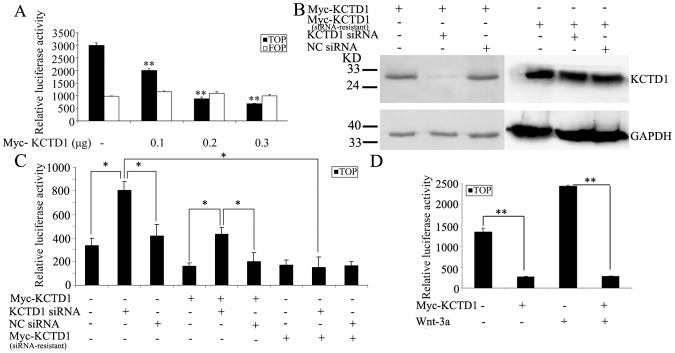
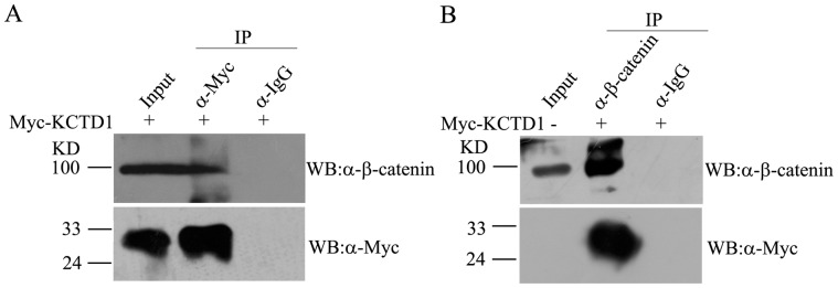
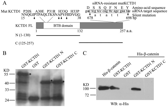
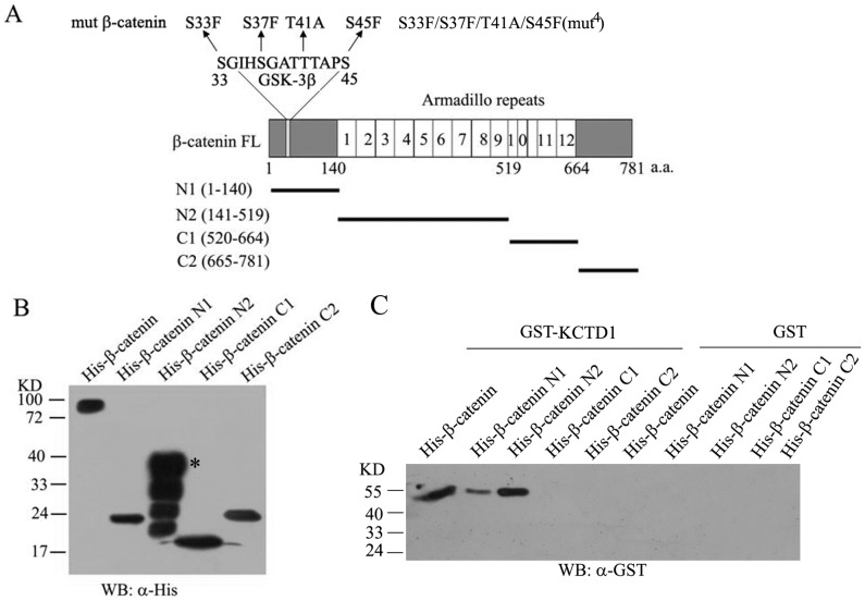
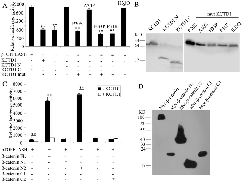
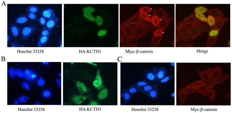
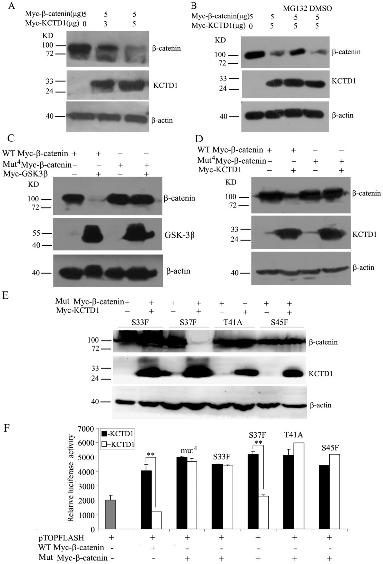
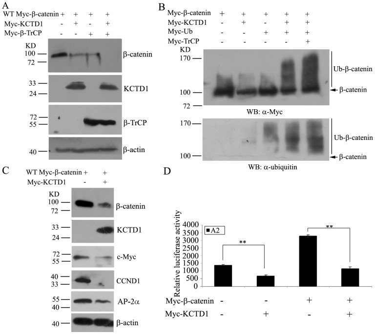
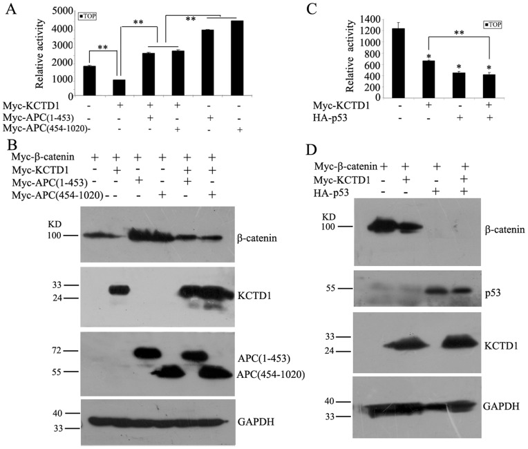
The canonical Wnt signaling pathway controls normal embryonic development, cellular proliferation and growth, and its aberrant activity results in human carcinogenesis. The core component in regulation of this pathway is β-catenin, but molecular regulation mechanisms of β-catenin stability are not completely known. Here, our recent studies have shown that KCTD1 strongly inhibits TCF/LEF reporter activity. Moreover, KCTD1 interacted with β-catenin both in vivo by co-immunoprecipitation as well as in vitro through GST pull-down assays. We further mapped the interaction regions to the 1-9 armadillo repeats of β-catenin and the BTB domain of KCTD1, especially Position Ala-30 and His-33. Immunofluorescence analysis indicated that KCTD1 promotes the cytoplasmic accumulation of β-catenin. Furthermore, protein stability assays revealed that KCTD1 enhances the ubiquitination/degradation of β-catenin in a concentration-dependent manner in HeLa cells. And the degradation of β-catenin mediated by KCTD1 was alleviated by the proteasome inhibitor, MG132. In addition, KCTD1-mediated β-catenin degradation was dependent on casein kinase 1 (CK1)- and glycogen synthase kinase-3β (GSK-3β)-mediated phosphorylation and enhanced by the E3 ubiquitin ligase β-transducin repeat-containing protein (β-TrCP). Moreover, KCTD1 suppressed the expression of endogenous Wnt downstream genes and transcription factor AP-2α. Finally, we found that Wnt pathway member APC and tumor suppressor p53 influence KCTD1-mediated downregulation of β-catenin. These results suggest that KCTD1 functions as a novel inhibitor of Wnt signaling pathway.


Conflict of interest statement

Competing Interests: The authors have declared that no competing interests exist.

Figures

Figure 1
Figure 1. Effects of KCTD1 on the TOPFLASH reporter activity.
(A) HEK293 cells were transfected with a TOPFLASH or FOPFLASH reporter plasmid, and different amounts of pCMV-Myc-KCTD1 plasmids. (B) HEK293 cells were transiently transfected with pCMV-Myc-KCTD1, siRNA-resistant pCMV-Myc-KCTD1, KCTD1 siRNA or negative control siRNA as indicated for 24 h, cell extracts were detected with mouse monoclonal antibodies against Myc-tag and GAPDH. (C) HEK293 cells were transiently transfected with a TOPFLASH reporter plasmid, pCMV-Myc-KCTD1, siRNA-resistant pCMV-Myc-KCTD1, KCTD1 siRNA or negative control siRNA or in combination. (D) HEK293 cells were transfected with a TOPFLASH reporter plasmid and pCMV-Myc-KCTD1 for 24 h and then treated with 100 ng/ml of Wnt-3a for 36 h. The amount of DNA in each transfection was kept constant by the addition of control empty vectors. Luciferase and β-galactosidase activities were measured 24 h after transfection. Relative reporter activity was presented as mean ±SD from three independent transfection experiments performed in triplicate. *, P<0.05; **, P<0.01 compared with controls.
Figure 2
Figure 2. Interaction of β-catenin and KCTD1 in vivo.
HeLa cells were only transfected with pCMV-Myc-KCTD1 plasmids and harvested 30 h after transfection. Cell extracts were incubated with rabbit polyclonal antibodies against Myc-tag (A) or β-catenin (B), precipitated by ProteinA/G beads and detected by western blots using mouse monoclonal antibodies against β-catenin and Myc-tag. Pre-immune IgG was used as negative control.
Figure 3
Figure 3. Identification of β-catenin binding domain in KCTD1.
(A) Schematic representation of KCTD1 domains, deletion and mutant constructs used for pull-down analysis and luciferase assays. And the five silent mutations in the siRNA-target sequence of wild-type KCTD1 cDNA were shown, the resulting protein mutKCTD1 confers resistance to the siRNA and no change in the amino acid sequence compared with the wild-type. (B) The full-length and truncated proteins of GST-KCTD1 were bacterially expressed, purified and detected with Western blots using mouse monoclonal anti-GST antibodies. (C) GST pull-down experiments were performed with GST, GST fusion proteins above and His-β-catenin recombinant proteins analyzed by immunoblots with mouse monoclonal antibodies against His-tag.
Figure 4
Figure 4. Identification of KCTD1 binding domain in β-catenin.
(A) Schematic representation of protein domain structure of β-catenin and its deletion constructs used for pull-down and luciferase assays. The mutant phosphorylation sites (the Ser45 and Ser33/37/Thr41 sites) in β-catenin mutations were shown. (B) Bacterially expressed and purified His-β-catenin fusion proteins were detected with Western blots using mouse monoclonal antibodies against His-tag. (C) His-tag pull-down experiments were performed with GST-KCTD1 and the full-length or truncated proteins of His-β-catenin analyzed by Western blots using mouse monoclonal anti-GST antibodies. GST proteins were used as negative control.
Figure 5
Figure 5. Effects of various truncations of KCTD1 and β-catenin on TOPFLASH reporter activity.
(A) HEK293 cells were transfected with a TOPFLASH reporter plasmid and the indicated KCTD1 plasmids. (B) Protein expression of various truncations and mutations of KCTD1 was demonstrated by Western blot with mouse monoclonal anti-Myc antibodies. (C) HEK293 cells were transfected with a TOPFLASH reporter plasmid and expression plasmids encoding full-length β-catenin or truncations of β-catenin in the absence and presence of KCTD1. (D) Protein expression of various truncations of β-catenin was confirmed by immunoblots with mouse monoclonal anti-Myc antibodies. Relative luciferase activities represent mean ±SD from at least three independent experiments after normalization to β-galactosidase activities. **, P<0.01 compared with controls.
Figure 6
Figure 6. Localization analysis of KCTD1 and β-catenin proteins.
(A) HeLa cells were transfected with HA-KCTD1 and Myc-β-catenin. HA-KCTD1 was detected with rabbit polyclonal anti-HA antibodies and Alexa 488 goat anti-rabbit secondary antibodies, while Myc-β-catenin was detected with mouse monoclonal anti-Myc antibodies and Alexa 594 goat anti-mouse secondary antibodies. (B) HeLa cells were transfected with HA-KCTD1 alone, which was detected as described above. (C) HeLa cells were only transfected with Myc-β-catenin detected as described in (A). Nuclei were stained with Hoechst 33258.
Figure 7
Figure 7. Effects of KCTD1 on the expression of β-catenin protein.
(A) HeLa cells were transfected with various amounts of pCMV-Myc-KCTD1 and the same amount of pCMV-Myc-β-catenin. Total cell extracts were prepared and analyzed by immunoblotting using mouse monoclonal anti-Myc antibodies. β-actin was used as the internal control. (B) HeLa cells were transfected with pCMV-Myc-KCTD1 and pCMV-Myc-β-catenin, then treated with 20 µM of MG132 or DMSO for 10 h before harvest followed by immunoblot analysis using mouse monoclonal anti-Myc antibodies. (C) HeLa cells were transfected with pCMV-Myc-GSK-3β and pCMV-Myc-β-catenin (WT) or with pCMV-Myc-GSK-3β and pCMV-Myc-β-catenin (Mut4) followed by Western blots with mouse monoclonal anti-Myc antibodies. (D) HeLa cells were transfected with pCMV-Myc-KCTD1 and pCMV-Myc-β-catenin (WT) or pCMV-Myc-β-catenin (Mut4) followed by Western blots using mouse monoclonal anti-Myc antibodies. (E) HeLa cells were transfected with pCMV-Myc-KCTD1 and pCMV-Myc-β-catenin mutations with S33F, S37F, T41A and S45F followed by Western blots using mouse monoclonal anti-Myc antibodies. (F) HEK293 cells were transfected with TOPFLASH reporter plasmid and expression plasmids encoding KCTD1 and WT β-catenin or Mut β-catenin. Luciferase and β-galactosidase activities were measured 24 h after transfection. Relative luciferase activities represent mean ±SD from at least three independent experiments after normalization to β-galactosidase activities. **, P<0.01 compared with controls.
Figure 8
Figure 8. Effects of KCTD1 on β-catenin ubiquitination and the expression of Wnt/β-catenin downstream genes and AP-2α.
(A) HeLa cells were transfected with either pCMV-Myc-β-catenin alone or with pCMV-Myc-KCTD1 or with pCMV-Myc-TrCP or with both pCMV-Myc-KCTD1 and pCMV-Myc-TrCP. Total cell extracts were analyzed by immunoblotting using mouse monoclonal anti-Myc antibodies. β-actin was used as the internal control. (B) HeLa cells were transfected with either pCMV-Myc-β-catenin alone or with pCMV-Myc-KCTD1, pCMV-Myc-ubiquitin or pCMV-Myc-TrCP or in combination as indicated. 24 h after transfection, cell lysates were immunoprecipitated with rabbit polyclonal anti-β-catenin antibodies followed by immunoblotting with mouse monoclonal antibodies against Myc-tag and ubiquitin to detect ubiquitin conjugation. (C) HeLa cells were transfected with either pCMV-Myc-β-catenin alone or with pCMV-Myc-KCTD1 for 24 h. The whole cell extracts were valuated by Western blotting using mouse monoclonal antibodies against Myc-tag, CCND1 and AP-2α. β-actin was used as a loading control for total lysate samples. (D) HEK293 cells were transfected with A2-Luc reporter plasmid and expression plasmids encoding KCTD1 and β-catenin. Relative luciferase activities represent mean ±SD from at least three independent experiments after normalization to β-galactosidase activities. **, P<0.01 compared with controls.
Figure 9
Figure 9. Effects of APC and p53 on KCTD1-mediated β-catenin degradation.
(A) HEK293 cells were transfected with TOPFLASH reporter plasmid either alone or with pCMV-Myc-KCTD1 or with pCMV-Myc-APC truncations or with both pCMV-Myc-KCTD1 and pCMV-Myc-APC truncations. (B) HeLa cells were transfected with either pCMV-Myc-β-catenin alone or with pCMV-Myc-KCTD1 or with pCMV-Myc-APC trucations or in combination as indicated. 24 h after transfection, cell lysates were detected by Western blots with mouse monoclonal anti-Myc antibodies. GAPDH was used as a loading control. (C) HEK293 cells were transfected with TOPFLASH reporter plasmid either alone or with pCMV-Myc-KCTD1 or with pCMV-HA-p53 or with both pCMV-Myc-KCTD1 and pCMV-HA-p53. (D) HeLa cells were transfected with either pCMV-Myc-β-catenin alone or with pCMV-Myc-KCTD1 or with pCMV-HA-p53 or in combination as indicated for 24 h, cell lysates were detected by immunoblotting with mouse monoclonal antibodies against Myc-tag and HA-tag. GAPDH was used as the internal control. Relative luciferase activities represent mean ±SD from at least three independent experiments after normalization to β-galactosidase activities. *, P<0.05 and **, P<0.01 compared with controls.

References

    1. Logan CY, Nusse R (2004) The Wnt signaling pathway in development and disease. Annu Rev Cell Dev Biol 20: 781–810. - PubMed
    1. Moon RT, Kohn AD, De Ferrari GV, Kaykas A (2004) WNT and beta-catenin signalling: diseases and therapies. Nat Rev Genet 5: 691–701. - PubMed
    1. Willert K, Brink M, Wodarz A, Varmus H, Nusse R (1997) Casein kinase 2 associates with and phosphorylates dishevelled. Embo J 16: 3089–3096. - PMC - PubMed
    1. Sakanaka C, Leong P, Xu L, Harrison SD, Williams LT (1999) Casein kinase iepsilon in the wnt pathway: regulation of beta-catenin function. Proc Natl Acad Sci U S A 96: 12548–12552. - PMC - PubMed
    1. He TC, Sparks AB, Rago C, Hermeking H, Zawel L, et al. (1998) Identification of c-MYC as a target of the APC pathway. Science 281: 1509–1512. - PubMed

Publication types

MeSH terms