. 2014 Jun;3(6):734-44.
doi: 10.5966/sctm.2013-0189. Epub 2014 Apr 15.

Cobalt protoporphyrin pretreatment protects human embryonic stem cell-derived cardiomyocytes from hypoxia/reoxygenation injury in vitro and increases graft size and vascularization in vivo

Affiliations

Cobalt protoporphyrin pretreatment protects human embryonic stem cell-derived cardiomyocytes from hypoxia/reoxygenation injury in vitro and increases graft size and vascularization in vivo

Jun Luo et al. Stem Cells Transl Med. 2014 Jun.

Abstract

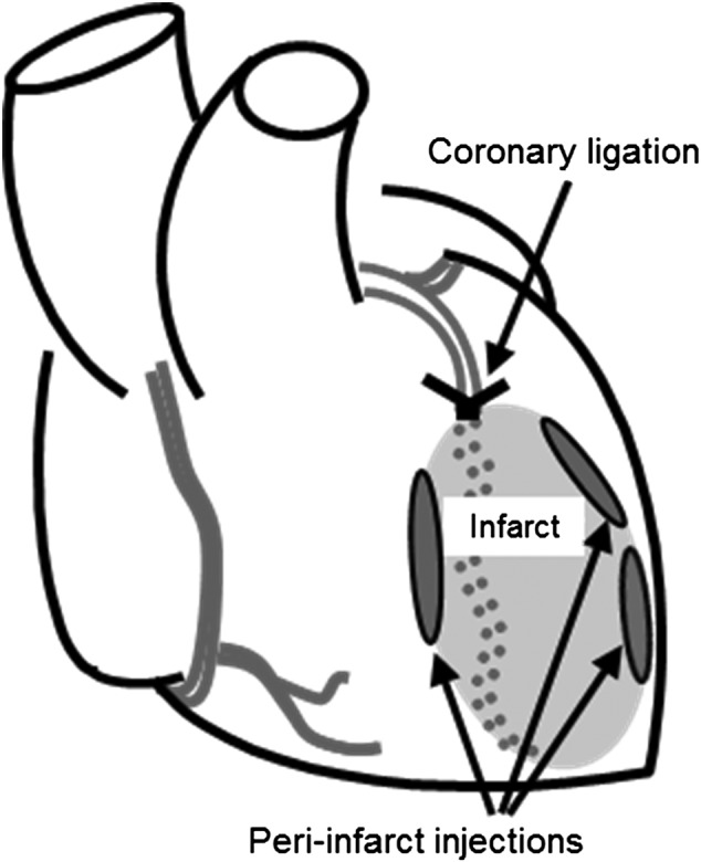
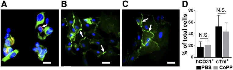
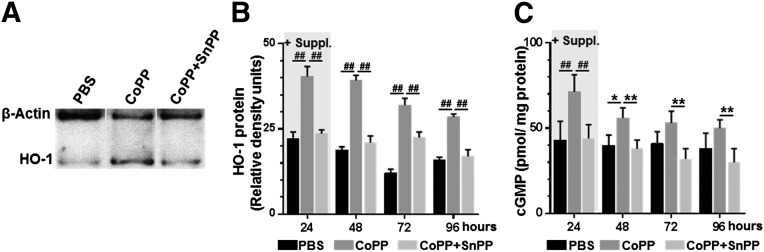
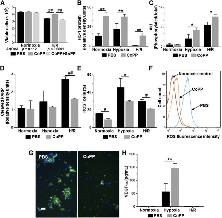
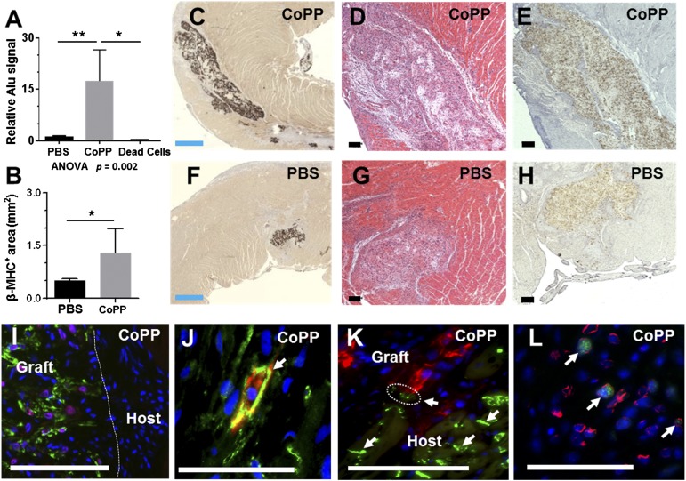
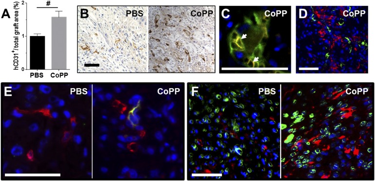
Human embryonic stem cell-derived cardiomyocytes (hESC-CMs) can regenerate infarcted myocardium. However, when implanted into acutely infarcted hearts, few cells survive the first week postimplant. To improve early graft survival, hESC-CMs were pretreated with cobalt protoporphyrin (CoPP), a transcriptional activator of cytoprotective heme oxygenase-1 (HO-1). When hESC-CMs were challenged with an in vitro hypoxia/reoxygenation injury, mimicking cell transplantation into an ischemic site, survival was significantly greater among cells pretreated with CoPP versus phosphate-buffered saline (PBS)-pretreated controls. Compared with PBS-pretreated cells, CoPP-pretreated hESC-CM preparations exhibited higher levels of HO-1 expression, Akt phosphorylation, and vascular endothelial growth factor production, with reduced apoptosis, and a 30% decrease in intracellular reactive oxygen species. For in vivo translation, 1 × 10(7) hESC-CMs were pretreated ex vivo with CoPP or PBS and then injected intramyocardially into rat hearts immediately following acute infarction (permanent coronary ligation). At 1 week, hESC-CM content, assessed by quantitative polymerase chain reaction for human Alu sequences, was 17-fold higher in hearts receiving CoPP- than PBS-pretreated cells. On histomorphometry, cardiomyocyte graft size was 2.6-fold larger in hearts receiving CoPP- than PBS-pretreated cells, occupying up to 12% of the ventricular area. Vascular density of host-perfused human-derived capillaries was significantly greater in grafts composed of CoPP- than PBS-pretreated cells. Taken together, these experiments demonstrate that ex vivo pretreatment of hESC-CMs with a single dose of CoPP before intramyocardial implantation more than doubled resulting graft size and improved early graft vascularization in acutely infarcted hearts. These findings open the door for delivery of these, or other, stem cells during acute interventional therapy following myocardial infarction or ischemia.

Keywords: Acute myocardial infarction; Cell therapy; Heme oxygenase-1; Human embryonic stem cell; Infarct repair; Preconditioning.


Figures

Figure 1.
Figure 1.
Heart diagram illustrating peri-infarct injection sites surrounding the acute infarct.
Figure 2.
Figure 2.
Histologic characterization of human embryonic stem cell-derived cardiomyocyte preparations after directed differentiation. Aliquots were immunostained with antibodies to cTnI (green) and hCD31 (red, arrow) (A), pan-cadherin (green, arrows) (B), and connexin 43 (green, arrows) (C). Hoechst nuclear stain (blue). Scale bars = 25 μm. (D): Percentages of total cells positively immunostained for hCD31 and cTnI following the 24-hour pretreatment with CoPP or PBS (p = N.S. for both; n = 4 per group). Abbreviations: CoPP, cobalt protoporphyrin; cTnI, cardiac troponin I; hCD31, human CD31; N.S., not significant; PBS, phosphate-buffered saline.
Figure 3.
Figure 3.
HO-1 induction in human embryonic stem cell-derived cardiomyocytes after CoPP exposure. (A): Representative immunoblot illustrating HO-1 expression after 24-hour exposure to PBS, CoPP, or CoPP plus SnPP. (B): HO-1 protein quantified by Western blot densitometry following treatment with PBS, CoPP, or CoPP plus SnPP as culture medium supplements (+Suppl). After 24 hours, cells were switched to supplement-free media. (C): Levels of cGMP measured by enzyme-linked immunosorbent assay to indicate HO-1 enzymatic activity. ##, p < .0001, ∗∗, p < .01, ∗, p < .05, two-way analyses of variance with Tukey-Kramer corrections. Abbreviations: cGMP, cyclic GMP; CoPP, cobalt protoporphyrin; HO-1, heme oxygenase-1; PBS, phosphate-buffered saline; SnPP, tin protoporphyrin.
Figure 4.
Figure 4.
Effects of CoPP pretreatment on human embryonic stem cell-derived cardiomyocyte (hESC-CM) preparations subjected to hypoxia/reoxygenation injury. (A): Numbers of viable cells by trypan blue exclusion counts. (B): HO-1 expression. (C): Phosphorylated Akt/total Akt. (D): Cleaved PARP generation by Western blot densitometry. (E–G): ROS generation in response to H/R injury. (E): Percentage of cells exhibiting ROS positivity as indicated by staining intensity on flow cytometry. (F): Fluorescence intensity at the end of reoxygenation in CoPP- versus PBS-pretreated hESC-CMs. (G): Representative examples of ROS fluorescence (green) imaged in PBS- and CoPP-pretreated hESC-CMs after H/R injury. Hoechst nuclear stain (blue). Scale bar = 50 μm. (H): VEGF165 production in cell supernatants by enzyme-linked immunosorbent assay. In all graphs, mean values ± SD represent at least three independent experiments with four replicates per experiment. ##, p < .0001, #, p ≤ .001, ∗∗, p ≤ .01, ∗, p < .05 versus PBS-treated hESC-CMs. Abbreviations: ANOVA, analyses of variance; CoPP, cobalt protoporphyrin; H/R, hypoxia/reoxygenation; HO-1, heme oxygenase-1; PARP, poly-ADP ribose polymerase; PBS, phosphate-buffered saline; ROS, reactive oxygen species; SnPP, tin protoporphyrin; VEGF, vascular endothelial growth factor.
Figure 5.
Figure 5.
Graft size at 1 week after human embryonic stem cell-derived cardiomyocyte (hESC-CM) transplantation into acutely infarcted rat ventricles. (A): Graft sizes quantified by quantitative polymerase chain reaction for human Alu sequences in whole hearts. The column labeled “Dead Cells” represents hearts injected with nonviable hESC-CMs; actual column height is .006 ± .004 (bar height not to scale). (B): Mean graft size quantified morphometrically as the total β-MHC+ areas across all surveyed sections/heart. (C, F): Representative sections immunostained for β-MHC (brown chromogen, 3, 3′-diaminobenzidine [DAB]) to delineate human cardiomyocyte-containing areas, demonstrating the relative size of CoPP- and PBS-pretreated grafts. (D, G): Serial sections stained with hematoxylin and eosin. (E, H): In situ hybridization with a human-specific pan-centromeric probe to confirm the human origin of graft cells (brown, DAB). (I): Human cardiomyocyte grafts doubly stained for β-MHC (green) and Nkx2.5 (red), demonstrating expression of the early cardiomyocyte marker, Nkx2.5, in graft, but not host, myocardium. (J): Double staining for pan-cadherin (green, arrow) and β-MHC (red), illustrating a β-MHC+ graft cardiomyocyte with N cadherin expression among host myocytes, also expressing cadherins. (K): Connexin 43 staining (green, arrows) illustrates a close juxtaposition (circled) between gap junctions on host cardiomyocytes and graft cardiomyocytes, identified by β-MHC staining (red). (L): Ki-67 staining (green) showed that hESC-CMs retained proliferative capacity 1 week after implantation. Costaining with human-specific lamin A/C (red) confirmed the human origin of imaged graft cells. (I–L): Hoechst nuclear stain (blue). Blue scale bars = 500 µm. Black and white scale bars = 100 µm. ∗∗, p < .01, ∗, p < .05. Abbreviations: ANOVA, analyses of variance; β-MHC, β-myosin heavy chain; CoPP, cobalt protoporphyrin; PBS, phosphate-buffered saline.
Figure 6.
Figure 6.
Vascularization of human embryonic stem cell-derived cardiomyocyte (hESC-CM) grafts in acutely infarcted ventricles at 1 week. (A): Vascular density within grafts. (B): Immunostaining for human endothelium (hCD31, brown, 3, 3′-diaminobenzidine) illustrating the prevalence of human-derived vascular structures. (C): Dual staining for hCD31 (green) and red blood cells (TER-199, red, arrows) demonstrating host blood cells present within human-derived vasculature at 1 week. (D): Dual staining for hCD31 (green) and β-myosin heavy chain (β-MHC) (red) shows the prevalence of formed human vascular structures within the β-MHC+ human cardiomyocyte grafts. (E): Dual staining for rat endothelium (rat endothelial cell antigen-1, green) and human endothelium (hCD31, red) reveals that host-derived endothelial cells had penetrated within hESC-CM grafts by 1 week in CoPP-, but not PBS-pretreated grafts. (F): Dual staining for c-Kit (red) and human lamin A/C (green) illustrates a greater prevalence of c-Kit+ cells, presumably host-derived, within CoPP- than PBS-pretreated grafts. (D–F): Hoechst nuclear stain (blue). Scale bars = 50 µm. #, p < .001 versus PBS-pretreated grafts. Abbreviations: CoPP, cobalt protoporphyrin; hCD31, human CD31; PBS, phosphate-buffered saline.

References

    1. Zhang M, Methot D, Poppa V, et al. Cardiomyocyte grafting for cardiac repair: Graft cell death and anti-death strategies. J Mol Cell Cardiol. 2001;33:907–921. - PubMed
    1. Laflamme MA, Chen KY, Naumova AV, et al. Cardiomyocytes derived from human embryonic stem cells in pro-survival factors enhance function of infarcted rat hearts. Nat Biotechnol. 2007;25:1015–1024. - PubMed
    1. Li RK, Mickle DA, Weisel RD, et al. Optimal time for cardiomyocyte transplantation to maximize myocardial function after left ventricular injury. Ann Thorac Surg. 2001;72:1957–1963. - PubMed
    1. Terrovitis JV, Smith RR, Marbán E. Assessment and optimization of cell engraftment after transplantation into the heart. Circ Res. 2010;106:479–494. - PMC - PubMed
    1. Murry CE, Jennings RB, Reimer KA. Preconditioning with ischemia: A delay of lethal cell injury in ischemic myocardium. Circulation. 1986;74:1124–1136. - PubMed

Publication types

MeSH terms