. 2014 Apr 15;9(4):e94386.
doi: 10.1371/journal.pone.0094386. eCollection 2014.

Real time live imaging of phytopathogenic bacteria Xanthomonas campestris pv. campestris MAFF106712 in 'plant sweet home'

Affiliations

Real time live imaging of phytopathogenic bacteria Xanthomonas campestris pv. campestris MAFF106712 in 'plant sweet home'

Chiharu Akimoto-Tomiyama et al. PLoS One. .

Abstract

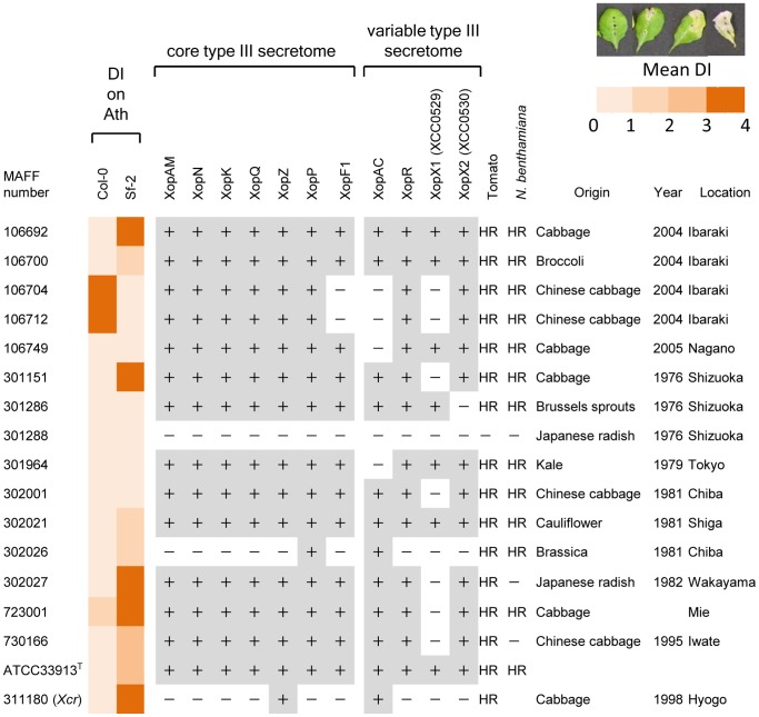
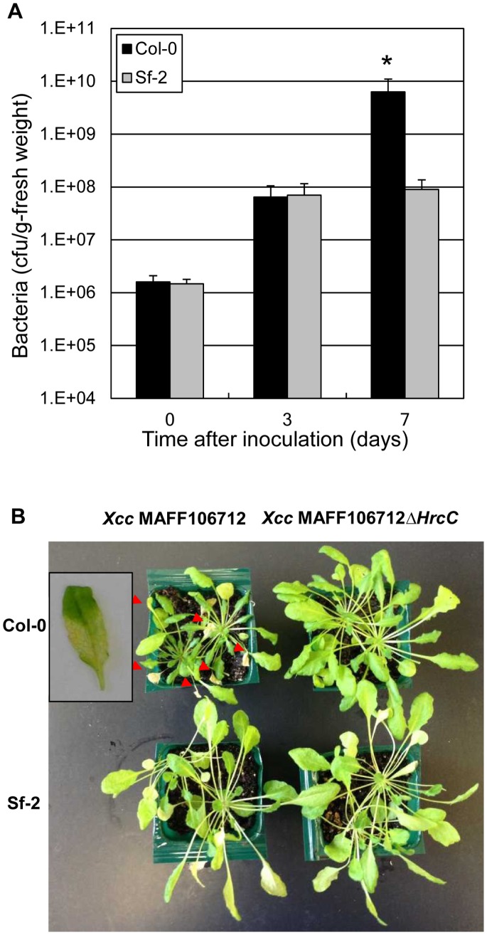
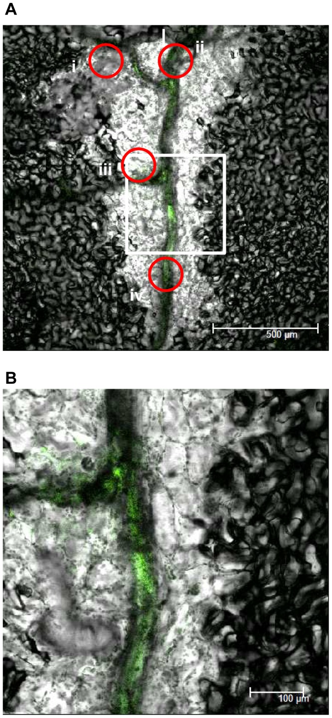
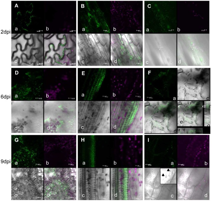
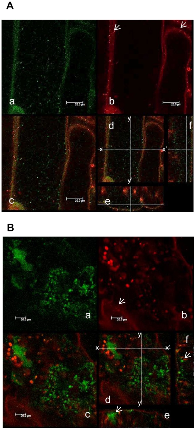
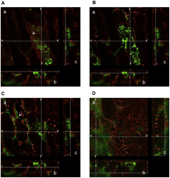
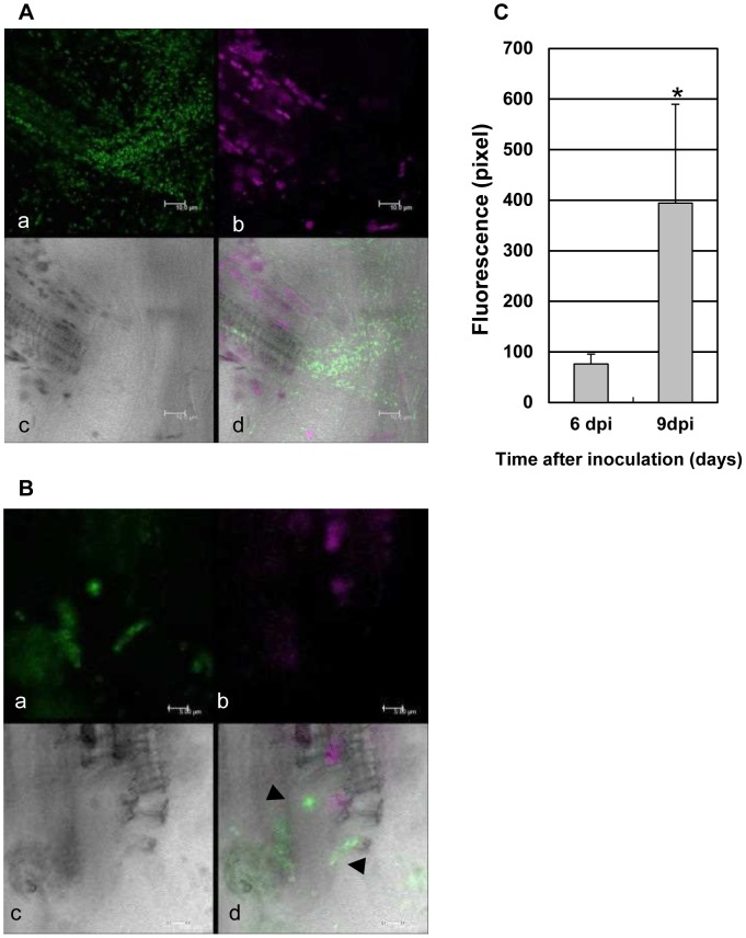
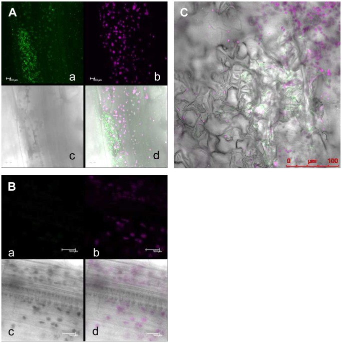
Xanthomonas is one of the most widespread phytobacteria, causing diseases on a variety of agricultural plants. To develop novel control techniques, knowledge of bacterial behavior inside plant cells is essential. Xanthomonas campestris pv. campestris, a vascular pathogen, is the causal agent of black rot on leaves of Brassicaceae, including Arabidopsis thaliana. Among the X. campestris pv. campestris stocks in the MAFF collection, we selected XccMAFF106712 as a model compatible pathogen for the A. thaliana reference ecotype Columbia (Col-0). Using modified green fluorescent protein (AcGFP) as a reporter, we observed real time XccMAFF106712 colonization in planta with confocal microscopy. AcGFP-expressing bacteria colonized the inside of epidermal cells and the apoplast, as well as the xylem vessels of the vasculature. In the case of the type III mutant, bacteria colonization was never detected in the xylem vessel or apoplast, though they freely enter the xylem vessel through the wound. After 9 days post inoculation with XccMAFF106712, the xylem vessel became filled with bacterial aggregates. This suggests that Xcc colonization can be divided into main four steps, (1) movement in the xylem vessel, (2) movement to the next cell, (3) adhesion to the host plant cells, and (4) formation of bacterial aggregates. The type III mutant abolished at least steps (1) and (2). Better understanding of Xcc colonization is essential for development of novel control techniques for black rot.


Conflict of interest statement

Competing Interests: The authors have declared that no competing interests exist.

Figures

Figure 1
Figure 1. Interactions between two Arabidopsis ecotypes vs. 16 X. campestris pv. campestris isolates.
All strains are shown by their MAFF collection number except for ATCC33913 type strain (ATCC33913T), and the distribution of genes coding for type III secreted proteins is indicated. The pathogenicity (mean disease index at 7 dpi) of these X. campestris pv. campestris strains on Arabidopsis ecotypes Col-0 and Sf-2 is indicated by color coding (0 to 1 indicates no symptoms, 1 to 2 indicates weak chlorosis, 2 to 3 indicates strong chlorosis, and 3 to 4 indicates necrosis as shown in the upper panel). Bacteria were inoculated by piercing the central leaf vein three times with a D200 pipette tip that had been dipped in a bacterial suspension (109 cfu mL−1). The presence or absence of a homologous T3SP gene sequence was determined by PCR. Strains were also inoculated on nonhost tomato (momotaro) and N. benthamiana. HR, hypersensitive response; -, no HR. Dark-gray squares and+represent the presence of the corresponding genes with both primer combinations at the expected sizes, whereas white squares and - represent the absence of PCR amplification or PCR with a different amplicon size.
Figure 2
Figure 2. XccMAFF106712 is virulent for Arabidopsis ecotypes Col-0 plants in a type III dependent manner.
(A) In planta bacterial growth of XccMAFF106712 after inoculation on Arabidopsis ecotypes Col-0 (solid) and Sf-2 (gray). Twenty-four leaves from 8 different plants were inoculated by piercing the leaves with an XccMAFF106712 suspension of 109 cfu mL−1. Enhanced bacterial multiplication in Col-0 compared to Sf-2 was significant (*P<0.001, Student’s t test). Results are representative of at least three experiments per condition. (B) Xcc MAFF106712 pathogenicity on Col-0 is dependent on the type III secretion system. Bacteria (XccMAFF106712 and MAFF106712ΔhrcC) were inoculated on Col-0 and Sf-2 plants and photographs were taken 15 days post inoculation. Black rot phenotype of Col-0 plant 7 days post XccMAFF106712 inoculation is shown in the window. At 7 dpi, some infected leaves are still green and some of the leaves start showing the disease phenotype. We used mixed up them for the growth assay shown in the panel A. Red arrows indicate necrotic leaves caused by bacteria inoculation.
Figure 3
Figure 3. An AcGFP-expressing XccMAFF106712 bacteria colonized distal to the inoculated site at 6 days post inoculation.
An inoculated leaf was detached from the plant and observed by a CLSM. Shown is the merged image of green fluorescence (green) and bright field. (A) Image shown around the inoculation site (I) in a low magnification. The red circles indicates the approximate location of the images taken in this manuscript. i, around (I); ii, central vein around (I); iii, distal to (I); iv, central vein distal to (I). White square showed the image shown in B Bar, 500 μm. (B) Image shown at the white square part of panel A in a higher magnification. Bar, 100 μm.
Figure 4
Figure 4. Time course extension of AcGFP-expressing XccMAFF106712 in Col-0 plant.
Localization of AcGFP-expressing XccMAFF106712 (A to H) and XccMAFF106712 (I) in an Arabidopsis Col-0 plant. Leaf surface (A, D and G), central vein (B, C and I), the xylem vessel of the central vein (E, H) and mesophyll cells (F) were observed by confocal microscopy. Photographs were taken at 2 days (A, B and C), 6 days (D, E and F) and 9 days (G, H and I) after inoculation. Location indicated in Figure 2 : i,(A, I); ii, (B, C); iii, (D, F and G); iv, (E, H). Green fluorescent bacteria spread both in the cytosol of leaf epidermal cells, apoplast and the xylem vessel of the vein. a, green fluorescence (green); b, chlorophyll autofluorescence (red); c, bright field; d, merged image; e, same as d in panel F showing cross sectional line; f, cross-sectional view of c along the line x-x′; g, cross-sectional view of c along the line y-y′. X-x′, 158 μm; y-y′, 158 μm; z-z′, 32.8 μm. Bars, 10 μm (ACDE); 20 μm (BFI); 50 μm(G); 5 μm(H). Whole z series photos for panel F available at supplemental movie S5.
Figure 5
Figure 5. AcGFP-expressing XccMAFF106712 colonized in both leaf epidermal cells and the intercellular space in a Col-0 plant.
An inoculated leaf was detached at the indicated time point and plasma membrane was stained with FM4-64. a, green fluorescence (green); b, FM4-64 and chlorophyll auto fluorescence are shown in red; c, merged image; d, same as panel c showing cross sectional line for e and f; e, cross-sectional view of c along the line x-x′ on d; f, cross-sectional view of c along the line y-y′ on d. (A) Epidermal cells of the central vein approximately 100 μm away from the initial inoculation edge in 3 dpi plant. The white arrow in panel b shows bacteria in plant surface that were stained with FM4-64. The bottom of the panel e and right side of the panel f shows plant surface. X-x′, 117 μm; y-y′, 117 μm; z-z′, 44 μm. Bars, 20 μm. (B) Epidermal cells approximately 600 μm away from the initial inoculation edge in the 9 dpi plant. The white arrow in panel b shows the stained bacteria on the surface of the epidermal cells. The white arrow in panel e shows aggregated cells in the intercellular space. The white arrow in panel f shows bacteria colonizing the epidermal cells. The upper side of the panel e and left side of the panel f shows the plant surface. X-x′, 180 μm; y-y′, 180 μm; z-z′, 20.5 μm. Bars, 20 μm.
Figure 6
Figure 6. AcGFP-expressing XccMAFF106712 colonized the bundle sheath cells in a Col-0 plant.
An inoculated leaf was detached at 9 days post inoculation and the plasma membrane was stained with FM4-64. Images shown are from the central vein approximately 3 mm away from the initial inoculation edge. a, merged image of the green fluorescence (green) and FM4-64 and chlorophyll auto fluorescence (red); b, cross-sectional view of a along the line x-x′ on a; c, cross-sectional view of a along the line y-y′ on a. X-x′, 240 μm; y-y′, 240 μm; z-z′, 80 μm. Upper side of the panel e and left side of the panel f showing the plant surface. A z-stack CLMS image is shown in supplemental Movie S7. (A) Cell surface. The white arrow in panel a shows bacteria at the surface that were stained with FM4-64. (B) Bacteria colonizing the epidermal cell. (C) Bacteria colonizing the apoplast. (D) Bacteria colonizing bundle sheath cells.
Figure 7
Figure 7. AcGFP-expressing XccMAFF106712 proliferated and agglomerated inside the xylem vessel of a compatible plant.
The leaf surface layer was peeled off and the xylem vessel was extracted; bacteria which escaped the vessel were imaged. (A) Confocal image taken 6 days after inoculation. AcGFP-expressing XccMAFF106712 proliferated, but no bacterial aggregates formed. Bars, 10 μM. (B) Confocal image taken 9 days after inoculation. Black arrows indicate large bacterial aggregates in a vessel. Bars, 5 μM. (A, B) a, green fluorescence; b, chlorophyll autofluorescence; c, bright field; d, merged image. (C) Size of aggregates at the indicated time was estimated by fluorescence area using ImageJ. More than 30 aggregates from a vessel (n = 8) were measured and the difference between 6 and 9 dpi was significant (*P<0.01, Student’s t test).
Figure 8
Figure 8. AcGFP-expressing XccMAFF106712ΔhrcC do not colonize a Col-0 plant.
Localization of AcGFP-expressing XccMAFF106712ΔhrcC (A to F) in an Arabidopsis Col-0 plant. Leaf surface (A, C and E), central vein 100 μm away from the inoculation site (B) and the xylem vessel of the central vein (D, F) were observed by CLSM. Photographs were taken at 2 days (A, B), 6 days (C, D) and 9 days (E, F) after inoculation. Bacteria only proliferated inside cells adjacent to the inoculation site (I in panel C-d and E-d). Location indicated in Figure 2; i, (A, C and E); ii, (B, D and F). a, green fluorescence; b, chlorophyll autofluorescence; c, bright field; d, merged image. Bars, 10 μM (A, C, D and F); 20 μM (E); 50 μM (B).
Figure 9
Figure 9. AcGFP-expressing XccMAFF106712 do not colonize a Sf-2 plant.
Localization of AcGFP-expressing XccMAFF106712 in the incompatible Sf-2 plant was determined by green fluorescence 6 days after inoculation of epidermal cells at the central vein (A), xylem vessel (B) epidermal cells (C). Location indicated in Figure 2; i, (C); ii, (A, B). Bacteria did not proliferate in the xylem vessel (B). Epidermal cell shrinkage was observed where bacteria localized (C). a, green fluorescence; b, chlorophyll autofluorescence; c, bright field; d, merged image. Bars, 5 μM (AB); 100 μM(C).
Figure 10
Figure 10. Visualization of cell death.
Trypan blue staining of Col-0 (A, E and H) and Sf-2 (B, C, F and I) plants 8 days after AcGFP-tagged XccMAFF106712 inoculation. XccMAFF106712ΔhrcC in a Col-0 plant 8 days after inoculation (D, G). Shown are: Whole leaf observed by stereomicroscope (A, B and D), an infected area at high magnification (C showing a zoom of the black rectangle in B) by optical microscope, the inoculation wound (E, F and G) by optical microscope, xylem vessel (H, I) by an optical microscope. Bars, 5 mm (A, B and D); 20 μM (C); 1 mm (E, F and G); 10 μM (H, I).

References

    1. Das A, Rangaraj N, Sonti VR (2009) Multiple adhesin-like functions of Xanthomonas oryzae pv. oryzae are involved in promoting leaf attachment, entry, and virulence on rice. Mol plant-microbe Interact Interact 22: 73–85. - PubMed
    1. Dow J, Daniels M (1994) Pathogenicity determinants and global regulation of pathogenicity of Xanthomonas campestris pv. campestris . Curr Top Microbiol Immunol 192: 29–41. - PubMed
    1. Büttner D, Bonas U (2010) Regulation and secretion of Xanthomonas virulence factors. FEMS Microbiol Rev 34: 107–133. - PubMed
    1. Kamoun S, Clarence LK (1990) A Plant-Inducible Gene of Xanthomonas campestrispv. campestrisencodes an Exocellular component required for growth in the host and hypersensitivity on Nonhosts. J Bacteriol: 5165–5172. - PMC - PubMed
    1. Ray S, Rajeshwari R, Sonti R (2000) Mutants of Xanthomonas oryzae pv. oryzae deficient in general secretory pathway are virulence deficient and unable to secrete xylanase. Mol Plant-Microbe Interact 13: 394–401. - PubMed

Publication types

LinkOut - more resources