

The KIT gene is associated with the english spotting coat color locus and congenital megacolon in Checkered Giant rabbits (Oryctolagus cuniculus)
- PMID: 24736498
- PMCID: PMC3988019
- DOI: 10.1371/journal.pone.0093750
The KIT gene is associated with the english spotting coat color locus and congenital megacolon in Checkered Giant rabbits (Oryctolagus cuniculus)
Abstract
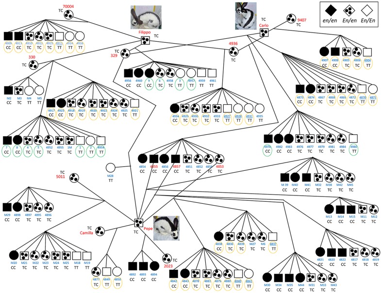
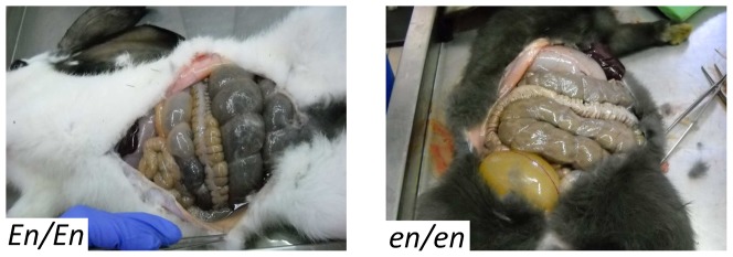
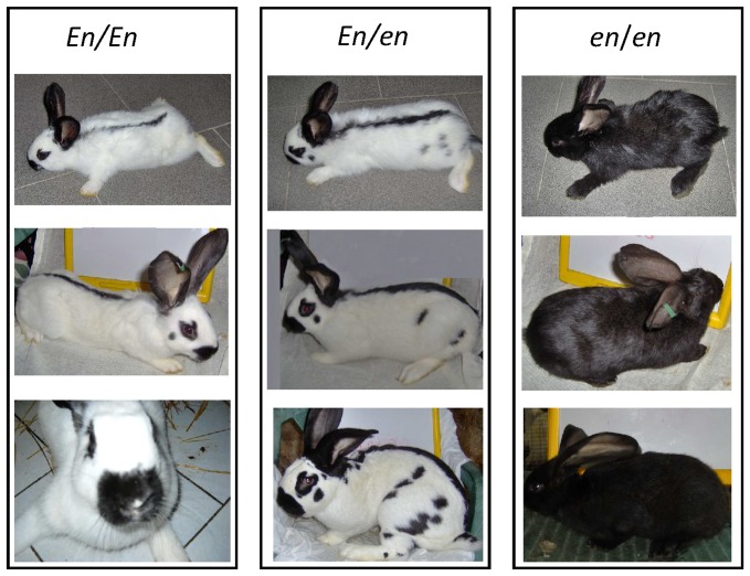
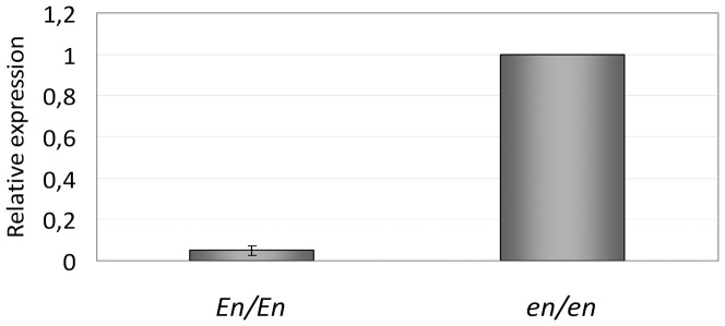
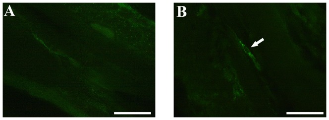
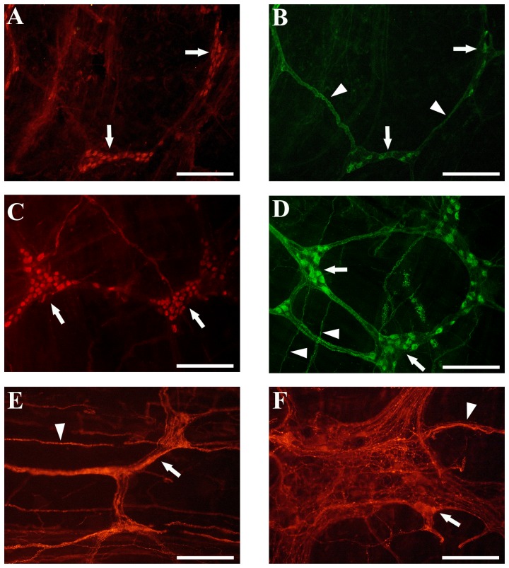
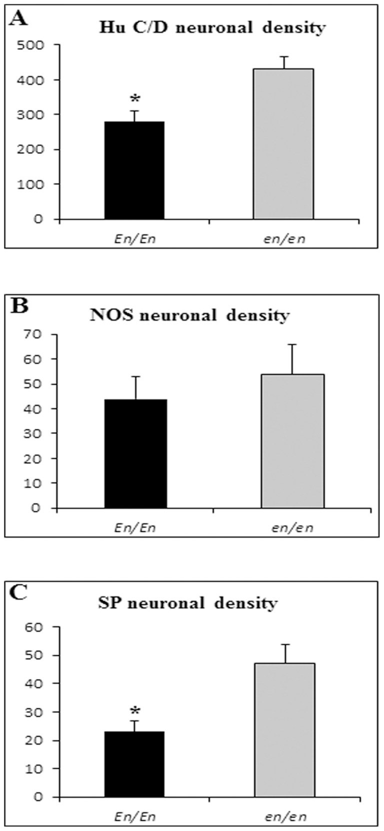
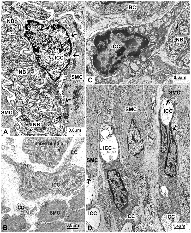
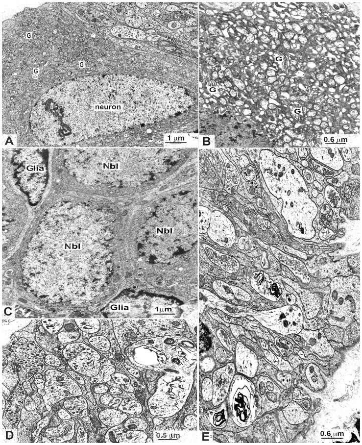
The English spotting coat color locus in rabbits, also known as Dominant white spotting locus, is determined by an incompletely dominant allele (En). Rabbits homozygous for the recessive wild-type allele (en/en) are self-colored, heterozygous En/en rabbits are normally spotted, and homozygous En/En animals are almost completely white. Compared to vital en/en and En/en rabbits, En/En animals are subvital because of a dilated ("mega") cecum and ascending colon. In this study, we investigated the role of the KIT gene as a candidate for the English spotting locus in Checkered Giant rabbits and characterized the abnormalities affecting enteric neurons and c-kit positive interstitial cells of Cajal (ICC) in the megacolon of En/En rabbits. Twenty-one litters were obtained by crossing three Checkered Giant bucks (En/en) with nine Checkered Giant (En/en) and two en/en does, producing a total of 138 F1 and backcrossed rabbits. Resequencing all coding exons and portions of non-coding regions of the KIT gene in 28 rabbits of different breeds identified 98 polymorphisms. A single nucleotide polymorphism genotyped in all F1 families showed complete cosegregation with the English spotting coat color phenotype (θ=0.00 LOD =75.56). KIT gene expression in cecum and colon specimens of En/En (pathological) rabbits was 5-10% of that of en/en (control) rabbits. En/En rabbits showed reduced and altered c-kit immunolabelled ICC compared to en/en controls. Morphometric data on whole mounts of the ascending colon showed a significant decrease of HuC/D (P<0.05) and substance P (P<0.01) immunoreactive neurons in En/En vs. en/en. Electron microscopy analysis showed neuronal and ICC abnormalities in En/En tissues. The En/En rabbit model shows neuro-ICC changes reminiscent of the human non-aganglionic megacolon. This rabbit model may provide a better understanding of the molecular abnormalities underlying conditions associated with non-aganglionic megacolon.
Conflict of interest statement
Figures
Similar articles
-
A frameshift mutation in the melanophilin gene causes the dilute coat colour in rabbit (Oryctolagus cuniculus) breeds.Anim Genet. 2014 Apr;45(2):248-55. doi: 10.1111/age.12104. Epub 2013 Dec 10. Anim Genet. 2014. PMID: 24320228
-
Mutations in the melanocortin 1 receptor (MC1R) gene are associated with coat colours in the domestic rabbit (Oryctolagus cuniculus).Anim Genet. 2006 Oct;37(5):489-93. doi: 10.1111/j.1365-2052.2006.01494.x. Anim Genet. 2006. PMID: 16978179
-
Platinum coat color in red fox (Vulpes vulpes) is caused by a mutation in an autosomal copy of KIT.Anim Genet. 2015 Apr;46(2):190-9. doi: 10.1111/age.12270. Epub 2015 Feb 6. Anim Genet. 2015. PMID: 25662789
-
[Effects of Kit gene on coat depigmentation in white horses].Yi Chuan. 2011 Nov;33(11):1171-8. doi: 10.3724/sp.j.1005.2011.01171. Yi Chuan. 2011. PMID: 22120071 Review. Chinese.
-
Rabbit Pediatrics.Vet Clin North Am Exot Anim Pract. 2024 May;27(2):171-191. doi: 10.1016/j.cvex.2023.11.003. Epub 2023 Nov 22. Vet Clin North Am Exot Anim Pract. 2024. PMID: 37993319 Review.
Cited by
-
The Genetics of Deafness in Domestic Animals.Front Vet Sci. 2015 Sep 8;2:29. doi: 10.3389/fvets.2015.00029. eCollection 2015. Front Vet Sci. 2015. PMID: 26664958 Free PMC article. Review.
-
A Frameshift Mutation in KIT is Associated with White Spotting in the Arabian Camel.Genes (Basel). 2017 Mar 9;8(3):102. doi: 10.3390/genes8030102. Genes (Basel). 2017. PMID: 28282952 Free PMC article.
-
Endothelin Receptor B2 (EDNRB2) Gene Is Associated with Spot Plumage Pattern in Domestic Ducks (Anas platyrhynchos).PLoS One. 2015 May 8;10(5):e0125883. doi: 10.1371/journal.pone.0125883. eCollection 2015. PLoS One. 2015. PMID: 25955279 Free PMC article.
-
Genomic diversity and signatures of selection in meat and fancy rabbit breeds based on high-density marker data.Genet Sel Evol. 2022 Jan 21;54(1):3. doi: 10.1186/s12711-022-00696-9. Genet Sel Evol. 2022. PMID: 35062866 Free PMC article.
-
TAD border deletion at the Kit locus causes tissue-specific ectopic activation of a neighboring gene.Nat Commun. 2024 May 28;15(1):4521. doi: 10.1038/s41467-024-48523-7. Nat Commun. 2024. PMID: 38806452 Free PMC article.
References
-
- Castle WE (1930) The genetics of domestic rabbit. Cambridge: Harvard University Press. p. 31.
-
- Robinson R (1958) Genetic studies of the rabbit. Bibl Genet 17: 229–558.
-
- Searle AG (1968) Comparative genetics of coat colour in mammals. London, UK: Logos Press. 308 p.
-
- Aigner B, Besenfelder U, Müller M, Brem G (2000) Tyrosinase gene variants in different rabbit strains. Mamm Genome 11: 700–702. - PubMed
-
- Fontanesi L, Tazzoli M, Beretti F, Russo V (2006) Mutations in the melanocortin 1 receptor (MC1R) gene are associated with coat colours in the domestic rabbit (Oryctolagus cuniculus). Anim Genet 37: 489–493. - PubMed
Publication types
MeSH terms
Substances
LinkOut - more resources
Full Text Sources
Other Literature Sources
Research Materials
