. 2014 Apr 15;9(4):e94238.
doi: 10.1371/journal.pone.0094238. eCollection 2014.

Expression of a truncated ATHB17 protein in maize increases ear weight at silking

Affiliations

Expression of a truncated ATHB17 protein in maize increases ear weight at silking

Elena A Rice et al. PLoS One. .

Abstract

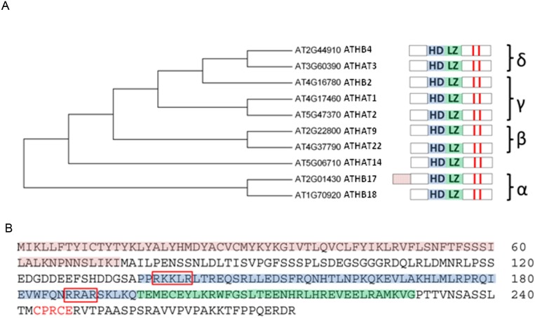
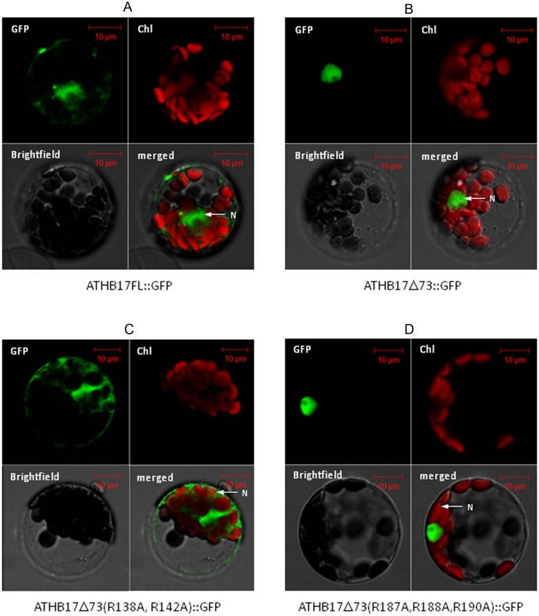
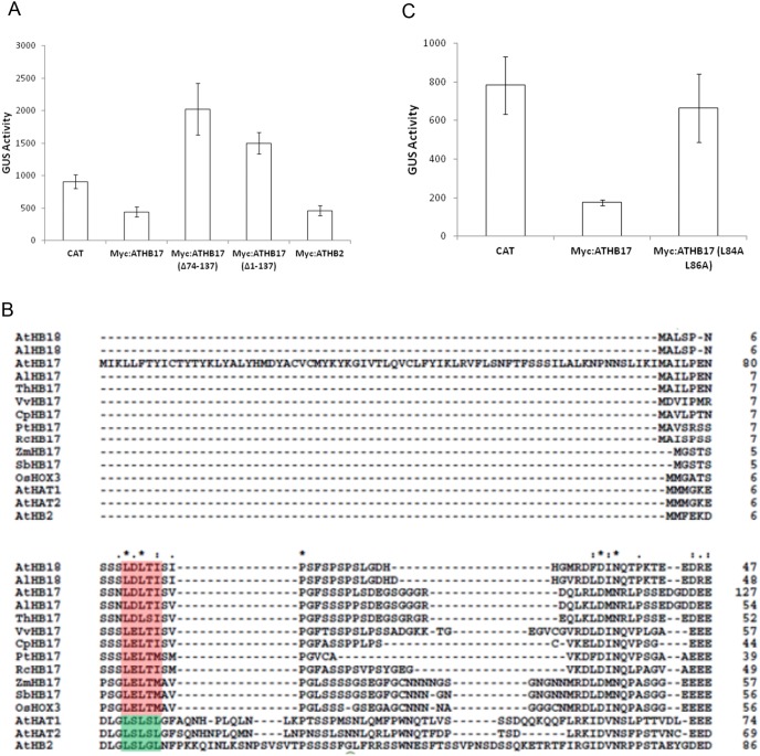
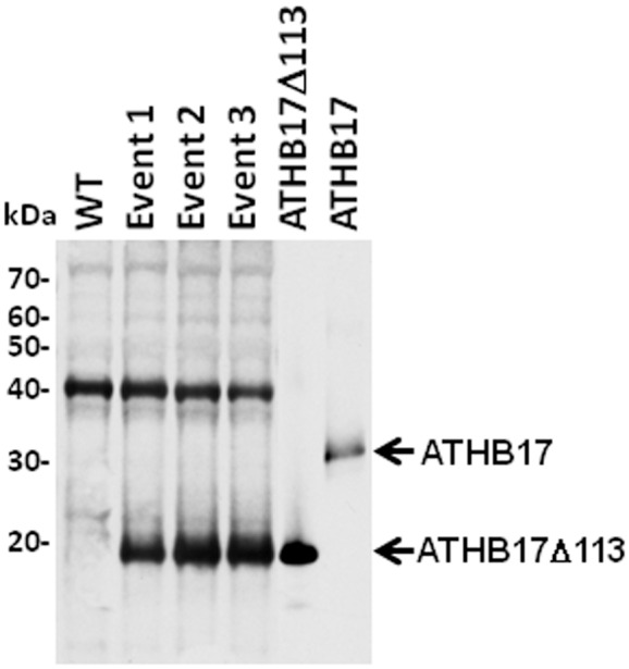
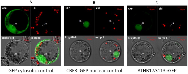
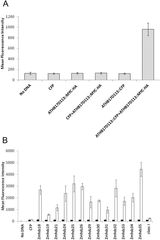
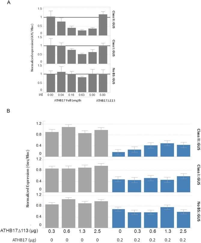
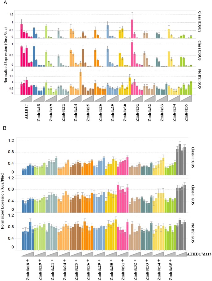
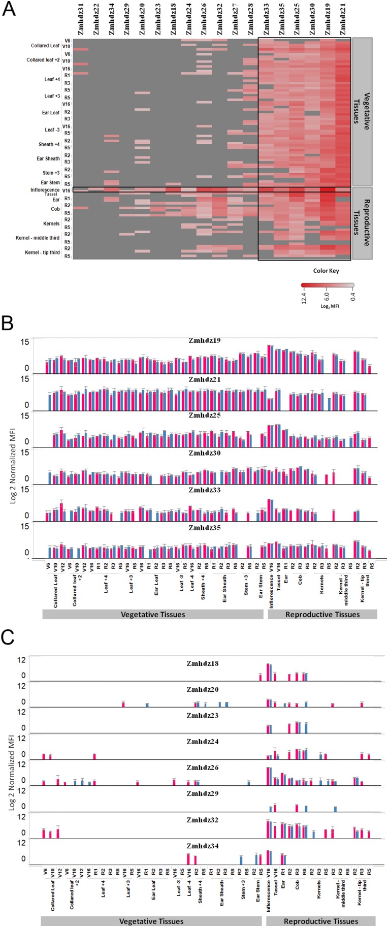
ATHB17 (AT2G01430) is an Arabidopsis gene encoding a member of the α-subclass of the homeodomain leucine zipper class II (HD-Zip II) family of transcription factors. The ATHB17 monomer contains four domains common to all class II HD-Zip proteins: a putative repression domain adjacent to a homeodomain, leucine zipper, and carboxy terminal domain. However, it also possesses a unique N-terminus not present in other members of the family. In this study we demonstrate that the unique 73 amino acid N-terminus is involved in regulation of cellular localization of ATHB17. The ATHB17 protein is shown to function as a transcriptional repressor and an EAR-like motif is identified within the putative repression domain of ATHB17. Transformation of maize with an ATHB17 expression construct leads to the expression of ATHB17Δ113, a truncated protein lacking the first 113 amino acids which encodes a significant portion of the repression domain. Because ATHB17Δ113 lacks the repression domain, the protein cannot directly affect the transcription of its target genes. ATHB17Δ113 can homodimerize, form heterodimers with maize endogenous HD-Zip II proteins, and bind to target DNA sequences; thus, ATHB17Δ113 may interfere with HD-Zip II mediated transcriptional activity via a dominant negative mechanism. We provide evidence that maize HD-Zip II proteins function as transcriptional repressors and that ATHB17Δ113 relieves this HD-Zip II mediated transcriptional repression activity. Expression of ATHB17Δ113 in maize leads to increased ear size at silking and, therefore, may enhance sink potential. We hypothesize that this phenotype could be a result of modulation of endogenous HD-Zip II pathways in maize.


Conflict of interest statement

Competing Interests: All authors are or were affiliated with the Monsanto Company or Mendel Biotechnology, who were also funders of this study. This study was partly funded by BASF. Monsanto Company has filed the following United States patent applications on certain aspects of this study: “Transgenic Plants with Enhanced Agronomic Traits” (11/311,920); “Interfering with HD-ZIP Transcription Factor Repression of Gene Expression to Produce Plants with Enhanced Traits” (61/888,980); “Transgenic Corn Event MON87403 and Methods for Detection Thereof” (61/888,978); and “Plant Regulatory Elements and Uses Thereof” (61/888,972). Mendel Biotechnology has obtained the following United States patent on certain aspects of this study: “Polynucleotides and Polypeptides in Plants” (US 7,511,190) and has filed the following United States patent application on certain aspects of this study: “Transcription Factor Sequences for Conferring Advantageous Properties to Plants” (12/573,311). This does not alter the authors’ adherence to all the PLoS ONE policies on sharing data and materials.

Figures

Figure 1
Figure 1. ATHB17 is a member of the α subclass within the HD-Zip II protein family.
(A) represents the dendrogram and the domain architecture of the ATHB17 homologs. ATHB17 contains a typical homeodomain (HD; blue shading) and a leucine zipper motif (LZ; green shading) adjacent to the C-terminus of the HD. Red bars indicate conserved cysteines in the C-terminus. (B) shows the protein sequence of ATHB17. ATHB17 contains a unique N-terminal extension (red shading) rich in cysteines and tyrosines. Additional structural feature identified for ATHB17 is a nuclear localization signal (red boxes). Downstream of the LZ motif is a putative redox sensing motif (CPXCE; red letters).
Figure 2
Figure 2. Representative fluorescence micrographs of Arabidopsis mesophyll protoplasts.
Purified protoplasts were transformed with (A) full length ATHB17:GFP or (B) a variant lacking the first 73 amino acids (ATHB17Δ73:GFP). The full length ATHB17:GFP fusion protein localizes to the nucleus and cytosol (A) in Arabidopsis protoplasts and ATHB17Δ73:GFP is localized exclusively in the nucleus when the unique N- terminus is removed (B). Two regions with a cluster of basic (K and R) residues were identified as putative nuclear localization signals. The arginine residues were mutated to alanines in these regions in the Δ73 variant to disrupt translocation. (C) R138A and R142A mutations in ATHB17 Δ73 disrupted nuclear retention while (D) R187A, R188A and R190A mutations did not alter the nuclear localization of the Δ73 variant.
Figure 3
Figure 3. Effect of various mutations on the transcriptional repression and identification of EAR-like motif.
(A) Arabidopsis mesophyll protoplasts were co-transformed with the pHAT1::GUS reporter gene and the following effector constructs: chloramphenicol acetyltransferase (CAT), ATHB17, ATHB17 with amino acids 74–137 deleted, ATHB17 with amino acids 1–137 deleted and ATHB2. Data are mean fluorescence readings measuring GUS-mediated substrate (4-methylumbelliferyl-beta-D-glucuronide) conversion. Error bars represent ± SD of three replicates. (B) Multiple alignment of the protein sequences similar to the amino terminus of ATHB17 from publically available HD-Zip II α subfamily proteins. Gaps are indicated by dashes. A putative EAR motif is shaded in red for the α-subclass, while the EAR motif identified in is shaded in green. Abbreviations for species are as follows: At Arabidopsis thaliana; Al Arabidopsis lyrata; Th Thellungiella halophila; Vv Vitis vinifera; Cp Carica papaya; Pt Populus trichocarpa; Rc Ricinus communis; Zm Zea mays; Sb Sorghum bicolor; Os Oryza sativa. (C) ATHB17, ATHB17 with the EAR motif mutated (ATHB17 L84A L86A), or CAT effector constructs were co-transformed into Arabidopsis mesophyll protoplasts with the pHAT1::GUS reporter gene. Data are mean fluorescence readings measuring GUS-mediated substrate (4-methylumbelliferyl-beta-D-glucuronide) conversion. Error bars represent ± SD of three replicates.
Figure 4
Figure 4. Western blot analysis showing truncated ATHB17 protein in transgenic maize events.
Extracts from WT and transgenic maize events were separated on SDS-PAGE, transferred on to nitrocellulose membrane, and probed with ATHB17 antibody. An E. coli purified preparation of full-length and truncated protein was also analyzed for comparison with plant expressed protein as a control. Molecular weight markers are shown on the left.
Figure 5
Figure 5. ATHB17Δ113 is localized to nucleus.
Fluorescence micrographs of maize leaf protoplasts transformed with (A) GFP alone, (B) AtCBF3 fused to GFP as positive control for nuclear localization or (C) ATHB17Δ113 fused with GFP. Protoplasts from each construct were analyzed by laser scanning microscopy for GFP and chlorophyll fluorescence and acquired images were merged on bright-field image using the Image examiner (See Materials and Methods for details).
Figure 6
Figure 6. ATHB17Δ113 forms homodimers and makes heterodimers with maize HD-Zip II proteins.
Maize leaf protoplasts were (A) mock- transformed or transformed with constructs expressing CFP, MYC::HA and CFP tagged ATHB17Δ113 alone or CFP and ATHB17Δ113::CFP in combination with ATHB17Δ113::MYC construct, (B) transformed (filled bars) with constructs expressing CFP or each of the CFP tagged HD-Zip IIs alone or co-transformed (empty bars) with the construct expressing ATHB17Δ113::MYC-HA. Cellular extracts were co-immunoprecipitated using anti-MYC antibody and the immunoprecipitated complex was probed with anti-CFP antibody and visualized using phycoerythrin-conjugated reporter molecules. One HD-Zip I, Zmhdz3, was used to evaluate interaction with ATHB17Δ113. Nomenclature of HD-Zip II and HD-Zip I constructs is based on Zhao et al..
Figure 7
Figure 7. Full- length ATHB17 protein functions as transcriptional repressor and ATHB17Δ113 can relieve repression caused by full-length ATHB17 protein.
Maize mesophyll protoplasts were transformed (A) with 4 µg cells of reporter (Class II::GUS, Class I::GUS or No BS::GUS) and 0–5 µg cells of effector (Full-length ATHB17) or 5 µg of ATHB17Δ113 and Renilla luciferase (B) with 4 µg reporter (Class II::GUS, Class I::GUS or No BS::GUS), 0–5 µg ATHB17Δ113, and 0 (grey bars) or 0.2 µg (blue bars) of ATHB17 full length. DNA amounts are per 320,000 cells. After 18 h, cells were assayed for GUS and luciferase expression. GUS values were divided by luciferase internal control values for each well and normalized to respective GFP samples. Bars are means and error bars represent 1 SD.
Figure 8
Figure 8. Maize HD-Zip II proteins function as transcriptional repressors and ATHB17Δ113 relieves the repression caused by endogenous maize HD-Zip II proteins.
(A) Cells were co-transformed with 4 µg/320,000 cells of each reporter (right side label) and an increasing amount of each HD-Zip II family member (left-to-right for each color: 0.008; 0.04; 0.2; 1; 5 µg/320,000 cells). Error bars are 1 SD. (B) Cells were triple-transformed with 4 µg/320,000 cells of each reporter (right side label), 20 ng of each HD-Zip family member and an increasing amount of ATHB17Δ113 (left-to-right for each color: 0.0, 0.3; 0.6; 1.3; 2.5 µg/320,000 cells). For a given reporter, HD-Zip II, and concentration of ATHB17Δ113, the calculation of standard error relies on the number of biological replicates and the estimated error variance derived from a one-way ANOVA.
Figure 9
Figure 9. Expression pattern of endogenous HD-Zip II transcripts in maize.
(A) Heat map representation of endogenous HD-Zip II transcript expression across tissues and developmental stages in two maize hybrids. The red color gradient shows expression values represented on a Log2 scale normalized median fluorescence intensity (MFI); a darker shade indicates a higher expression level. Grey color represents that expression was below background. For each of the listed tissue and development stages two hybrids (NH6214 and NN6306) are shown in the order that hybrid NN6306 is the first one. (B) Constitutive expression profiles of maize HD-Zip II genes. (C) Reproductive tissue specific profiles of maize HD-Zip II genes. Pink bar represents NN6306 and blue bar represents NH6214. Each panel shows the expression profile for the indicated HD-Zip II gene. The error bars are +/−1 stderr.

Similar articles

Cited by

References

    1. Ariel FD, Manavella PA, Dezar CA, Chan RL (2007) The true story of the HD-Zip family. Trends in Plant Science 12: 419–426. - PubMed
    1. Ruberti I, Sessa G, Lucchetti S, Morelli G (1991) A novel class of plant proteins containing a homeodomain with a closely linked leucine zipper motif. EMBO J 10: 1787–1791. - PMC - PubMed
    1. Mukherjee K, Brocchieri L, Burglin TR (2009) A Comprehensive Classification and Evolutionary Analysis of Plant Homeobox Genes. Molecular Biology and Evolution 26: 2775–2794. - PMC - PubMed
    1. Sessa G, Morelli G, Ruberti I (1993) The Athb-1 and -2 HD-Zip domains homodimerize forming complexes of different DNA binding specificities. EMBO J 12: 3507–3517. - PMC - PubMed
    1. Steindler C, Matteucci A, Sessa G, Weimar T, Ohgishi M, et al. (1999) Shade avoidance responses are mediated by the ATHB-2 HD-Zip protein, a negative regulator of gene expression. Development 126: 4235–4245. - PubMed

Publication types

MeSH terms