. 2014 Apr 15;9(4):e94982.
doi: 10.1371/journal.pone.0094982. eCollection 2014.

Activated macrophage survival is coordinated by TAK1 binding proteins

Affiliations

Activated macrophage survival is coordinated by TAK1 binding proteins

September R Mihaly et al. PLoS One. .

Abstract

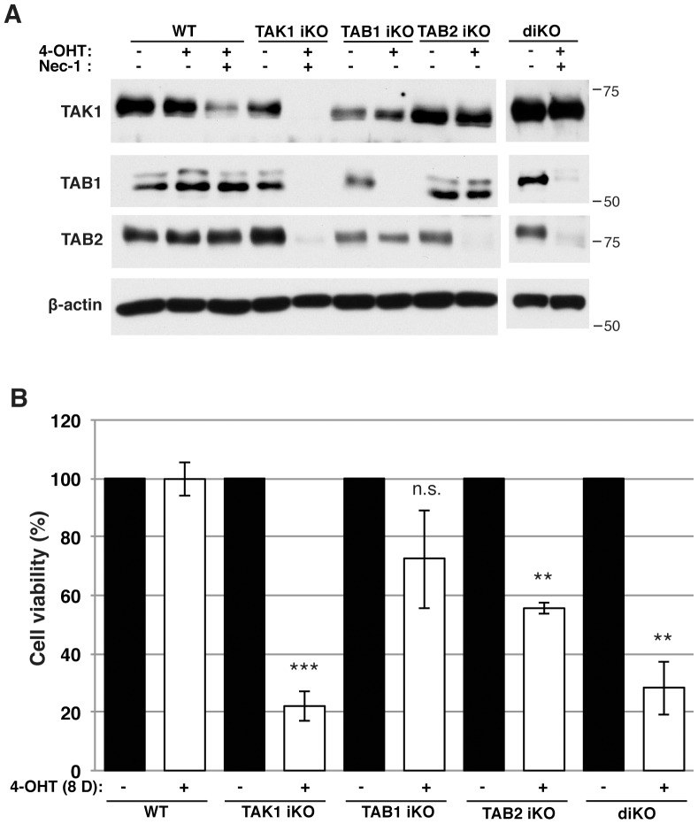
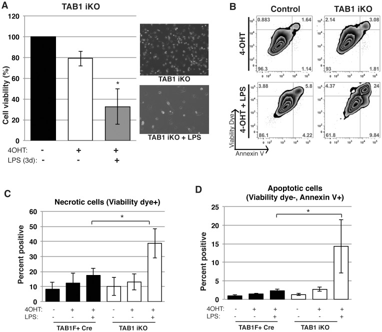
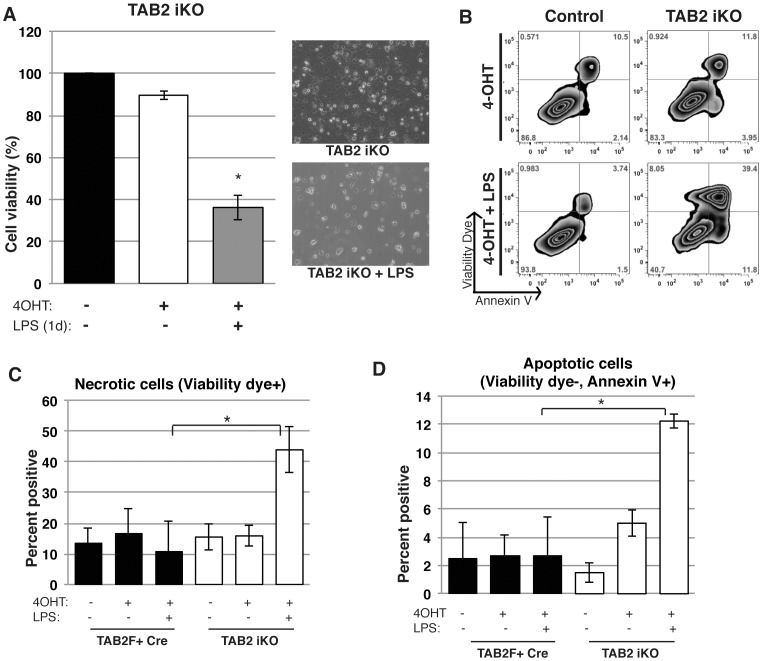
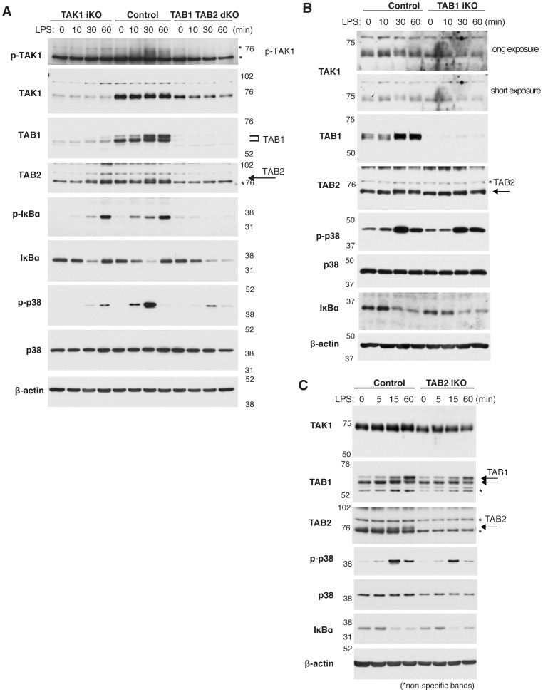
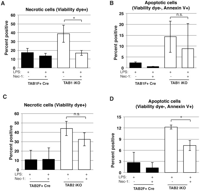
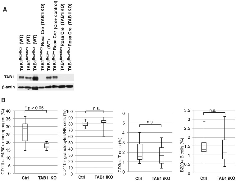
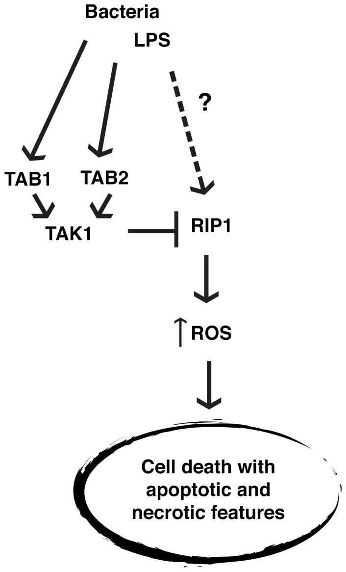
Macrophages play diverse roles in tissue homeostasis and immunity, and canonically activated macrophages are critically associated with acute inflammatory responses. It is known that activated macrophages undergo cell death after transient activation in some settings, and the viability of macrophages impacts on inflammatory status. Here we report that TGFβ- activated kinase (TAK1) activators, TAK1-binding protein 1 (TAB1) and TAK1-binding protein 2 (TAB2), are critical molecules in the regulation of activated macrophage survival. While deletion of Tak1 induced cell death in bone marrow derived macrophages even without activation, Tab1 or Tab2 deletion alone did not profoundly affect survival of naïve macrophages. However, in lipopolysaccharide (LPS)-activated macrophages, even single deletion of Tab1 or Tab2 resulted in macrophage death with both necrotic and apoptotic features. We show that TAB1 and TAB2 were redundantly involved in LPS-induced TAK1 activation in macrophages. These results demonstrate that TAK1 activity is the key to activated macrophage survival. Finally, in an in vivo setting, Tab1 deficiency impaired increase of peritoneal macrophages upon LPS challenge, suggesting that TAK1 complex regulation of macrophages may participate in in vivo macrophage homeostasis. Our results demonstrate that TAB1 and TAB2 are required for activated macrophages, making TAB1 and TAB2 effective targets to control inflammation by modulating macrophage survival.


Conflict of interest statement

Competing Interests: The authors have declared that no competing interests exist.

Figures

Figure 1
Figure 1. TAK1 is required for macrophage survival.
(A) Western blotting analysis of TAK1, TAB1 and TAB2 in control, Tak1iKO, Tab1iKO, Tab2iKO and diKO BMDMs. Bone marrow cells were cultured in macrophage medium and treated with 0.3 µM 4-OHT or vehicle (ethanol) for 4 days. Tak1iKO and diKO BMDMs were additionally treated with 50 µM Necrostatin-1 (Nec-1). Anti-β-actin Western blotting was used as a loading control. The numbers beside each panel denote the size and the position of molecular weight markers. (B) Viability of WT, Tak1iKO, Tab1iKO, Tab2iKO, and inducible double-deficient (diKO) BMDMs. Cells were cultured for 8 days with 0.3 µM 4-OHT and stained with 0.1% Crystal Violet. Data are mean percentages of attached macrophages compared to ethanol-treated +/− SD for 3 independent experiments. Asterisks indicate p-values: **  =  P<0.005; ***  =  p<0.0005.
Figure 2
Figure 2. TAB1 is required for LPS-activated macrophage survival.
(A) Viability of LPS-treated Tab1iKO macrophages. Tab1iKO and control BMDMs were cultured for 8 days with 0.3 µM 4-OHT followed by 3 days 1 µg/ml LPS. Viability was measured by Crystal Violet Assay, and data shown are mean percentages of attached macrophages compared to 8 days treated with vehicle +/− SD of 3 independent experiments. (B) Flow cytometry analysis of Tab1iKO BMDMs. Tab1iKO or Tab1F+ Cre BMDMs were cultured in macrophage medium with 0.3 µM 4-OHT or vehicle (ethanol) for 4 days. All cells including attached and floating cells were collected and stained with annexin V-Pacific Blue and Fixable viability dye eFlour 780, then analyzed on flow cytometer. Events were gated to exclude dead cells and debris, then gated on events positive for annexin V and fixable viability dye compared with unstained controls. Shown is representative figure of 3 independent experiments. (C and D) Tab1iKO and controls including WT and F+Cre BMDMs were cultured 3 days in 0.3 µM 4-OHT-containing macrophage medium, then 4 days with the addition of 1 µg/ml LPS. (C) Viability Dye-positive cells as a percentage of total cells is shown. (D) Graph shows Annexin V-positive but viability dye-negative cells. Graph represents results of four independent experiments +/− SD.
Figure 3
Figure 3. TAB2 is required for LPS-activated macrophage survival.
(A) Viability of LPS-activated Tab2-deficient macrophages. Tab2iKO BMDMs were cultured for 8 days with 0.3 µM 4-OHT, then 1 µg/ml LPS was added to culture medium for 1 day. Data are mean percentages of attached macrophages compared to 8 days treated with 4-OHT alone +/− SD between 3 independent experiments as measured by Crystal Violet Assay. (B) Percentages of annexin V and viability dye positive cells. Tab2iKO and control (WT) BMDMs were cultured with 0.3 µM 4-OHT for 3 days then with 1 µg/ml LPS for 1 additional day in 3 independent experiments. Cells were stained with viability dye and annexin V and analyzed by flow cytometry. Percentages of single positive and double positive cells based on unstained controls in a representative experiment are shown. (C and D) Necrotic and apoptotic LPS-activated macrophages with Tab2 deficiency. Tab2iKO, and controls including WT and F+Cre BMDMs were cultured 3 days in 0.3 µM 4-OHT-containing macrophage medium, then 1 day in medium containing 0.3 µM 4-OHT and 1 µg/ml LPS. (C) Necrotic cells are shown as percentage positive for viability dye. (D) Annexin V positive but viability dye-negative cells as a percentage of total cells is shown. Graphs indicate results of four independent experiments +/− SD.
Figure 4
Figure 4. LPS activates TAK1 through TAB1 and TAB2.
(A) Western blotting analysis of Tak1iKO and Tab1Tab2diKO and control BMDMs. Cells were cultured for 4 days with 0.3 µM 4-OHT in the presence of 50 µM Nec-1, followed by 1 µg/ml LPS treatment for the indicated period of time. Anti-βactin was used as a loading control. Asterisks indicate non-specific bands. (B) Tab1iKO or control BMDMs were cultured with 0.3 µM 4-OHT for 3 days then treated with 1 µg/ml LPS for the indicated period of time. Whole cell extracts were analyzed by Western blotting using the indicated antibodies. Asterisks indicate non-specific bands. (C) Tab2iKO or control BMDMs were cultured with 0.3 µM 4-OHT for 3 days then treated with 1 µg/ml LPS for the indicated period of time. Whole cell extracts were analyzed by Western blotting using the indicated antibodies. Asterisks indicate non-specific bands.
Figure 5
Figure 5. TAB1 or TAB2 deletion causes RIP1-dependent cell death.
(A) Tab1iKO or control BMDMs were cultured with 0.3 µM 4-OHT with or without 50 µM Nec-1 for 8 days then treated with 1 µM LPS for 3 days, and viability was measured by Crystal Violet Assay. *  =  p<.05. (B) Viability of Tab2iKO BMDMs treated with RIP1 inhibitor. Tab2iKO BMDMs were treated with 0.3 µM 4-OHT for 8 days with our without 50 µM Necrostatin-1 (Nec-1), then treated with 1 µM LPS for one day. Viability was measured by Crystal Violet Assay. *  =  p<.05.
Figure 6
Figure 6. LPS treated Tab1iKO macrophages are reduced in vivo.
(A) Tab1iKO or control including Tab1F+ Cre mice were intraperitoneally injected with 50 mg/kg tamoxifen for 3 consecutive days. After 3–5 weeks, peripheral blood was collected and leukocyte extracts were tested for Tab1 deletion by Western blotting. (B) Tab1iKO (n = 6) and control mice (n = 9) were intraperitoneally injected with 8 mg/kg LPS. Peritoneal leukocytes were collected at 72 hours and stained with fluorophore-conjugated antibodies. Shown is percent positive, excluding dead cells and debris, for the indicated markers. Percentages of CD11b+ F4/80+, CD11b+, CD3e+ or B220+ cells of total cells +/− SD is shown.
Figure 7
Figure 7. TAB1 and TAB2 are essential for LPS-activated macrophage survival.
TAK1 binding proteins, TAB1 and TAB2 are essential for protecting BMDMs from LPS-induced cell death, which occurs downstream of RIP1, involves increased ROS, and shows features of both apoptosis and necrosis.

Similar articles

Cited by

References

    1. Wynn TA, Chawla A, Pollard JW (2013) Macrophage biology in development, homeostasis and disease. Nature 496: 445–455 10.1038/nature12034 - DOI - PMC - PubMed
    1. Weigert A, Johann AM, Knethen A von, Schmidt H, Geisslinger G, et al. (2006) Apoptotic cells promote macrophage survival by releasing the antiapoptotic mediator sphingosine-1-phosphate. Blood 108: 1635–1642 10.1182/blood-2006-04-014852 - DOI - PubMed
    1. He S, Liang Y, Shao F, Wang X (2011) Toll-like receptors activate programmed necrosis in macrophages through a receptor-interacting kinase-3-mediated pathway. Proc Natl Acad Sci U S A 108: 20054–20059 10.1073/pnas.1116302108 - DOI - PMC - PubMed
    1. Park JM, Greten FR, Wong A, Westrick RJ, Arthur JS, et al. (2005) Signaling pathways and genes that inhibit pathogen-induced macrophage apoptosis—CREB and NF-kappaB as key regulators. Immunity 23: 319–329 10.1016/j.immuni.2005.08.010 - DOI - PubMed
    1. Ma Y, Temkin V, Liu H, Pope RM (2005) NF-kappaB protects macrophages from lipopolysaccharide-induced cell death: the role of caspase 8 and receptor-interacting protein. J Biol Chem 280: 41827–41834 10.1074/jbc.M510849200 - DOI - PubMed

Publication types

MeSH terms