Review
. 2014 Apr 15;28(8):812-28.
doi: 10.1101/gad.234294.113.

DNA methylation dynamics during epigenetic reprogramming in the germline and preimplantation embryos

Affiliations
Review

DNA methylation dynamics during epigenetic reprogramming in the germline and preimplantation embryos

Daniel M Messerschmidt et al. Genes Dev. .

Abstract

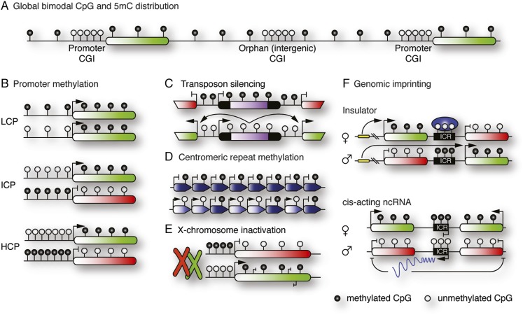
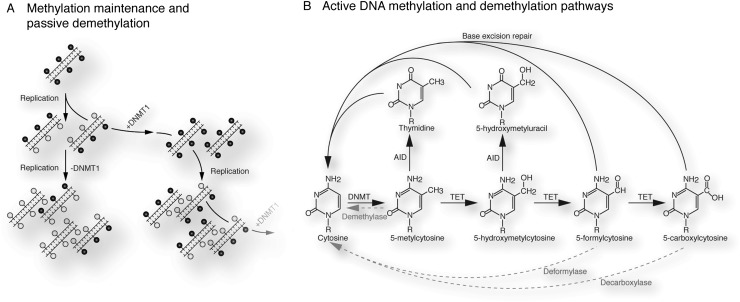
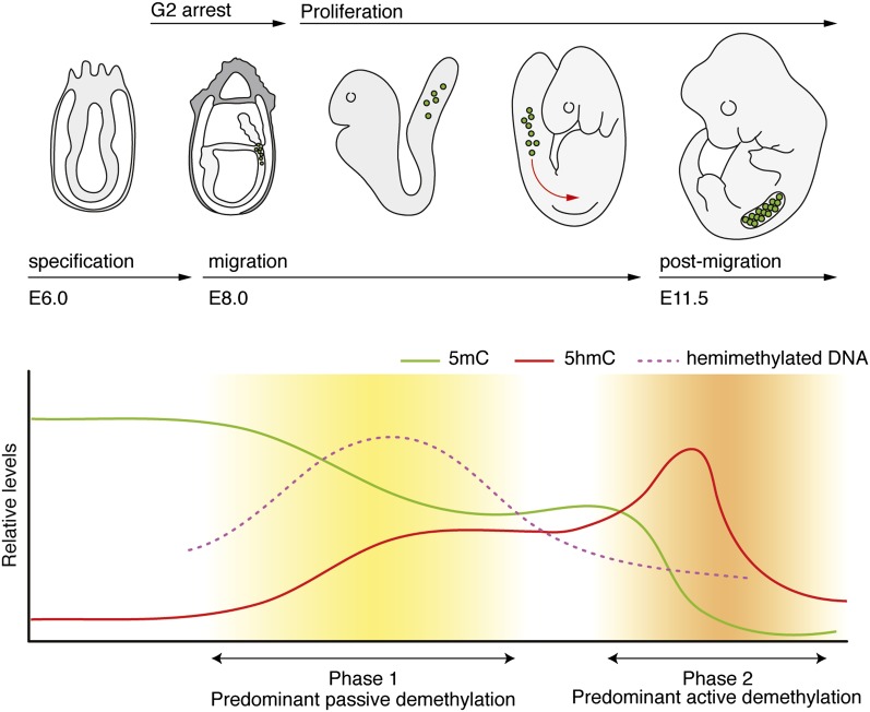
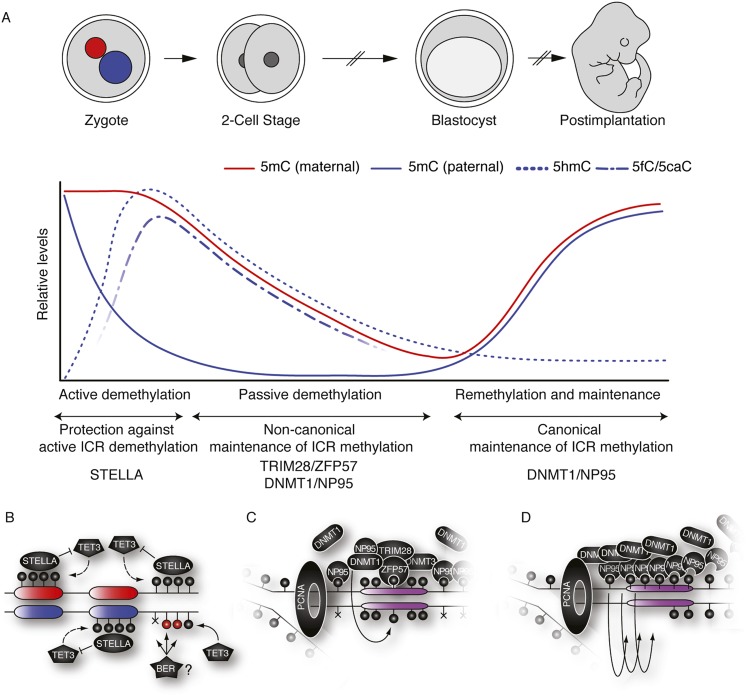
Methylation of DNA is an essential epigenetic control mechanism in mammals. During embryonic development, cells are directed toward their future lineages, and DNA methylation poses a fundamental epigenetic barrier that guides and restricts differentiation and prevents regression into an undifferentiated state. DNA methylation also plays an important role in sex chromosome dosage compensation, the repression of retrotransposons that threaten genome integrity, the maintenance of genome stability, and the coordinated expression of imprinted genes. However, DNA methylation marks must be globally removed to allow for sexual reproduction and the adoption of the specialized, hypomethylated epigenome of the primordial germ cell and the preimplantation embryo. Recent technological advances in genome-wide DNA methylation analysis and the functional description of novel enzymatic DNA demethylation pathways have provided significant insights into the molecular processes that prepare the mammalian embryo for normal development.

Keywords: DNA methylation; development; epigenetics; reprogramming.


Figures

Figure 1.
Figure 1.
DNA methylation in the mammalian genome. (A) Genomic CpG distribution: CGIs are generally hypomethylated and found at promoters or intergenic regions (orphan CpG). Non-CGI CpGs are generally hypermethylated. (B) Three categories of gene promoters according to CpG density respond differently to methylation. (LCP) Low CpG density promoter; (ICP) intermediate CpG density promoter; (HCP) high CpG density promoter. (C) Retrotransposons are repressed by DNA methylation. Derepression can result in coactivation of neighboring genes. (D) Basal transcription of centromeric repeats interferes with chromosome alignment and is repressed by DNA methylation. (E) DNA methylation reinforces gene silencing on the inactivated X chromosome. Transcribed genes on the active X chromosome (and globally) display gene body methylation, possibly repressing spurious expression from cryptic transcription start sites or aiding RNA processing. (F) Genomic imprinting drives allele-specific gene expression. DNA methylation at imprinting control regions (ICRs) regulates binding of insulator proteins or expression of cis-acting, noncoding RNAs.
Figure 2.
Figure 2.
DNA methylation and demethylation mechanisms. (A) The palindromic nature of the CpG DNA replication creates hemimethylated DNA. DNMT1 restores hemimethylated DNA to full methylation. Absence of the DNMT1 machinery produces unmethylated DNA after a subsequent round of cell division. (Black filled circles) Methylated CpG; (white filled circles) unmethylated CpG. (B) Possible active demethylation pathways: A direct demethylase converting 5mC to cytosine is, to date, speculative. TET enzymes oxidize 5mC to 5hmC, 5fC, and 5caC. Deamination of 5mC and 5hmC, potentially by AID, produces thymidine or hydroxymethyluracil, respectively. The base excision repair (BER) mechanism may target AID deamination products and possibly 5fC and 5caC directly. Direct deformylation or decarboxylation of 5fC and 5caC has been proposed but remains speculative.
Figure 3.
Figure 3.
Biphasic demethylation dynamics in mouse PGCs. PGCs are derived from the embryonic ectoderm of the E6.5 embryo and display high (somatic) 5mC levels (green lines) and low 5hmC levels (red lines). Upon migration, PGCs proliferate, and 5mC levels are passively diluted. Coincidently, hemimethylated DNA strands accumulate transiently and are subsequently lost (purple dashed line). Post-migratory PGCs enter a phase of active DNA demethylation, resulting in an almost complete loss of 5mC and a transient enrichment of 5hmC. At E13.5, both 5mC and 5hmC levels are low.
Figure 4.
Figure 4.
DNA demethylation dynamics and imprinting maintenance in preimplantation embryos. (A) Distinct characteristics of maternal and paternal genomes impose an epigenetic asymmetry in the zygote. The maternal genome (red pronucleus; red line) undergoes passive DNA demethylation throughout several rounds of DNA replication. The paternal genome (blue pronucleus; blue lines) undergoes active demethylation before DNA replication in the zygote ensues. Concomitant with global loss of paternal 5mC, 5hmC (blue dotted line) and the further oxidation derivatives (5fC and 5caC; blue dashed line) are enriched. Although selected loci are restored to unmodified cytosines, the bulk of paternal 5hmC is passively diluted, paralleling demethylation of the maternal genome. (B) In the zygote, STELLA prevents TET3-dependent oxidation of 5mC through binding to H3K9me2-marked chromatin (maternal genome and paternally imprinted regions) and subsequent active restoration of cytosine by BER (or other pathways). (C) Throughout early cleavage stages, DNMT1 is largely excluded from the nucleus and requires noncanonical targeting to imprinted regions by the ZFP57/TRIM28 complex binding to its methylated consensus sequence found at most ICRs. Nonimprinted regions are efficiently demethylated through replication, while ICRs are maintained by DNMT1 and DNMT3A/B. (D) At later stages of embryogenesis and in adult tissues, high DNMT1/NP95 levels during replication maintain DNA methylation by targeting hemimethylated DNA in a canonical fashion.

References

    1. Abdalla H, Yoshizawa Y, Hochi S 2009. Active demethylation of paternal genome in mammalian zygotes. J Reprod Dev 55: 356–360 - PubMed
    1. Anastasiadou C, Malousi A, Maglaveras N, Kouidou S 2011. Human epigenome data reveal increased CpG methylation in alternatively spliced sites and putative exonic splicing enhancers. DNA Cell Biol 30: 267–275 - PubMed
    1. Aran D, Sabato S, Hellman A 2013. DNA methylation of distal regulatory sites characterizes dysregulation of cancer genes. Genome Biol 14: R21. - PMC - PubMed
    1. Arand J, Spieler D, Karius T, Branco MR, Meilinger D, Meissner A, Jenuwein T, Xu G, Leonhardt H, Wolf V, et al. 2012. In vivo control of CpG and non-CpG DNA methylation by DNA methyltransferases. PLoS Genet 8: e1002750. - PMC - PubMed
    1. Bartolomei MS 2009. Genomic imprinting: employing and avoiding epigenetic processes. Genes Dev 23: 2124–2133 - PMC - PubMed

Substances