. 2014 Jul;34(1):92-102.
doi: 10.3892/ijmm.2014.1745. Epub 2014 Apr 16.

The green tea extract epigallocatechin-3-gallate inhibits irradiation-induced pulmonary fibrosis in adult rats

Affiliations

The green tea extract epigallocatechin-3-gallate inhibits irradiation-induced pulmonary fibrosis in adult rats

Hua You et al. Int J Mol Med. 2014 Jul.

Abstract

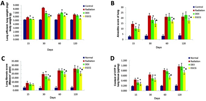
The present study evaluated the effect of epigallocatechin-3-gallate (EGCG), the most abundant catechin in green tea, on irradiation-induced pulmonary fibrosis and elucidated its mechanism of action. A rat model of irradiation-induced pulmonary fibrosis was generated using a (60)Co irradiator and a dose of 22 Gy. Rats were intraperitoneally injected with EGCG (25 mg/kg) or dexamethasone (DEX; 5 mg/kg) daily for 30 days. Mortality rates and lung index values were calculated. The severity of fibrosis was evaluated by assaying the hydroxyproline (Hyp) contents of pulmonary and lung tissue sections post-irradiation. Alveolitis and fibrosis scores were obtained from semi-quantitative analyses of hematoxylin and eosin (H&E) and Masson's trichrome lung section staining, respectively. The serum levels of transforming growth factor β1 (TGF-β1), interleukin (IL)-6, IL-10, and tumor necrosis factor-α (TNF-α) were also measured. Surfactant protein-B (SPB) and α-SMA expression patterns were evaluated using immunohistochemistry, and the protein levels of nuclear transcription factor NF-E2-related factor 2 (Nrf-2) and its associated antioxidant enzymes heme oxygenase-1 enzyme (HO-1) and

Nad(p)h: quinone oxidoreductase-1 (NQO-1) were examined via western blot analysis. Treatment with EGCG, but not DEX, reduced mortality rates and lung index scores, improved histological changes in the lung, reduced collagen depositions, reduced MDA content, enhanced SOD activity, inhibited (myo)fibroblast proliferation, protected alveolar epithelial type II (AE2) cells, and regulated serum levels of TGF-β1, IL-6, IL-10, and TNF-α. Treatment with EGCG, but not DEX, activated Nrf-2 and its downstream antioxidant enzymes HO-1 and NQO-1. Taken together, these results showed that EGCG treatment significantly inhibits irradiation-induced pulmonary fibrosis. Furthermore, the results suggested promising clinical EGCG therapies to treat this disorder.


Figures

Figure 1
Figure 1
Effect of epigallocatechin-3-gallate (EGCG) on the lung appearance at 120 days post gamma-ray irradiation. (A) Hematoxylin and eosin (H&E) staining of lung tissue from non-irradiated normal control, (B) untreated irradiated, but (C) dexamethasone (DEX)-treated irradiated and (D) EGCG-treated irradiated rats. Untreated irradiated and DEX-treated irradiated animals show marked lung collapse, rough surfaces and gray fibrous nodule development. EGCG treatment animals show lung tissue edema, rough surfaces, scattered punctate bleeding, but no lung collapse and gray fibrous nodules. Bar, 0.5 cm.
Figure 2
Figure 2
(A) The effect of epigallocatechin-3-gallate (EGCG) on the lung index score, (B) combined alveolitis score, (C) combined fibrosis score, and (D) hydroxyproline (Hyp) content at 15, 30, 60 and 120 days post-irradiation. (A) Lung index values were significantly lower (p<0.05) among the EGCG-treated animals compared with those of the DEX-treated animals at 30 and 0 days post-irradiation but significantly higher (p<0.05) at 120 days post-irradiation. Lung index values were significantly lower (p<0.05) among dexamethasone (DEX)-treated animals compared with those of untreated animals at 15, 30 and 60 days post-irradiation but similar at 120 days post-irradiation. (B) The combined alveolitis score was significantly lower (p<0.05) among EGCG-treated animals than that among DEX-treated animals at 30, 60 and 120 days post-irradiation. This score was also significantly lower (p<0.05) among DEX-treated animals compared with that of untreated animals at 15 and 30 days post-irradiation but similar at 60 and 120 days post-irradiation. (C) The combined fibrosis score was significantly lower (p<0.05) among EGCG-treated animals compared with that of DEX-treated animals at 30, 60 and 120 days post-irradiation. The fibrosis score was significantly lower (p<0.05) among DEX-treated rats compared with that of untreated rats at 30 days post-irradiation but similar at 60 and 120 days post-irradiation. (D) The Hyp content was significantly lower (p<0.05) among EGCG-treated animals compared with that of DEX-treated animals at 60 and 120 days post-irradiation. This parameter was significantly lower (p<0.05) among the DEX-treated rats relative to that of the untreated animals at 15 and 30 days post-irradiation but similar at 60 and 120 days post-irradiation. The bars in the graph are the standard deviations (SDs). Asterisks show significance (p<0.05) compared with the untreated radiation-only rats, and stars show significance (p<0.05) compared with DEX-treated animals.
Figure 3
Figure 3
Effect of epigallocatechin-3-gallate (EGCG) on the histological changes in lung tissue at 120 days post-irradiation. Photomicrographs show staining of rat lung tissue sections with hematoxylin and eosin (H&E) (left column), Masson’s trichrome (middle column) and Sirius red (right column) from animals in the non-irradiated control (normal), irradiated but untreated (radiation) dexamethasone (DEX)-treated, and EGCG-treated (EGCG) groups. Note that inflammatory cell infiltration, fibrotic lesions and collagen fiber deposition were significantly improved in EGCG-treated animals. Bar, 100 μm.
Figure 4
Figure 4
(A) The effect of epigallocatechin-3-gallate (EGCG) on serum malondialdehyde (MDA) concentrations, (B) superoxide dismutase (SOD) activity, (C) α smooth muscle actin (α-SMA) levels and (D) surfactant protein-B (SPB) levels at 15, 30, 60 and 120 days post-irradiation. (A) The MDA concentration in serum was significantly lower (p<0.05) among the EGCG-treated animals compared with that of the dexamethasone (DEX)-treated animals at 60 and 120 days post-irradiation. Conversely, the MDA concentrations of the DEX-treated and untreated animals were comparable at 15, 30, 60 and 120 days post-irradiation. (B) Serum SOD activity was significantly higher (p<0.05) among the EGCG-treated animals compared with the DEX-treated animals at 15, 30, 60 and 120 days post-irradiation. The SOD activity of the latter group was significantly higher (p<0.05) than that of the radiation-only animals at 30 days post-irradiation but similar at 15, 60 and 120 days post-irradiation. (C) The OD value of α-SMA immunohistochemical (IHC) was significantly lower (p<0.05) among EGCG-treated animals than that of the DEX-treated animals at 60 and 120 days post-irradiation. Conversely, the OD value of α-SMA IHC among the DEX-treated animals was comparable to that of the untreated animals at 15, 30, 60 and 120 days post-irradiation (p>0.05). (D) The OD value of SPB IHC was significantly higher (p<0.05) among the EGCG-treated animals relative to the DEX-treated animals at 15, 30, 60 and 120 days post-irradiation. The OD value of SPB IHC among the DEX-treated animals was similar to that of the untreated animals at 15 days post-irradiation but significantly higher (p<0.05) at 30, 60 and 120 days post-irradiation. The bars in the graph are the standard deviations (SDs). Asterisks show significance (p<0.05) compared with the untreated radiation-only animals, and stars show significance (p<0.05) compared with the DEX-treated animals.
Figure 5
Figure 5
Effect of epigallocatechin-3-gallate (EGCG) on the α smooth muscle actin (α-SMA) and surfactant protein-B (SPB) immunohistochemistry at 120 days post-irradiation. Photomicrographs show the immunohistochemical (IHC) staining with antibodies detecting α-SMA (left column) or SPB (right column) in lung tissue from rats in the non-irradiated (normal), irradiated but untreated (Radiation), dexamethasone (DEX)-treated, and EGCG-treated (EGCG) groups. Myofibroblast accumulation hyperplasia as stained by α-SMA IHC was significantly reduced, and alveolar type II cells (stained by SPB IHC) were significantly increased in EGCG-treated animals. Bar, 100 μm.
Figure 6
Figure 6
(A) The effect of epigallocatechin-3-gallate (EGCG) on the serum levels of transforming growth factor β1 (TGF-β1), (B) interleukin (IL)-6, (C) IL-10, and (D) tumor necrosis factor α (TNF-α) at 15, 30, 60 and 120 days post-irradiation. (A) Serum TGF-β1 levels were significantly lower (p<0.05) in the EGCG-treated animals compared with those in the dexamethasone (DEX)-treated animals at 30, 60 and 120 days post-irradiation. The serum TGF-β1 levels in the DEX-treated animals were similar to those of the untreated animals at 30, 60 and 120 days post-irradiation. (B–D) The serum levels of IL-6, IL-10, and TNF-α were significantly lower (p<0.05) in the EGCG-treated animals compared with the DEX-treated animals at 60 and 120 days post-irradiation. The serum levels of IL-6 and TNF-α were significantly lower (p<0.05) in the DEX-treated animals compared with those in the untreated animals at 15 and 30 days post-irradiation. The serum levels of IL-10 were significantly lower (p<0.05) in the DEX-treated animals than those in the radiation-only animals at 30 days post-irradiation. The serum levels of IL-6, IL-10, and TNF-α in the DEX-treated animals were similar to those in the radiation-only animals at 60 and 120 days post-irradiation. The bars in each graph are the standard deviations (SDs). Asterisks show significance (p<0.05) compared with the untreated animals and stars show significance (p<0.05) compared with the DEX-treated animals.
Figure 7
Figure 7
(A) Epigallocatechin-3-gallate (EGCG) activates nuclear transcription factor NF-E2-related factor 2 (Nrf-2), (B) heme oxygenase-1 (HO-1) and (C) NAD(P)H:quinone oxidoreductase-1 (NQO-1) protein expression as detected by western blot analysis of lung tissue extracts at 15 days post-irradiation. Immunoblot analysis revealed that the protein expression of Nrf-2, HO-1, and NQO-1 was strongly activated by EGCG administration, while Nrf-2 was weakly activated by dexamethasone (DEX).
Figure 8
Figure 8
(A) The protein levels of nuclear transcription factor NF-E2-related factor 2 (Nrf-2), (B) heme oxygenase-1 (HO-1) and (C) NAD(P)H:quinone oxidoreductase-1 enzyme (NQO-1) were compared using western blot analysis of lung tissue extracts at 15, 30, 60 and 20 days post-irradiation. The relative protein expression levels of Nrf-2, HO-1, and NQO-1 in lung homogenates were significantly higher (p<0.05) in the epigallocatechin-3-gallate (EGCG)-treated animals than the dexamethasone (DEX)-treated animals at 15, 30, 60 and 120 days post-irradiation. The expression levels of these proteins in the latter group were significantly higher (p<0.05) than those of the radiation-only animals at 15 and 30 days post-irradiation but similar at 60 and 120 days post-irradiation. Experiments were performed with n=6 rats per group, and the western blot analysis OD data were normalized to OD values for β-actin. Data are presented as the means ± standard deviations (SDs). Asterisks show significance (p<0.05) compared with the untreated animals, and stars show significance (p<0.05) compared with the DEX-treated animals.

Similar articles

Cited by

References

    1. Almeida C, Nagarajan D, Tian J, et al. The role of alveolar epithelium in radiation-induced lung injury. PLoS One. 2013;8:e53628. - PMC - PubMed
    1. Epperly MW, Guo H, Gretton JE, Greenberger JS. Bone marrow origin of myofibroblasts in irradiation pulmonary fibrosis. Am J Respir Cell Mol Biol. 2003;29:213–224. - PubMed
    1. Matsuo Y, Shibuya K, Nakamura M, et al. Dose-volume metrics associated with radiation pneumonitis after stereotactic body radiation therapy for lung cancer. Int J Radiat Oncol Biol Phys. 2012;83:e545–e549. - PubMed
    1. Minor GI, Yashar CM, Spanos WJ, Jr, et al. The relationship of radiation pneumonitis to treated lung volume in breast conservation therapy. Breast J. 2006;12:48–52. - PubMed
    1. Rosenzweig KE, Zauderer MG, Laser B, et al. Pleural intensity-modulated radiotherapy for malignant pleural mesothelioma. Int J Radiat Oncol Biol Phys. 2012;83:1278–1283. - PMC - PubMed

Publication types

MeSH terms