

Multiple orthokeratinized odontogenic cysts: a case report
- PMID: 24737103
- PMCID: PMC4382487
- DOI: 10.1007/s12105-014-0545-5
Multiple orthokeratinized odontogenic cysts: a case report
Abstract
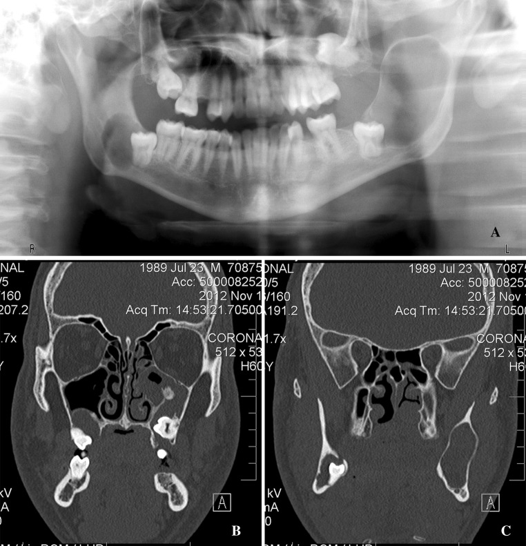
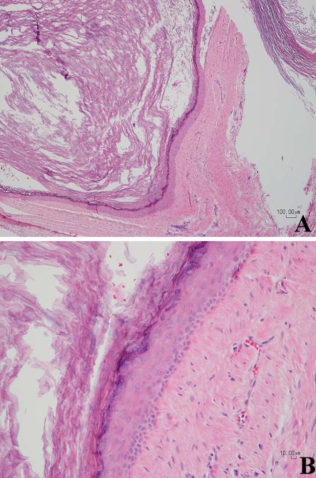
The purpose of this report is to document the clinical, radiographic, pathological and molecular findings of the first case of multiple orthokeratinized odontogenic cysts (OOCs). Multiple odontogenic keratocysts are one of the major features of nevoid basal cell carcinoma syndrome (NBCCS), and loss of heterozygosity in the PTCH gene, the culprit gene for NBCCS, has recently been found in sporadic OOC cases. Therefore, in this presenting case, we also investigated the possibility that this patient might also have NBCCS, by comparing the available clinical information and the molecular findings of this case to the diagnostic criteria for NBCCS (as proposed by the First International Colloquium on NBCCS in 2011). However, this patient with multiple OOCs showed no evidence of having NBCCS. This conclusion supports the findings from previous case series based on sporadic cases that OOC does not appear to be associated with NBCCS.
Figures
Similar articles
-
Multiple orthokeratinized odontogenic cysts: clinical, pathological, and genetic characteristics.Diagn Pathol. 2022 Oct 14;17(1):82. doi: 10.1186/s13000-022-01261-0. Diagn Pathol. 2022. PMID: 36242048 Free PMC article.
-
Multiple Orthokeratinized Odontogenic Cysts: A Report of Two Cases and Review of the Literature.Head Neck Pathol. 2020 Jun;14(2):381-385. doi: 10.1007/s12105-019-01042-0. Epub 2019 May 22. Head Neck Pathol. 2020. PMID: 31119532 Free PMC article. Review.
-
Podoplanin Expression in Odontogenic Keratocysts Associated or not Associated With Nevoid Basal Cell Carcinoma Syndrome.Appl Immunohistochem Mol Morphol. 2020 Aug;28(7):513-517. doi: 10.1097/PAI.0000000000000785. Appl Immunohistochem Mol Morphol. 2020. PMID: 31241560
-
Evidence of loss of heterozygosity of the PTCH gene in orthokeratinized odontogenic cyst.J Oral Pathol Med. 2011 Mar;40(3):277-80. doi: 10.1111/j.1600-0714.2010.00977.x. Epub 2010 Dec 8. J Oral Pathol Med. 2011. PMID: 21138481
-
Orthokeratinized odontogenic cyst: A large series and comprehensive literature review with emphasis on synchronous multiple occurrence and neoplastic transformation.Oral Surg Oral Med Oral Pathol Oral Radiol. 2022 Mar;133(3):e72-e82. doi: 10.1016/j.oooo.2021.07.009. Epub 2021 Jul 21. Oral Surg Oral Med Oral Pathol Oral Radiol. 2022. PMID: 34511349 Review.
Cited by
-
Orthokeratinized odontogenic cysts: a Spanish tertiary care centre study based on HPV DNA detection.Head Face Med. 2018 Jul 13;14(1):10. doi: 10.1186/s13005-018-0167-3. Head Face Med. 2018. PMID: 30005670 Free PMC article.
-
Multiple orthokeratinized odontogenic cysts: clinical, pathological, and genetic characteristics.Diagn Pathol. 2022 Oct 14;17(1):82. doi: 10.1186/s13000-022-01261-0. Diagn Pathol. 2022. PMID: 36242048 Free PMC article.
-
Developmental odontogenic cysts with special focus on the occurrence of multiple cysts and syndromic association: a single-centre cross-sectional study from the Czech Republic.Orphanet J Rare Dis. 2025 Mar 4;20(1):103. doi: 10.1186/s13023-025-03623-5. Orphanet J Rare Dis. 2025. PMID: 40038706 Free PMC article.
-
New tumour entities in the 4th edition of the World Health Organization Classification of Head and Neck tumours: odontogenic and maxillofacial bone tumours.Virchows Arch. 2018 Mar;472(3):331-339. doi: 10.1007/s00428-017-2182-3. Epub 2017 Jul 3. Virchows Arch. 2018. PMID: 28674741 Free PMC article. Review.
-
Developmental Odontogenic Lesions Associated with the Crown of an Impacted Tooth: A Guide to the Distinct Histologic Features Required for Classification.Head Neck Pathol. 2021 Mar;15(1):107-112. doi: 10.1007/s12105-020-01279-0. Epub 2021 Mar 15. Head Neck Pathol. 2021. PMID: 33723765 Free PMC article.
References
-
- Dong Q, Pan S, Sun LS, Li TJ. Orthokeratinized odontogenic cyst: a clinicopathologic study of 61 cases. Arch Pathol Lab Med. 2010;134(2):271–275. - PubMed
-
- Neville BW, Damm DD, Allen CM, Bouquot JE. Orthokeratinized odontogenic cyst. In: Neville BW, Damm DD, Allen CM, Bouquot JE, editors. Oral and maxillofacial pathology. St. Louis: Saunders Elsevier; 2009. pp. 687–688.
Publication types
MeSH terms
LinkOut - more resources
Full Text Sources
Other Literature Sources
