. 2014 May 30;289(22):15363-73.
doi: 10.1074/jbc.M114.562785. Epub 2014 Apr 15.

Reciprocal changes in phosphorylation and methylation of mammalian brain sodium channels in response to seizures

Affiliations

Reciprocal changes in phosphorylation and methylation of mammalian brain sodium channels in response to seizures

Je-Hyun Baek et al. J Biol Chem. .

Abstract

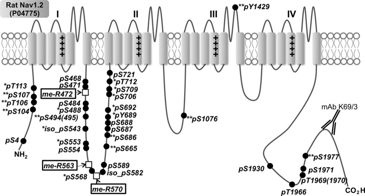
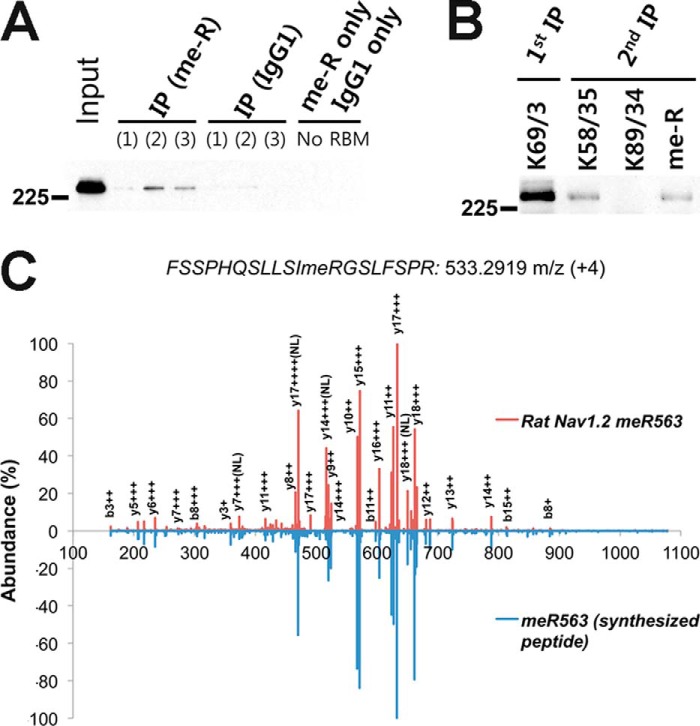
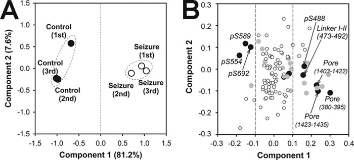
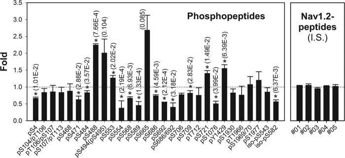
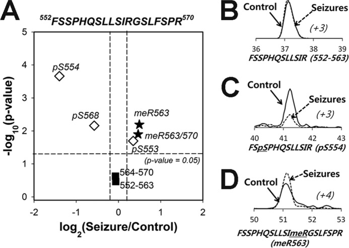
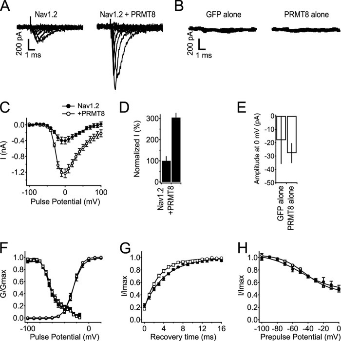
Voltage-gated sodium (Nav) channels initiate action potentials in brain neurons and are primary therapeutic targets for anti-epileptic drugs controlling neuronal hyperexcitability in epilepsy. The molecular mechanisms underlying abnormal Nav channel expression, localization, and function during development of epilepsy are poorly understood but can potentially result from altered posttranslational modifications (PTMs). For example, phosphorylation regulates Nav channel gating, and has been proposed to contribute to acquired insensitivity to anti-epileptic drugs exhibited by Nav channels in epileptic neurons. However, whether changes in specific brain Nav channel PTMs occur acutely in response to seizures has not been established. Here, we show changes in PTMs of the major brain Nav channel, Nav1.2, after acute kainate-induced seizures. Mass spectrometry-based proteomic analyses of Nav1.2 purified from the brains of control and seizure animals revealed a significant down-regulation of phosphorylation at nine sites, primarily located in the interdomain I-II linker, the region of Nav1.2 crucial for phosphorylation-dependent regulation of activity. Interestingly, Nav1.2 in the seizure samples contained methylated arginine (MeArg) at three sites. These MeArgs were adjacent to down-regulated sites of phosphorylation, and Nav1.2 methylation increased after seizure. Phosphorylation and MeArg were not found together on the same tryptic peptide, suggesting reciprocal regulation of these two PTMs. Coexpression of Nav1.2 with the primary brain arginine methyltransferase PRMT8 led to a surprising 3-fold increase in Nav1.2 current. Reciprocal regulation of phosphorylation and MeArg of Nav1.2 may underlie changes in neuronal Nav channel function in response to seizures and also contribute to physiological modulation of neuronal excitability.

Keywords: Electrophysiology; Epilepsy; Ion Channels; Mass Spectrometry (MS); Proteomics.


Figures

FIGURE 1.
FIGURE 1.
Immunopurification of rat brain Nav1.2 from control and seizure animals. A, immunoblot analysis of Nav1.2 and Kv2.1 in control (C) and seizure (S) RBM samples (10 μg of protein/lane). B, solubilization of Nav1.2 in 1% Triton X-100 detergent extract. T, total RBM starting material; S, detergent-soluble fraction; I, detergent-insoluble fraction. C, immunoblot analysis of Nav1.2 IP. Input, 20 μg of protein; FT, flow-through fraction from immunopurification; IP, IP fraction. Fractions were each 10 μg of protein. D, colloidal Coomassie Blue staining of IP products from control and seizure brain. Nav, Nav channel band excised for analysis. Hspa5, IgG binding protein. mAb, mAb IgG heavy chain. Numbers to the left of panels refer to Mr standards.
FIGURE 2.
FIGURE 2.
Mapping of in vivo multiple PTMs on rat brain Nav1.2. Closed circles, phosphosites on Ser/Thr/Tyr; open boxes, MeArg. **, novel phosphorylation sites identified here; *, phosphorylation sites known previously only in mice and identified here in rats.
FIGURE 3.
FIGURE 3.
Biochemical confirmation of Arg methylation on Nav1.2. A, IP of Nav1.2 from three independent RBM samples using anti-MeArg mAb. B, sequential IP of purified Nav1.2 with anti-pan-Nav (K58/35), anti-Kv2.1 (K89/34; negative control), or anti-MeArg (me-R) IgG1 mAbs. All immunoblots were probed for Nav1.2. C, mirrored mass spectra of seizure rat brain Nav1.2 peptide with MeArg at MeArg-563 (red) and corresponding synthetic peptide (blue).
FIGURE 4.
FIGURE 4.
Principle component analyses of Nav1.2 peptides quantified by label-free quantification. A, sample group separation. Closed circles, control samples; open circles, seizure samples. B, peptide component separation. Black circles (n = 10), ANOVA p value < 0.001; gray circles (n = 18), 0.001 ≤ p value < 0.01; white circles (n = 79), p value ≥ 0.01.
FIGURE 5.
FIGURE 5.
Stoichiometry of Nav1.2 phosphopeptides observed by label-free quantification. Bars indicate fold changes of phosphopeptides in seizure samples relative to control samples, normalized against unmodified internal standard (I.S.) peptide levels shown on the right. *, significant (p < 0.05) changes by ANOVA analysis with individual p values shown. Error bars, S.E.
FIGURE 6.
FIGURE 6.
Phosphorylation (pS554) and Arg methylation (MeR563) in the Nav1.2 ID I-II linker region are reciprocally regulated in control and seizure samples. A, volcano plot of Nav1.2 peptides from the region 552–570 containing phosphorylation and MeArg. B–D, examples of label-free quantification of peptides that are either unmodified (B), phosphorylated at Ser(P)-554 (C), or methylated at MeArg-563 (D) between control (solid line) and seizure (dashed line) samples. The x axis shows the retention time (min) of peptides on C18-column liquid chromatography, and parentheses with italic type indicate the charge state of each peptide.
FIGURE 7.
FIGURE 7.
Coexpression of PRMT8 increases Nav1.2 currents without effects on voltage-dependent properties. A, representative families of Nav1.2 current without and with coexpression of PRMT8. Currents in response to steps from −100 to +100 mV in 20-mV increments are shown. B, average currents from tsA-201 cells expressing GFP alone or PRMT8 alone at 0 mV, n = 5 for each. C, mean current-voltage (I-V) relationships of peak currents. Currents were elicited in response to voltage steps to the indicated potentials in 10-mV steps from a holding potential of −100 mV. Peak current is plotted as a function of test pulse potential. Note that the y axis is in nA. D, percentage change in peak amplitude of Nav1.2 current at 0 mV associated with coexpression of PRMT8. E, average of maximal inward currents from tsA-201 cells expressing GFP alone or PRMT8 alone at 0 mV. Note that the y axis is in pA. F, voltage dependence of the activation and voltage dependence of fast inactivation for cells expressing Nav1.2 alone or with coexpression of PRMT8. Conductance-voltage relationships (circles) were determined from I-V relationships as in B as G = I/(VVRev), where V is the test potential, and VRev is the extrapolated reversal potential. The voltage dependence of inactivation (squares) was determined using 100-ms-long prepulses to the indicated potentials, followed by a test pulse to 0 mV. Normalized peak test pulse current is plotted as a function of prepulse potential. G, recovery from fast inactivation. Nav1.2, τ = 4.59 ± 0.289 ms, n = 10; Nav1.2 + PRMT8, τ = 3.05 ± 0.284 ms, n = 12; p = 0.001. H, voltage dependence of slow inactivation (Nav1.2, n = 6; Nav1.2 with PRMT8, n = 10). Error bars, S.E.

References

    1. Catterall W. A., Goldin A. L., Waxman S. G. (2005) International Union of Pharmacology. XLVII. Nomenclature and structure-function relationships of voltage-gated sodium channels. Pharmacol. Rev. 57, 397–409 - PubMed
    1. Cantrell A. R., Catterall W. A. (2001) Neuromodulation of Na+ channels: an unexpected form of cellular plasticity. Nat. Rev. Neurosci. 2, 397–407 - PubMed
    1. Berendt F. J., Park K. S., Trimmer J. S. (2010) Multisite phosphorylation of voltage-gated sodium channel α subunits from rat brain. J. Proteome Res. 9, 1976–1984 - PMC - PubMed
    1. Vacher H., Mohapatra D. P., Trimmer J. S. (2008) Localization and targeting of voltage-dependent ion channels in mammalian central neurons. Physiol. Rev. 88, 1407–1447 - PMC - PubMed
    1. Scheuer T. (2011) Regulation of sodium channel activity by phosphorylation. Semin. Cell Dev. Biol. 22, 160–165 - PMC - PubMed

Publication types

MeSH terms