. 2014 Apr;26(4):1712-1728.
doi: 10.1105/tpc.114.123216. Epub 2014 Apr 15.

Arabidopsis Class I α-Mannosidases MNS4 and MNS5 Are Involved in Endoplasmic Reticulum-Associated Degradation of Misfolded Glycoproteins

Affiliations

Arabidopsis Class I α-Mannosidases MNS4 and MNS5 Are Involved in Endoplasmic Reticulum-Associated Degradation of Misfolded Glycoproteins

Silvia Hüttner et al. Plant Cell. 2014 Apr.

Abstract

To ensure that aberrantly folded proteins are cleared from the endoplasmic reticulum (ER), all eukaryotic cells possess a mechanism known as endoplasmic reticulum-associated degradation (ERAD). Many secretory proteins are N-glycosylated, and despite some recent progress, little is known about the mechanism that selects misfolded glycoproteins for degradation in plants. Here, we investigated the role of Arabidopsis thaliana class I α-mannosidases (MNS1 to MNS5) in glycan-dependent ERAD. Our genetic and biochemical data show that the two ER-resident proteins MNS4 and MNS5 are involved in the degradation of misfolded variants of the heavily glycosylated brassinosteroid receptor, BRASSINOSTEROID INSENSITIVE1, while MNS1 to MNS3 appear dispensable for this ERAD process. By contrast, N-glycan analysis of different mns mutant combinations revealed that MNS4 and MNS5 are not involved in regular N-glycan processing of properly folded secretory glycoproteins. Overexpression of MNS4 or MNS5 together with ER-retained glycoproteins indicates further that both enzymes can convert Glc0-1Man8-9GlcNAc2 into N-glycans with a terminal α1,6-linked Man residue in the C-branch. Thus, MNS4 and MNS5 function in the formation of unique N-glycan structures that are specifically recognized by other components of the ERAD machinery, which ultimately results in the disposal of misfolded glycoproteins.


Figures

Figure 1.
Figure 1.
MNS4 and MNS5 Encode Putative Orthologs of Human EDEMs and Yeast HTM1. (A) The precursor N-glycan, which is composed of three Glc, nine Man, and two GlcNAc residues, is shown. The three branches (A, B, and C) as well as the linkage between the individual sugar residues are indicated. (B) N-Glycan processing mediated by the subsequent action of MNS3 and MNS1/MNS2. (C) Comparison of the domain organization of S. cerevisiae HTM1, human EDEM1, EDEM2, and EDEM3, as well as MNS4 and MNS5. Predicted signal peptides are shown in gray, predicted transmembrane domains in black, and GH47 domains in dark gray. The protease-associated domain of EDEM3 is crosshatched. Positions of putative N-glycosylation sites are indicated (Y). Signal peptides, transmembrane domains, and N-glycosylation sites are depicted according to UniProt/GenBank entries or derived from predictions using SignalP (http://www.cbs.dtu.dk/services/SignalP). aa, amino acids. [See online article for color version of this figure.]
Figure 2.
Figure 2.
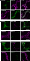
MNS4-GFP and MNS5-mRFP Are ER-Resident Proteins. Images were taken 2 to 3 d after infiltration (DAI). Bars = 20 µm. (A) Confocal images (2 DAI) of N. benthamiana leaf epidermal cells transiently coexpressing MNS4-GFP or MNS5-mRFP and the ER marker protein OS9-mRFP. (B) Confocal images (2 DAI) of N. benthamiana leaf epidermal cells transiently coexpressing MNS4-GFP or MNS5-mRFP and the Golgi marker MNS1-mRFP. (C) Confocal images (3 DAI) of N. benthamiana leaf epidermal cells transiently coexpressing MNS4-GFP or MNS5-mRFP and MNS3-mRFP.
Figure 3.
Figure 3.
MNS4 and MNS5 Deficiency Does Not Enhance the mns1 mns2 mns3 Root Growth Phenotype. (A) Schematic representation of the MNS4 and MNS5 genes and the identified alleles. Boxes represent exons (the black areas represent the coding regions), and vertical lines indicate the positions of T-DNA insertions. Primers used for the characterization of transcript expression are shown by arrows. (B) RT-PCR analysis of mns mutants. RT-PCR was performed on RNA isolated from rosette leaves of the indicated lines. Wild-type (Columbia-0 [Col-0]) was used as a control, and primers specific for the indicated transcripts were used for amplification. UBQ5 served as a positive control. (C) Phenotypes of the indicated seedlings (mns1 mns2 mns3 = 123; mns1 mns2 mns3 mns4-1 mns5-1 = 12345) grown on 0.5× MS medium plus 1.5% Suc for 14 d under 16 h of light or for 17 d in the dark on 0.5× MS medium plus 4% Suc. Images of 4-week-old soil-grown wild-type (Col-0) plants as well as mns triple and quintuple mutants are shown.
Figure 4.
Figure 4.
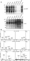
mns4-1 and mns5-1 Mutants Display No Changes in N-Glycosylation. (A) Protein gel blot analysis. Proteins were extracted from leaves of the indicated mutants and subjected to SDS-PAGE under reducing conditions. Detection was performed using anti-horseradish peroxidase (anti-HRP) antibodies, which recognize β1,2-Xyl and core α1,3-Fuc residues on N-glycans, and the lectin concanavalin A (ConA). (B) Matrix-assisted laser desorption ionization mass spectra of total N-glycans extracted from leaves of wild-type (Col-0), mns1 mns2 mns3 (mns123), mns4-1 mns5-1 (mns45), mns1 mns2 mns3 mns4-1 mns5-1 (mns12345), mns3, and mns3 mns4-1 mns5-1 (mns345) plants.
Figure 5.
Figure 5.
The mns4-1 mns5-1 Double Mutant Displays UPR Induction and Is Sensitive to Salt Stress. (A) Leaves of 3-week-old wild-type (Col-0) and mns4-1 mns5-1 (mns45) plants were treated for 5 h with 5 µg/mL−1 tunicamycin (TM) or with 0.25% DMSO (control). RNA was subjected to qRT-PCR using primers for the indicated transcripts. Values are normalized means ± sd from two independent biological samples. ACTIN2 expression was used as a control. RT-PCR analysis of BiP3 expression is shown for wild-type (Col-0) and mns4-1 mns5-1 leaves. UBQ5 served as a positive control. (B) qRT-PCR analysis of MNS4, MNS5, and PDI5 expression in the presence or absence of tunicamycin. Treatment and quantification were done as described in (A). (C) Phenotypes of 10-d-old seedlings grown on 0.5× MS agar plates plus 2% Suc and 0.3 µg/mL−1 tunicamycin. (D) Phenotypes of 12-d-old seedlings grown on 0.5× MS agar plates supplemented with 1.5% Suc and 120 mM NaCl where indicated. The percentages of seedlings with green leaves (black bars), small yellow leaves (white bars), and nongerminated seedlings (gray bars) are shown in the right panel (n > 120).
Figure 6.
Figure 6.
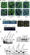
Combined Deficiency of MNS4 and MNS5 Suppresses the bri1-5 Growth Phenotype. (A) Shoot phenotypes of 19-d-old wild-type (Col-0), mns4-1 mns5-1, mns4-1, mns5-1, bri1-5, mns4-1 mns5-1 bri1-5, mns4-1 bri1-5, and mns5-1 bri1-5 plants grown under long-day conditions. (B) Phenotypes of 12-d-old mns3 bri1-5 and mns1 mns2 mns3 bri1-5 (mns123 bri1-5) seedlings and 23-d-old mns3 bri1-5 plants grown on soil. (C) Protein gel blot analysis. Microsomal preparations of 10-d-old seedlings were subjected to SDS-PAGE under reducing conditions. Detection was performed using anti-BRI1 antibodies. An unspecific band (asterisk) served as a loading control. (D) Fourteen-day-old seedlings were incubated for the indicated time in 0.5× MS medium supplemented with 1.5% Suc and 200 µg/mL−1 cycloheximide (CHX). Microsomal preparations were then analyzed with anti-BRI1 and anti-PDI (control) antibodies. (E) Endo H digestion of microsomal preparations from 10-d-old seedlings followed by immunoblotting with anti-BRI1 antibodies. Total protein staining with amido black (amido b.) was used as a loading control.
Figure 7.
Figure 7.
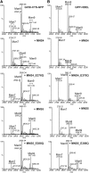
MNS4 or MNS5 Expression Results in Enhanced Removal of Man Residues from ER-Resident Glycoproteins in Planta. (A) Liquid chromatography–electrospray ionization–mass spectrometry (LC-ESI-MS) of the GCSI-CTS-GFPglyc reporter. Mass spectra of the glycopeptide EEQYNSTYR are shown. GCSI-CTS-GFPglyc was transiently expressed in N. benthamiana leaves (top panel) and coexpressed with MNS4-GFP, MNS4-E376Q-GFP, MNS5-mRFP, or MNS5-E388Q-mRFP, as indicated. (B) LC-ESI-MS of the GFPglyc-HDEL reporter. Mass spectra of the glycopeptide EEQYNSTYR are shown. GFPglyc-HDEL was transiently expressed in N. benthamiana leaves (top panel) and coexpressed with MNS4-GFP, MNS4-E376Q-GFP, MNS5-mRFP, or MNS5-E388Q-mRFP, as indicated.
Figure 8.
Figure 8.
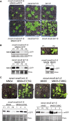
Reconstitution of MNS4 or MNS5 Expression Reverts the Suppression of the bri1-5 Phenotype in mns4-1 mns5-1 bri1-5 Plants. (A) Complementation of mns4-1 mns5-1 bri1-5 with MNS4-GFP and MNS5-mRFP. Images of 4-week-old soil-grown plants are shown. (B) Protein gel blot analysis of transgenic Arabidopsis expressing MNS4-GFP or MNS5-GFP. Crude protein extracts from leaves were subjected to immunoblotting with anti-GFP antibodies. The enhanced bri1-5 phenotype is marked as P1, while a bri1-5–like phenotype is marked as P2. Total protein staining with Ponceau S (Ponc.) was used as a loading control. (C) Complementation of mns4-1 mns5-1 bri1-5 with MNS4-E376Q-GFP and MNS5-E388Q-mRFP. Images show the phenotypes of 4-week-old plants. Immunoblot analysis of transgenic Arabidopsis expressing MNS4-GFP, MNS4-E376Q-GFP, MNS5-mRFP, and MNS5-E388Q-mRFP is also shown.
Figure 9.
Figure 9.
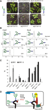
The BRI1-5 ERAD Signal Very Likely Consists of a Terminal α1,6-Linked Man Residue. (A) Shoot phenotypes of 4-week-old soil-grown mutants. The major oligomannosidic N-glycans that are present on ER-resident glycoproteins from alg3 and alg12 mutants are indicated. (B) BRI1-GFP and BRI1-5-GFP were transiently expressed in N. benthamiana. The N-glycans liberated from the purified proteins were reduced and then analyzed by porous graphitic carbon–liquid chromatography–electrospray ionization–mass spectrometry (PGC-LC-ESI-MS). Selected ion chromatograms (SIC) of Hex8GlcNAc2 and Hex9GlcNAc2 alditols are shown. The Hex9GlcNAc2 and Hex8GlcNAc2 isomers from RNase B were used as standards. (C) The values shown represent the relative amounts derived from PGC-LC-ESI-MS analysis (mean ± sd from two independent experiments) for each glycoform (see Supplemental Figure 17 for N-glycan structures). (D) Proposed model for BRI1-5 ERAD. Monoglucosylated N-glycans from BRI1-5 (C69Y indicates the amino acid change causing the folding defect) interact with CNX during folding attempts mediated by the CNX/CRT cycle. MNS4 or MNS5 removes a single Man from the C-branch of distinct N-glycans and diverts BRI1-5 to ERAD. MNS3 may remove an additional Man residue from the B-branch, but this trimming is dispensable for BRI1-5 degradation. The terminal α1,6-Man residue exposed by MNS4 or MNS5 and the non-native protein conformation are recognized by the substrate receptors OS9 and SEL1L that deliver BRI1-5 to HRD1 for ubiquitylation and subsequent degradation.

Similar articles

Cited by

References

    1. Abas L., Luschnig C. (2010). Maximum yields of microsomal-type membranes from small amounts of plant material without requiring ultracentrifugation. Anal. Biochem. 401: 217–227 - PMC - PubMed
    1. Aebi M., Bernasconi R., Clerc S., Molinari M. (2010). N-Glycan structures: Recognition and processing in the ER. Trends Biochem. Sci. 35: 74–82 - PubMed
    1. Avezov E., Frenkel Z., Ehrlich M., Herscovics A., Lederkremer G.Z. (2008). Endoplasmic reticulum (ER) mannosidase I is compartmentalized and required for N-glycan trimming to Man5-6GlcNAc2 in glycoprotein ER-associated degradation. Mol. Biol. Cell 19: 216–225 - PMC - PubMed
    1. Brandizzi F., Hanton S., DaSilva L.L., Boevink P., Evans D., Oparka K., Denecke J., Hawes C. (2003). ER quality control can lead to retrograde transport from the ER lumen to the cytosol and the nucleoplasm in plants. Plant J. 34: 269–281 - PubMed
    1. Brodsky J.L. (2012). Cleaning up: ER-associated degradation to the rescue. Cell 151: 1163–1167 - PMC - PubMed