. 2014 Jun;113(7):1155-73.
doi: 10.1093/aob/mcu045. Epub 2014 Apr 15.

Comparative anatomy of floral elaiophores in Vitekorchis Romowicz & Szlach., Cyrtochilum Kunth and a florally dimorphic species of Oncidium Sw. (Orchidaceae: Oncidiinae)

Affiliations

Comparative anatomy of floral elaiophores in Vitekorchis Romowicz & Szlach., Cyrtochilum Kunth and a florally dimorphic species of Oncidium Sw. (Orchidaceae: Oncidiinae)

Kevin L Davies et al. Ann Bot. 2014 Jun.

Abstract

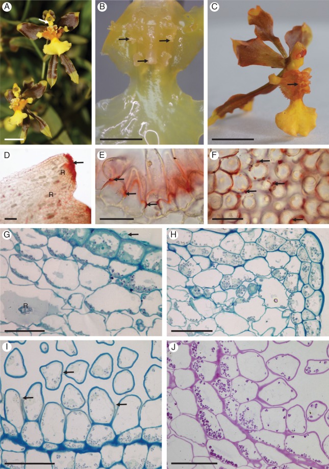
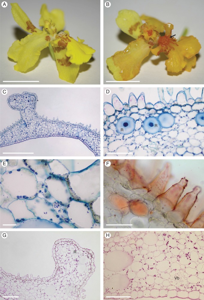
Background and aims: Recently, molecular approaches have been used to investigate the phylogeny of subtribe Oncidiinae, resulting in the re-alignment of several of its genera. Here, a description is given of the structure of the floral elaiophores (oil glands) of four species formerly assigned to Oncidium Sw. Those of Vitekorchis excavata (Lindl.) Romowicz & Szlach., Cyrtochilum meirax (Rchb.f.) Dalström and a species of Oncidium displaying floral dimorphism, namely O. heteranthum Poepp. & Endl. var. album, are compared with that of Gomesa longipes (Lindl.) M.W. Chase & N.H. Williams, whose epithelial elaiophores are typical of many Oncidiinae, in order to extend our understanding of elaiophore diversity within this subtribe.

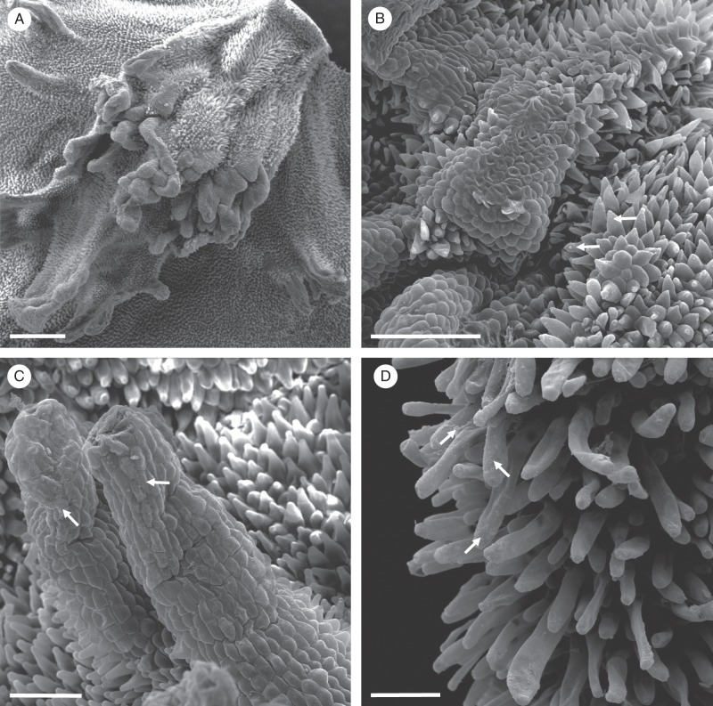
Methods: Floral elaiophore structure was examined and compared at anthesis for all four species using light microscopy, scanning electron microscopy, transmission electron microscopy and histochemistry.

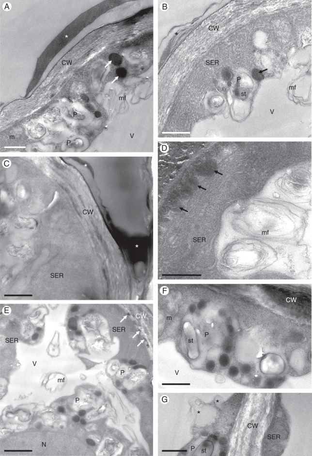
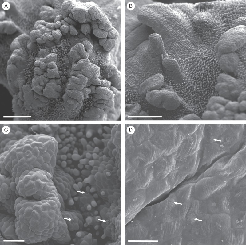
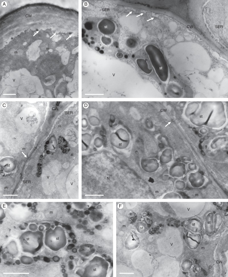
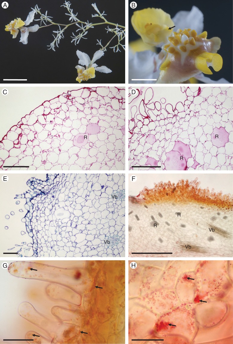
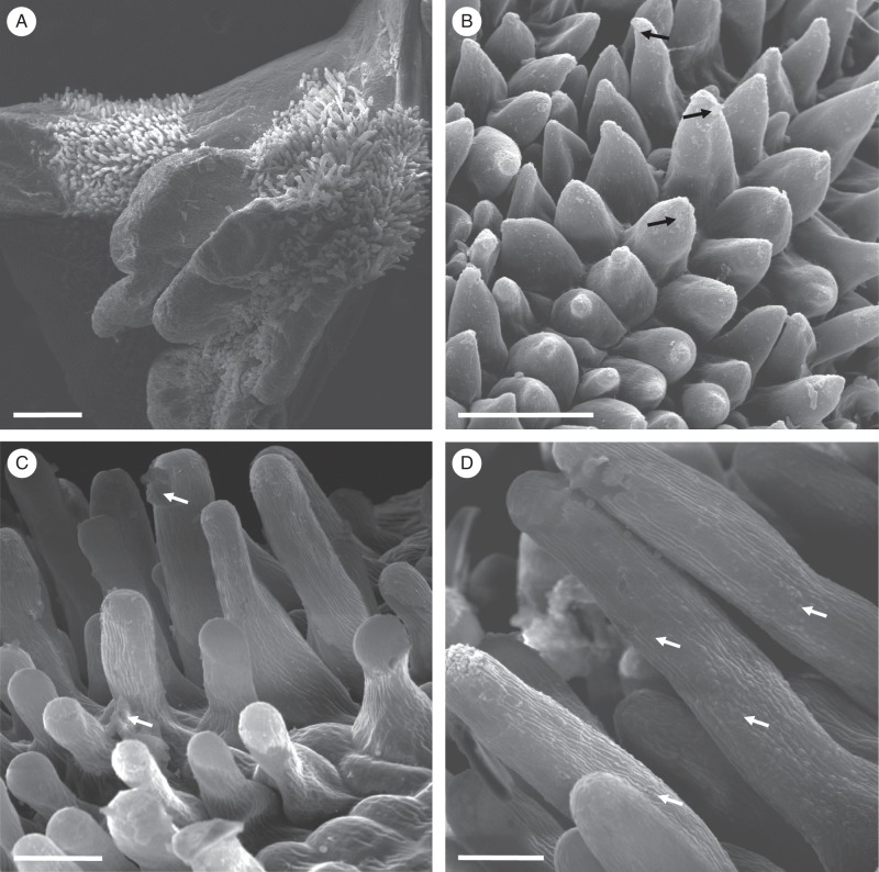
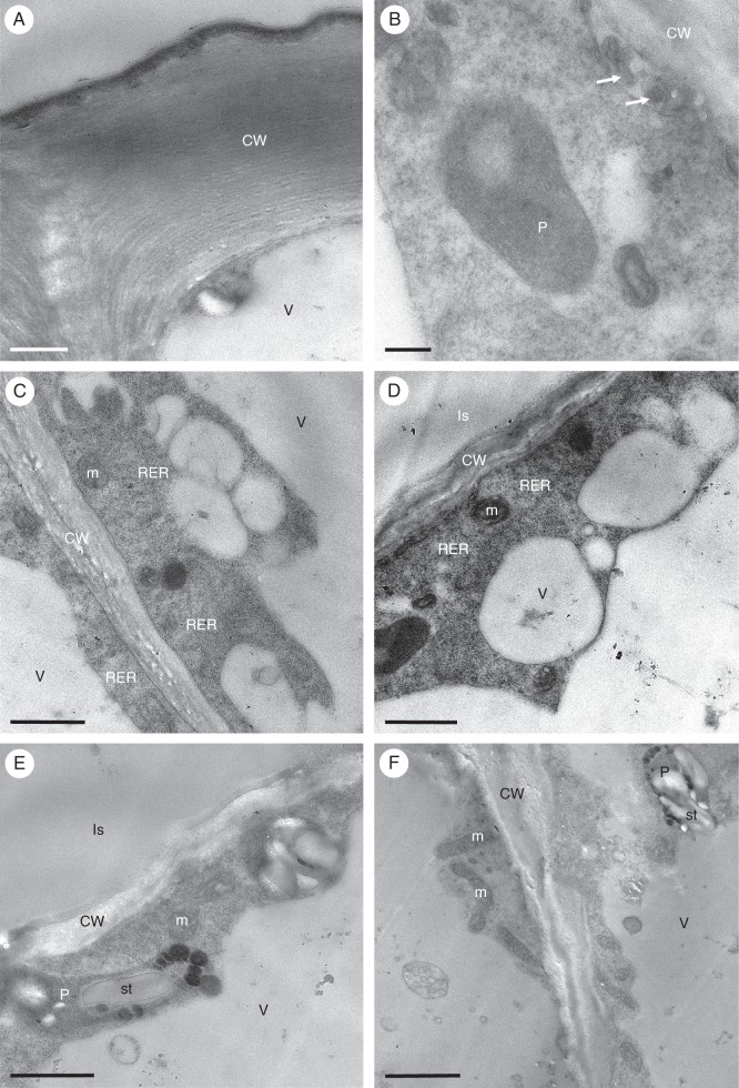
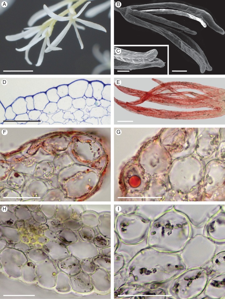
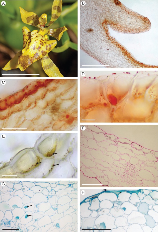
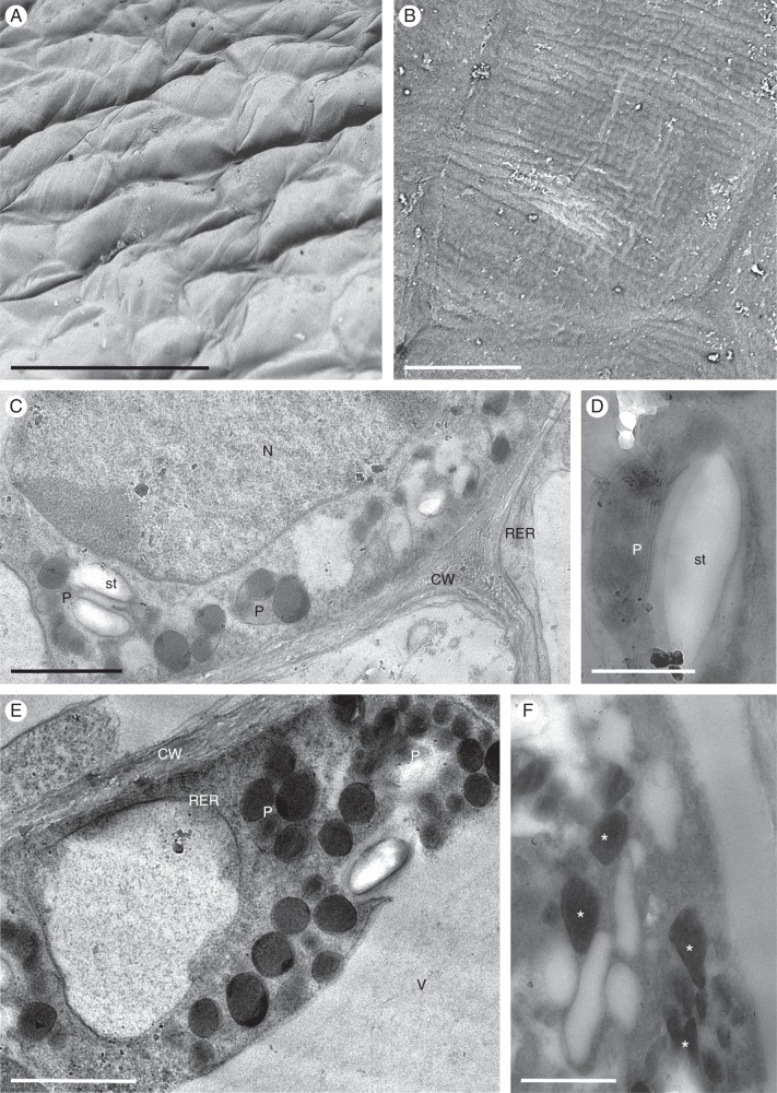
Key results: In all species investigated, with the exception of C. meirax, the floral elaiophore occurs on the labellar callus and is of the intermediate type, possessing both glabrous and trichomatous regions. By contrast, although all four species produce lipid secretions, C. meirax lacks an obvious elaiophore. In each case, the secretory tissue is represented by a single-layered epidermis of cuboidal cells (trichomatous and/or atrichomatous). Palisade cells are absent. The secretion may be wax- or oil-like and is usually produced by smooth endoplasmic reticulum (SER). However, in C. meirax, where rough endoplasmic reticulum (RER) predominates, oil accumulates as plastoglobuli within elaioplasts. These plastoglobuli are then discharged into the cytoplasm, forming oil bodies. In some species, oil usually accumulates within vesicles at the plasmalemma or in the periplasmic space before traversing the cell wall and accumulating beneath the cuticle, sometimes with distension of the latter. Gomesa longipes is unusual in its production of a heterogeneous secretion, whereas Vitekorchis excavata is equally remarkable for the protuberances found on the walls of its secretory cells.

Conclusions: Anatomically, the secretory tissues of all four species, despite currently being assigned to four different genera, are remarkably similar and indicative of homoplasy. This supports previous investigations of the floral elaiophore in Oncidiinae, which showed that the same elaiophore characters may be shared by different clades, but not always by species of the same genus. Consequently, elaiophores are considered to be of limited value in investigating the phylogeny of this subtribe. Furthermore, floral dimorphism does not greatly modify elaiophore structure in the fertile flowers of Oncidium heteranthum var. album. Based on the presence or absence of well-defined elaiophores, the nature of the secretion and the cell ultrastructure, it is likely that floral oil may be produced in Oncidiinae in one of two ways: by the ER (mainly SER) or by plastids, most notably elaioplasts. Once the oil is discharged into the cytoplasm as oil bodies or oil droplets, there is little difference between the subsequent stages of oil secretion; the oil traversing the cytoplasm (often vesicle-mediated) and cell wall before accumulating beneath the cuticle.

Keywords: Anatomy; Cyrtochilum; Gomesa; Oncidiinae; Oncidium; Orchidaceae; Vitekorchis; floral elaiophore; lipids; micromorphology; oil glands; ultrastructure.


Figures

Fig. 1.
Fig. 1.
Gomesa longipes: habit and LM. (A) Habit of flower with pronounced callus and tabula infrastigmatica (arrow). (B) Callus showing projections (arrows). (C) Entire flower following immersion in alcoholic Sudan III solution clearly showing stained callus region (arrow). Note the pronounced tabula infrastigmatica. (D) Apical part of callus projection stains red for lipids (arrow) following treatment with Sudan III. Numerous idioblasts containing raphides occur in the ground parenchyma. (E) Lipid-rich material is also present towards the bases of trichomes (arrows). (F) Lipid droplets also occur on the surface of epidermal papillae (arrows). (G) Section of callus stained with MB/AII showing glabrous epidermis and residues of secreted material (arrow) on the callus surface. (H) Section of callus projection stained with MB/AII. Epidermal and ground parenchyma cells contain amyloplasts. (I, J) Section of callus epidermis with trichomes. Note the thick tangential cell walls of sub-epidermal parenchyma. (I) Secretory residues (arrows) following staining with MB/AII. (J) Starch grains within trichomatous and atrichomatous epidermal cells, together with ground parenchyma cells, following the PAS reaction. Scale bars = 1 cm, 3 mm, 1 cm, 100 μm, 50 μm, 50 μm, 50 μm, 50 μm, 50 μm, 50 μm, respectively. R, idioblasts with raphides.
Fig. 2.
Fig. 2.
Gomesa longipes: SEM. (A) Surface of labellar callus showing projections, papillae and trichomes. (B) Conical papillae with residues of secretory material (arrows). (C) Secreted material (arrows) on the surface of the glabrous epidermis overlying the callus projection. (D) Secreted material on the surface of trichomes (arrows). Scale bars = 500 μm, 200 μm, 100 μm, 100 μm, respectively.
Fig. 3.
Fig. 3.
Gomesa longipes: TEM. (A) Osmiophilic secretion (asterisk) on the surface of epidermal cell wall of callus. Large, osmiophilic droplets (arrows) occur in the parietal cytoplasm. (B) Heterogeneous secretion (asterisk) on the epidermal cell wall surface. Abundant smooth endoplasmic reticulum profiles, plastids and osmiophilic droplets (arrow) occur in the cytoplasm, together with intravacuolar myelin-like figures. (C) Cell wall of basal part of trichome coated with secretion (asterisks). Smooth endoplasmic reticulum predominates in the cytoplasm. (D) Osmiophilic material accumulates beneath the cell wall (arrows). (E) Osmiophilic droplets occur in the cytoplasm and between the structural elements of the cell wall (arrows). (F) Plastids containing starch grains and plastoglobuli. (G) Details of plastid with few internal membranes, starch and plastoglobuli. Two vacuoles coalesce (asterisks), resulting in the initiation of myelin-like figure formation. Scale bars = 1 μm, 1 μm, 1 μm, 1 μm, 2 μm, 1 μm, 1 μm, respectively. CW, cell wall; m, mitochondrion; mf, myelin-like figure; N, nucleus; P, plastid; SER, smooth endoplasmic reticulum; st, starch; V, vacuole.
Fig. 4.
Fig. 4.
Vitekorchis excavata: habit and LM. (A, B) Flower prior to (A) and following treatment with Sudan III (B) showing distinctly stained, lipid-rich callus region (arrow). (C) Section of callus showing glabrous, abaxial and papillose, adaxial epidermis. Note that the callus projection lacks papillae. (D) Epidermal papillae contain intravacuolar anthocyanins, whereas large idioblasts occurring in the ground parenchyma contain raphides. (E) Plastids in parietal cytoplasm following staining with MB/AII. (F) Surface and intracellular lipids stained with Sudan III. These compounds occur in both epidermal papillae and sub-epidermal cells. (G, H) Starch grains present in glabrous epidermal cells, papillae and parenchyma cells stain with the PAS reaction. Note the weak staining of large idioblasts and vascular bundles near the abaxial epidermis (H). Scale bars = 1 cm, 1 cm, 500 μm, 50 μm, 20 μm, 50 μm, 100 μm, 100 μm, respectively. R, idioblasts with raphides; Vb, vascular bundle.
Fig. 5.
Fig. 5.
Vitekorchis excavata: SEM. (A, B) Papillose labellar callus bearing glabrous rugae (A) and projections (B). (C) Detail of glabrous epidermal cells overlying rugae. Unicellular, conical papillae and trichomes occur between the projections and rugae. Stomata are indicated by arrows. (D) Residues of secreted material (arrows) on the cuticular surface of glabrous epidermal cells. Scale bars = 600 μm, 500 μm, 100 μm, 40 μm, respectively.
Fig. 6.
Fig. 6.
Vitekorchis excavata: TEM. (A) Protuberances arising from the inner surface of the trichome cell wall (arrows), the dense cytoplasm containing mitochondria. (B) Plastids of trichomes with large starch grains. Folding of the plasmalemma (arrows) results in the formation of small vesicles within the periplasmic space. (C) Plastid of irregular shape with starch and numerous plastoglobuli, together with vacuoles containing inconspicuous membranous enclaves. Plasmodesmata interconnect adjoining epidermal cells (arrow). (D) Plastids containing starch and plastoglobuli. The cell walls of adjoining epidermal cells have irregular outlines and are perforated by primary pit-fields with plasmodesmata (arrow). (E) Plastid lacking well-developed internal membranes and containing plastoglobuli that are located adjacent to the plastid envelope. (F) Vacuoles with fine flocculent contents, but lacking myelin-like structures. Scale bars = 1 μm, 1 μm, 1 μm, 1 μm, 1 μm, 1 μm, respectively. CW, cell wall; D, dictyosome; m, mitochondrion; N, nucleus; P, plastid; SER, smooth endoplasmic reticulum; st, starch; V, vacuole.
Fig. 7.
Fig. 7.
Oncidium heteranthum var. album: habit and LM. (A) Habit of paniculate inflorescence with branches bearing numerous sterile flowers and few terminal, fertile flowers. (B) Tuberculate callus of fertile flower with projections. Note the tabula infrastigmatica (arrow), which is located on the column. (C) Section of callus projection with overlying glabrous epidermis containing numerous stomata. Idioblasts containing raphides occur in the ground parenchyma. Small quantities of starch, mainly in the sub-epidermal parenchyma, stain with the PAS reaction. (D) Trichomes and papillae occur between the callus projections, whereas idioblasts and occasional starch grains occur in the ground parenchyma. (E) Section of tuberculate callus stained with MB/AII showing vascular bundles that supply the ground parenchyma. None of these vascular elements supplies the sub-epidermis. (F) Callus trichomes stain red with Sudan III, indicating the presence of lipid. Idioblasts with raphides, as well as vascular bundles occur in the ground parenchyma. (G) Small lipid droplets (arrows) occur in the trichomatous and atrichomatous epidermal cells, as well as the sub-epidermal cells. (H) Lipid droplets are also visible within glabrous epidermal cells (arrows). Scale bars = 1 cm, 3 mm, 100 μm, 100 μm, 100 μm, 500 μm, 50 μm, 50 μm, respectively. R, idioblasts with raphides; Vb, vascular bundle.
Fig. 8.
Fig. 8.
Oncidium heteranthum var. album: fertile flower, SEM. (A) Part of callus showing projections with overlying glabrous epidermis. Between these projections occur unicellular trichomes and papillae. (B) Residues of secreted material (arrows) occur on the surface of the papillae. (C) Secreted material (arrows) is also present on the surface of trichome cell walls. (D) Striate cuticle of callus trichomes with small blisters (arrows). Scale bars = 500 μm, 50 μm, 50 μm, 20 μm, respectively.
Fig. 9.
Fig. 9.
Oncidium heteranthum var. album: fertile flower, TEM. (A) Outer cell wall of glabrous epidermal cell of callus with associated osmiophilic cuticle. (B) Granular cytoplasm of epidermal cell with vesicles present in the periplasmic space (arrows) and a plastid lacking both starch and plastoglobuli. (C) Parietal cytoplasm with mitochondria and short profiles of rough endoplasmic reticulum. The vacuoles generally lack myelin-like figures. (D) Granular cytoplasm of sub-epidermal cell containing mitochondria and rough endoplasmic reticulum. (E) Sub-epidermal cell with plastids containing plastoglobuli and starch. (F) The inner tangential walls of epidermal cells, adjacent to those of the sub-epidermal tissue are relatively thick. The cytoplasm contains both plastids and mitochondria. Scale bars = 1 μm, 200 nm, 1 μm, 1 μm, 1 μm, 2 μm, respectively. CW, cell wall; Is, intercellular space; m, mitochondrion; P, plastid; RER, rough endoplasmic reticulum; st, starch; V, vacuole.
Fig. 10.
Fig. 10.
Oncidium heteranthum var. album: sterile flowers, habit, LM and SEM. (A, B) The white, sterile flowers have five, simple perianth lobes of uniform size and shape. (C) The perianth lobes have a glabrous epidermis lacking a striate cuticle. (D) Section of perianth lobe stained with MB/AII showing glabrous epidermis with cuticle, together with mesophyll (ground parenchyma) showing well-developed, intercellular spaces. Both tissues have thin cell walls and prominent vacuoles. (E) The entire perianth stains red following treatment with Sudan III for lipids. (F) Lipids are mainly present in epidermal cells and on the surface of their walls. (G) In some cells, however, large lipid droplets may be formed. (H) Starch, stained here with IKI, is present within amyloplasts of ground parenchyma cells. (I) Detail of ground parenchyma. Cells of this tissue have thin walls, large vacuoles and contain amyloplasts with starch. Scale bars = 3 mm, 500 μm, 200 μm, 100 μm, 500 μm, 50 μm, 50 μm, 50 μm, 50 μm, respectively.
Fig. 11.
Fig. 11.
Cyrtochilum meirax: habit and LM. (A) Yellow and brown flowers are borne on a simple, racemose inflorescence. The tabula infrastigmatica is indicated by an arrow. (B) Section of labellum with glabrous callus stained with Sudan III. (C) Lipids accumulate mainly in the adaxial epidermal cells of the labellum. (D) Large lipid droplets stained with Sudan III are present both within epidermal cells and upon their surface. (E) Epidermal and sub-epidermal cells contain small quantities of starch (arrows) that stain with IKI. (F) Section of labellum following the PAS reaction showing thin cellulose walls of epidermal and ground parenchyma cells, and occasional starch grains. (G) Vacuolar contents of some parenchyma cells only partly stain with MB/AII (arrows). (H) The secreted material (asterisks) present between the outer tangential wall of epidermal cells and the cuticle stains intensely with MB/AII. Scale bars = 1 cm, 500 μm, 50 μm, 20 μm, 20 μm, 50 μm, 50 μm, 100 μm, respectively.
Fig. 12.
Fig. 12.
Cyrtochilum meirax: SEM and TEM. (A) Cuticular surface of epidermal cells of the callus lacking obvious secreted material. (B) Detail of the finely striate cuticle lacking blisters and cracks. (C) Highly granular and vesiculate parietal cytoplasm containing a large nucleus and plastids. (D) Plastid containing few internal membranes, as well as a starch grain and numerous plastoglobuli. (E) Large plastoglobuli may occur within plastids. (F) Osmiophilic bodies (asterisks), not unlike plastoglobuli, may also occur in the cytoplasm. Scale bars = 100 μm, 10 μm, 2 μm, 1 μm, 2 μm, 500 nm, respectively. CW, cell wall; N, nucleus; P, plastid; RER, rough endoplasmic reticulum; st, starch; V, vacuole.

Similar articles

Cited by

References

    1. Aliscioni SS, Torretta JP, Bello ME, Galati BG. Elaiophores in Gomesa bifolia (Sims) M.W. Chase & N.H. Williams (Oncidiinae: Cymbidieae: Orchidaceae): structure and oil secretion. Annals of Botany. 2009;104:1141–1149. - PMC - PubMed
    1. Blanco MA, Davies KL, Stpiczyńska M, Carlsward BS, Ionta GM, Gerlach G. Floral elaiophores in Lockhartia Hook. (Orchidaceae: Oncidiinae): their distribution, diversity and anatomy. Annals of Botany. 2013;112:1775–1791. - PMC - PubMed
    1. Brummitt RK, Powell CE. Authors of plant names. Kew, UK: Royal Botanic Gardens; 1992.
    1. Buchmann SL. The ecology of oil flowers and their bees. Annual Review of Ecology and Systematics. 1987;18:343–369.
    1. Chase MW. Classification of Orchidaceae in the age of DNA data. Curtis's Botanical Magazine. 2005;22:2–7.

Publication types

MeSH terms