. 2014 May;6(5):590-603.
doi: 10.1002/emmm.201403841.

Monocyte-derived dendritic cells promote T follicular helper cell differentiation

Affiliations

Monocyte-derived dendritic cells promote T follicular helper cell differentiation

Svetoslav Chakarov et al. EMBO Mol Med. 2014 May.

Abstract

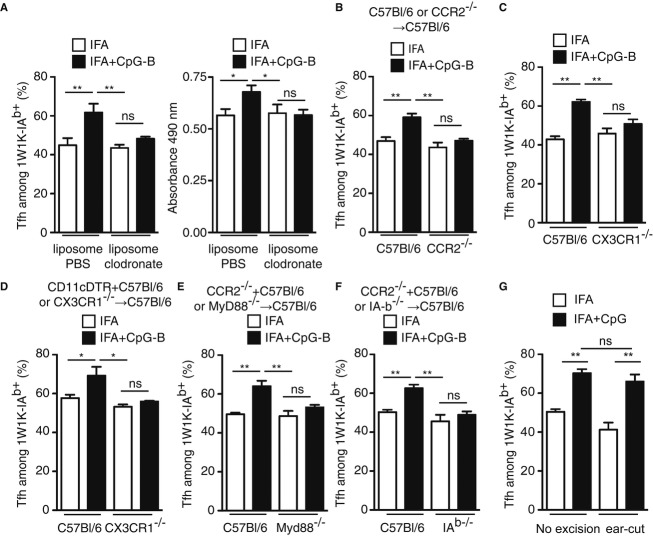
To be effective, protein priming must induce the development of a distinct lineage of CD4(+) T cells named T follicular helper (Tfh) cells, which regulate the differentiation of high-affinity memory B cells and long-lived plasma cells. In this context, we tested how adjuvantation with CpG, the Toll-like receptor 9 agonist used in clinics, contributes to antigen-specific T-cell-dependent B-cell responses in vivo. We found that addition of CpG to other vaccine adjuvant increased the differentiation of antigen-specific Tfh cells without changing the overall magnitude of the T-cell response. This phenomenon correlated with an enhancement of the germinal centre reaction, antigen-specific plasma cells and circulating antibodies. We comprehensively demonstrated that, in addition to the classical Tfh-cell differentiation mediated by conventional DC, the promoting effect due to CpG was orchestrated in vivo by antigen presentation and IL-6 secreted by monocyte-derived dendritic cells (DC) as shown in their absence. Thus, while conventional DC initiate T-cell responses, targeting monocyte-derived DC specifically enhances the Tfh programme needed to regulate high-affinity B-cell protection in vivo.


Figures

Figure 1
Figure 1
Adjuvantation with CpG-B of other vaccine adjuvant promotes Ag-specific Tfh-cell development. A–C Nine days after s.c. immunisation with 40 μg of 1W1K in IFA, dLN were analysed for the detection of 1W1K-specific activated CD4+ T cells (1W1K-IAb+CD44+) (A) and 1W1K-specific Tfh cells (CXCR5+CD62L) (B) as shown by Bcl-6 and PD-1 expression (C) (n = 5/condition, mean ± s.e.m.). D–F Frequency of CD4+ T cells (D), 1W1K-specific activated CD4+ T cells (E) and frequency of Tfh in 1W1K-specific CD4+ T cells (F) 9 days after s.c. immunisation with 40 μg of 1W1K in IFA or IFA+CpG-B (n = 5/group, mean ± s.e.m.). G, H Number of total 1W1K-specific CD4+ T cells (G) and frequency of 1W1K-specific Tfh (H) in dLN after s.c. immunisation with 1W1K in IFA (open circle) or IFA+CpG-B (solid circle) (n = 5/condition, mean ± s.e.m.). I–K Frequency of Tfh cells among 1W1K-specific CD4+ T cells in dLN 9 days after s.c. immunisation with IFA mixed with different dose of CpG-B (I), with other adjuvant (Alum, SAS) mixed with CpG-B (J) or with IFA mixed with different type of CpG (K) (n = 5/condition, mean ± s.e.m.). Data information: Data are representative of at least four independent experiments. ns, nonsignificant; *< 0.05; ** 0.01; *** 0.005; **** 0.001.
Figure 2
Figure 2
Addition of CpG-B boosts OVA-specific B-cell responses. A Fourteen days after s.c. immunisation with 100 μg of NP-OVA in IFA, dLN and sera were analysed to estimate NP-specific B cells and Ig levels, respectively. Representative example of FACS profiles: NP-specific B cells (left panel), among which plasma cells (PC: CD138+; middle panel) and GC-B cells (CD138B220+GL-7+CD95+; right panel). B Numbers of NP-specific GC-B cells and NP-specific PC in dLN after immunisation with IFA or IFA+CpG-B (n = 5/group; mean ± s.e.m.). C Levels of NP-specific IgG in sera after s.c. immunisation with NP-OVA in IFA or IFA+CpG-B (n = 5/group, mean ± s.e.m.). D 14 days after s.c. immunisation with 100 μg of OVA, numbers of OVA-specific GC-B cells and OVA-specific PC in dLN (n = 5/group; mean ± s.e.m.). E, F Levels of OVA-specific IgG in sera of mice 14 days after s.c. immunisation with OVA in adjuvant or adjuvant+CpG-B (n = 5/condition, mean ± s.e.m.) (E) and over time after s.c. immunisation using IFA (F). Data information: Data are representative of at least three independent experiments. ns, nonsignificant; *< 0.05; ** 0.01; *** 0.005; **** 0.001.
Figure 3
Figure 3
Impact of CpG-B on Tfh-cell differentiation relies on CD11c+ cells. A, BEight weeks after reconstitution, chimeric JHT (70%) + TLR9−/− (30%) → C57Bl/6 were s.c. immunised with 1W1K or OVA in IFA or IFA+CpG-B. Frequency of Tfh among 1W1K-specific CD4+ T cells in dLN 9 days after s.c. immunisation (n = 5/group, mean ± s.e.m.) (A) and levels of OVA-specific IgG, IgG1, IgG2b and IgG2c in the sera of mice 14 days after s.c. immunisation (n = 5/condition, mean ± s.e.m.) (B). C Five days after i.p. injection of JHT or C57Bl/6 littermates with 40 μg of 1W1K in Alum or Alum+CpG-B, spleens were collected and analysed. Frequency of Tfh among 1W1K-specific CD4+ T cells 9 days after i.p. injection is shown as well as serum concentrations of IL-6 estimated by ELISA 6 h after the i.p. injection (n = 5/group mean ± s.e.m.). D Eight weeks after reconstitution, chimeric CD11cDTR (70%) + (30%) C57Bl/6 or TLR9−/− → C57Bl/6 mice were treated every 2 days with DTx and s.c. immunised with 1W1K or OVA in IFA or IFA+CpG-B. Frequency of Tfh among 1W1K-specific CD4+ T cells in dLN 9 days after s.c. immunisation (n = 5/group, mean ± s.e.m.) and levels of OVA-specific IgG, in the sera of mice 14 days after s.c. immunisation (n = 5/group, mean ± s.e.m.). Data information: Data are representative of at least three independent experiments. ns, nonsignificant; *P < 0.05; ** 0.01; **** 0.001.
Figure 4
Figure 4
Ag-presenting DC in the dLN are CD11b+ conventional and monocyte-derived DC. A DC that captured and present Ag (CD11c+FITC+Y-Ae+) in dLN of mice 2 days after s.c. immunisation with Ea-FITC emulsified in IFA. B Histograms of Y-Ae staining correspond to mice s.c. immunised either with OVA-FITC or Ea-FITC in IFA. C Kinetics of Y-Ae+ FITC+ CD11c+ cells in the dLN after s.c. immunisation after IFA or IFA+CpG-B (IFA, open circle; IFA+CpG-B, solid circle) (n = 4/condition, mean ± s.e.m.). D, E Two days after s.c. immunisation with Ea-FITC in IFA or IFA+CpG-B, Y-Ae+ FITC+CD11c+ cells from dLN were analysed for CD8, CD11b expression (D) and for CD64 (E). F Kinetics of conventional DC (cDC, Y-Ae+ FITC+ CD11c+ CD11b+ CD64) and monocyte-derived DC (moDC, Y-Ae+ FITC+ CD11c+ CD11b+ CD64+) in the dLN after s.c. immunisation with IFA or IFA+CpG-B (IFA, open circle; IFA+CpG-B, solid circle) (n = 3/group, mean ± s.e.m.). Data information: Data are representative of at least five independent experiments.
Figure 5
Figure 5
Ag-presenting moDC produce IL-6 in response to CpG-B. IL-6 production in CD11c+ FITC+ cells from dLN 2 days after s.c. immunisation with Ea-FITC in IFA or IFA+CpG-B. Kinetics of IL-6+CD11c+FITC+ cells in dLN after Ea-FITC s.c. immunisation with IFA or IFA+CpG-B (IFA, open circle; IFA+CpG-B, solid circle) (n ≥ 4/condition, mean ± s.e.m.). Expression of CD11b and CD64 at the surface of CD11c+FITC, CD11c+FITC+IL6 and IL-6+ cells in dLN 24 h after s.c. immunisation with Ea-FITC in IFA or IFA+CpG-B. Frequency of IL-6+ cells in CD11c+Y-Ae+ cells in dLN 2 days after s.c. immunisation with 1010 Ea-coated beads in IFA complemented with different type of CpG (n = 4/condition, mean ± s.e.m.). Total CD11c+ beads+ Y-Ae+ in dLN 2 days after s.c. immunisation with 1010 Ea-coated beads in IFA or IFA+CpG-B were analysed. IL-6 production was assessed in CD11c+ beads+ cells from dLN 2 days after s.c. immunisation with 1010 Ea-coated beads in IFA or IFA+CpG-B (n = 6/group, mean ± s.e.m.). Total cell count numbers of CD11c+ beads+ IL-6+ cells in dLN of mice 2 days after s.c. immunisation with 1010 Ea-coated beads in IFA or IFA complemented 1, 2, 10, 50 or 100 μg of CpG-B (n ≥ 4/group, mean ± s.e.m.) (G). Data information: Data are representative of at least three independent experiments. ns, nonsignificant; *< 0.05; **≤ 0.01; ****≤ 0.001.
Figure 6
Figure 6
IL-6 produced by Ag-presenting DC in response to CpG-B adjuvantation promotes Tfh-cell differentiation. A Seven days after s.c. immunisation with 40 μg of 1W1K in IFA or IFA+CpG-B, dLN were collected and analysed for 1W1K-specific Tfh cells (1W1K-IAb+CD62Llo CXCR5+) after treatment with anti-IL-6Rα mAb or isotype control (Rat IgG2b) (n = 5/group, mean ± s.e.m.). Frequency of Tfh among 1W1K-specific CD4+ T cells was estimated. B, C Twenty-one days after s.c. immunisation with 100 μg of NP-OVA in IFA or IFA+CpG-B, dLN were collected and analysed for NP+ GC-B cells (NP+CD138B220+GL-7+CD95+) (B) and serum was collected for ELISA detection of NP8-specific IgG (C) (nd, not detected). D Eight weeks after reconstitution, chimeric CD11c-DTR (70%) + C57Bl/6 (30%) → C57Bl/6 (C57Bl/6) and CD11cDTR (70%) + IL-6−/− (30%) → C57Bl/6 (IL-6−/−) mice were s.c. immunised with 1W1K in IFA or IFA+CpG-B. Frequency of Tfh among 1W1K-specific CD4+ T cells in dLN 9 days after s.c. immunisation from IFA-immunised chimeric mice treated three times with DTx (n = 4/group, mean ± s.e.m.). Data are representative of at least three independent experiments. ns, nonsignificant; *< 0.05; ** 0.01; *** 0.005.
Figure 7
Figure 7
moDC drive the increase in Tfh-cell development due to CpG-B adjuvantation. A C57Bl/6 animals were s.c. immunised with 1W1K or OVA in IFA or IFA+CpG-B and treated twice with liposomes containing either PBS or clodronate. Nine days after s.c. immunisation with 1W1K, monocyte depletion was assessed in the blood of animals (data not shown) and the frequency of Tfh among 1W1K-specific CD4+ T cells was estimated in the corresponding animals (n = 5/group, mean ± s.e.m.). Fourteen days after s.c. immunisation with OVA, total OVA-specific IgG were estimated by ELISA (n = 5/group, mean ± s.e.m.). B–F Chimeric C57Bl/6 → C57Bl/6 (C57Bl/6) and CCR2−/− → C57Bl/6 (CCR2−/−) mice 8 weeks after reconstitution (B), CXC3CR1 and wild-type littermates (C), chimeric CD11c-DTR (70%) + C57Bl/6 (30%) → C57Bl/6 (C57Bl/6) and CD11cDTR (70%) + CX3CR1/− (30%) → C57Bl/6 (CX3CR1/−) mice treated three times with DTx (D), chimeric CCR2/− (70%) + C57Bl/6 (30%) → C57Bl/6 (C57Bl/6) and CCR2/− (70%) + MyD88−/− (30%) → C57Bl/6 (MyD88/−) mice (E) and chimeric CCR2/− (70%) + C57Bl/6 (30%) → C57Bl/6 (C57Bl/6) and CCR2/− (70%) + I-Ab/− (30%) → C57Bl/6 (I-Ab/−) mice (F) were s.c. immunised with 1W1K in IFA or IFA+CpG-B. Nine days after s.c. immunisation, the frequency of Tfh among 1W1K-specific CD4+ T cells was estimated in the corresponding animals (n = 5/group, mean ± s.e.m.). G C57Bl/6 animals were immunised s.c. in the ear with 1W1K in IFA or IFA+CpG-B. 1 h after immunisation, ear was resected and the frequency of Tfh among 1W1K-specific CD4+ T cells was estimated in the corresponding animals 9 days after (n = 5/group, mean ± s.e.m.). Data information: Data are representative of at least three independent experiments. ns, nonsignificant; *< 0.05; ** 0.01.

Comment in

References

    1. Baldridge JR, Crane RT. Monophosphoryl lipid A (MPL) formulations for the next generation of vaccines. Methods. 1999;19:103–107. - PubMed
    1. Banchereau J, Steinman RM. Dendritic cells and the control of immunity. Nature. 1998;392:245–252. - PubMed
    1. Beutler B, Jiang Z, Georgel P, Crozat K, Croker B, Rutschmann S, Du X, Hoebe K. Genetic analysis of host resistance: toll-like receptor signaling and immunity at large. Annu Rev Immunol. 2006;24:353–389. - PubMed
    1. Calabro S, Tortoli M, Baudner BC, Pacitto A, Cortese M, O'Hagan DT, De Gregorio E, Seubert A, Wack A. Vaccine adjuvants alum and MF59 induce rapid recruitment of neutrophils and monocytes that participate in antigen transport to draining lymph nodes. Vaccine. 2011;29:1812–1823. - PubMed
    1. Celli S, Lemaitre F, Bousso P. Real-time manipulation of T cell-dendritic cell interactions in vivo reveals the importance of prolonged contacts for CD4+ T cell activation. Immunity. 2007;27:625–634. - PubMed

Publication types

MeSH terms