Review
. 2014 Mar;11(1):34-43.
doi: 10.7497/j.issn.2095-3941.2014.01.003.

pH-responsive mesoporous silica nanoparticles employed in controlled drug delivery systems for cancer treatment

Affiliations
Review

pH-responsive mesoporous silica nanoparticles employed in controlled drug delivery systems for cancer treatment

Ke-Ni Yang et al. Cancer Biol Med. 2014 Mar.

Abstract

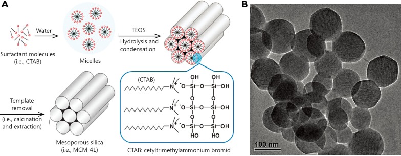
In the fight against cancer, controlled drug delivery systems have emerged to enhance the therapeutic efficacy and safety of anti-cancer drugs. Among these systems, mesoporous silica nanoparticles (MSNs) with a functional surface possess obvious advantages and were thus rapidly developed for cancer treatment. Many stimuli-responsive materials, such as nanoparticles, polymers, and inorganic materials, have been applied as caps and gatekeepers to control drug release from MSNs. This review presents an overview of the recent progress in the production of pH-responsive MSNs based on the pH gradient between normal tissues and the tumor microenvironment. Four main categories of gatekeepers can respond to acidic conditions. These categories will be described in detail.

Keywords: Mesoporous silica nanoparticles; antineoplastic protocols; controlled drug release; drug delivery systems; pH-responsive.


Conflict of interest statement

No potential conflicts of interest are disclosed.

Figures

Figure 1
Figure 1
Synthesis scheme for the preparation of MSNs (A) and transmission electron microscopy (TEM) images of MCM-41 (B). (Figure 1A is adapted from Ref. with permission of The Royal Society of Chemistry).
Figure 2
Figure 2
Graphical representation of pH-responsive MSNs with polyelectrolyte multilayers (A) and polyelectrolyte brushes (B). Release profiles of DOX from PEM-MENs (eight layers) in different pH media (C). DOX concentrations in plasma after DOX and DOX-loaded PEM-MSNs were injected intravenously through the vein for incremental time (D). Biodistribution of DOX in healthy SD rats at 2 h (E) and 24 h (F) after DOX and DOX-loaded PEM-MSNs at 2 mg/kg DOX equivalent were injected intravenously through the vein. *P<0.05 and **P<0.01 compared with free DOX group. (Figure 2C,D,E,F are adapted from Ref. with permission from The Royal Society of Chemistry).
Figure 3
Figure 3
Graphical representation of the pH-responsive MSNs with supramolecularnanovalves (A). Synthesis of the stalk on the surface of MSNs for further β-CD capping on the pore (B). Fluorescence intensity plots for the release of Hoechst dye, doxorubicin, and the pyrene-loaded cyclodextrin cap from MSNs (C) and release profiles of doxorubicin from ammonium-modified (7.5%, w/w) nanoparticles showing a faster and larger response compared with that of unmodified MSNs (D). Confocal images of KB-31 cells incubated with MSNs containing doxorubicin for the indicated times: KB-31 cancer cells effectively endocytosed the doxorubicin-loaded FITC-MSNs at 3 h. This action is followed by nuclear fragmentation after 80 h. However, with NH4Cl treatment, most of the doxorubicin was confined to nanoparticles, such that no observable cell death occurred (E). (Figure 3B,C,D,E are adapted with permission from Ref. . Copyright 2010, American Chemical Society).
Figure 4
Figure 4
Graphical representation of the pH-responsive MSNs capped with polymers (A) and nanoparticles (B) that linked to the surface of MSNs via pH-sensitive linkers.
Figure 5
Figure 5
(A) Graphical representation of pH-responsive MSNs with acid-decomposable inorganic gatekeepers. (B) DOX release profiles from DOX-Si-MP-UR and DOX-Si-MP-CaP under pH control. (C) Kinetics of calcium dissolution from DOX-Si-MP-CaP under pH control. (D) CLSM images of live MCF-7 cells treated with Lyso Tracker (50 nm), free DOX (5 µg/mL), and DOX-Si-MP-CaP (DOX =5 µg/mL), thereinto, (a) free DOX for 1 h exposure; (b) DOX-Si-MP-CaP for 1 h exposure; (c) DOX-Si-MP-CaP for 5 h exposure; (d) DOX-Si-MP-UR for 1 h exposure; and (e) DOX-Si-MP-UR for 5 h exposure. (Green fluorescence is associated with Lyso Tracker; the red fluorescence is expressed by free DOX, released DOX, and DOX retained within MSNs). Scale bar: 20 µm. (E) In vivo therapeutic efficacy after a single intratumoral injection of saline (●), free DOX (█), DOX-Si-MP-UR (▲), and DOX-Si-MP-CaP(◆) at a DOX-equivalent dose of 10 mg/kg. Inset: images of excised tumors at 16 days after treatment. I: saline, II: free DOX, III: DOX-Si-MP-UR, IV: DOX-Si-MP-CaP. (F) Tumor weights at 16 days after treatment. The results represent the means ± SDs (n=4); *P<0.05. (Figure 4B,C,D,E,F are adapted from Ref. with permission of John Wiley and Sons).

References

    1. Nazir S, Hussain T, Ayub A, Rashid U, MacRobert AJ. Nanomaterials in combating cancer: therapeutic applications and developments. Nanomedicine 2014;10:19-34 - PubMed
    1. Farokhzad OC, Langer R. Impact of nanotechnology on drug delivery. ACS Nano 2009;3:16-20 - PubMed
    1. Maeda H.The enhanced permeability and retention (EPR) effect in tumor vasculature: the key role of tumor-selective macromolecular drug targeting. Adv Enzyme Regul 2001;41:189-207 - PubMed
    1. Cho K, Wang X, Nie S, Chen ZG, Shin DM. Therapeutic nanoparticles for drug delivery in cancer. Clin Cancer Res 2008;14:1310-1316 - PubMed
    1. Davis ME, Chen ZG, Shin DM. Nanoparticle therapeutics: an emerging treatment modality for cancer. Nat Rev Drug Discov 2008;7:771-782 - PubMed

LinkOut - more resources