

Fear processing in dental phobia during crossmodal symptom provocation: an fMRI study
- PMID: 24738049
- PMCID: PMC3967629
- DOI: 10.1155/2014/196353
Fear processing in dental phobia during crossmodal symptom provocation: an fMRI study
Erratum in
-
Corrigendum to "Fear Processing in Dental Phobia during Crossmodal Symptom Provocation: An fMRI Study".Biomed Res Int. 2018 Nov 6;2018:6497672. doi: 10.1155/2018/6497672. eCollection 2018. Biomed Res Int. 2018. PMID: 30533437 Free PMC article.
Abstract
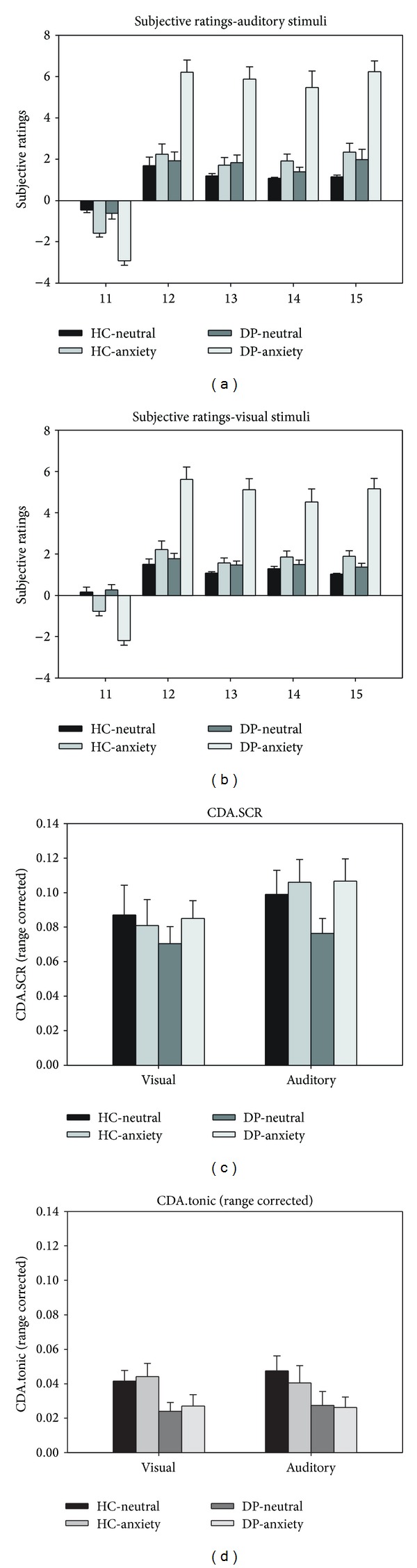
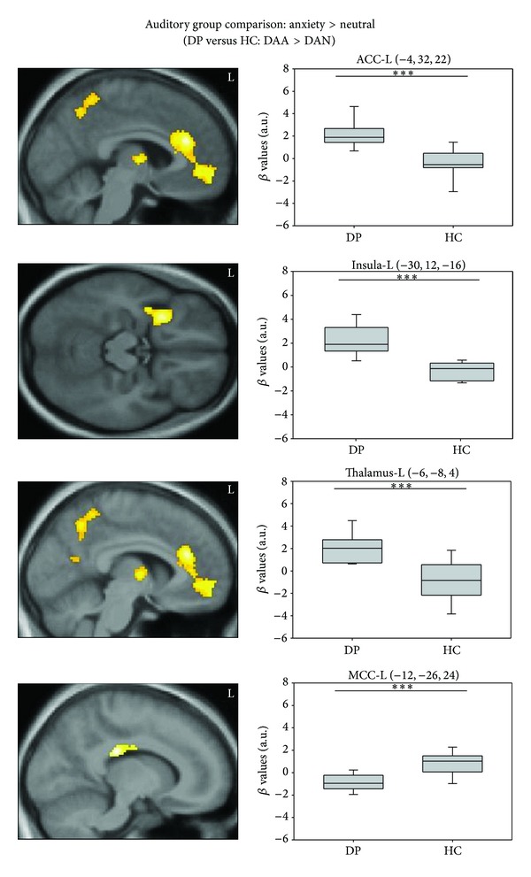
While previous studies successfully identified the core neural substrates of the animal subtype of specific phobia, only few and inconsistent research is available for dental phobia. These findings might partly relate to the fact that, typically, visual stimuli were employed. The current study aimed to investigate the influence of stimulus modality on neural fear processing in dental phobia. Thirteen dental phobics (DP) and thirteen healthy controls (HC) attended a block-design functional magnetic resonance imaging (fMRI) symptom provocation paradigm encompassing both visual and auditory stimuli. Drill sounds and matched neutral sinus tones served as auditory stimuli and dentist scenes and matched neutral videos as visual stimuli. Group comparisons showed increased activation in the insula, anterior cingulate cortex, orbitofrontal cortex, and thalamus in DP compared to HC during auditory but not visual stimulation. On the contrary, no differential autonomic reactions were observed in DP. Present results are largely comparable to brain areas identified in animal phobia, but also point towards a potential downregulation of autonomic outflow by neural fear circuits in this disorder. Findings enlarge our knowledge about neural correlates of dental phobia and may help to understand the neural underpinnings of the clinical and physiological characteristics of the disorder.
Figures
Similar articles
-
Networks of phobic fear: Functional connectivity shifts in two subtypes of specific phobia.Neurosci Lett. 2018 Jan 1;662:167-172. doi: 10.1016/j.neulet.2017.10.031. Epub 2017 Oct 18. Neurosci Lett. 2018. PMID: 29054435
-
How specific is specific phobia? Different neural response patterns in two subtypes of specific phobia.Neuroimage. 2011 May 1;56(1):363-72. doi: 10.1016/j.neuroimage.2011.02.015. Epub 2011 Feb 18. Neuroimage. 2011. PMID: 21316468
-
The functional neuroanatomy of blood-injection-injury phobia: a comparison with spider phobics and healthy controls.Psychol Med. 2010 Jan;40(1):125-34. doi: 10.1017/S0033291709005972. Epub 2009 May 13. Psychol Med. 2010. PMID: 19435544
-
Functional changes in brain activity after hypnosis in patients with dental phobia.J Physiol Paris. 2015 Dec;109(4-6):131-142. doi: 10.1016/j.jphysparis.2016.10.001. Epub 2016 Oct 6. J Physiol Paris. 2015. PMID: 27720948
-
Meta-analysis of functional brain imaging in specific phobia.Psychiatry Clin Neurosci. 2013 Jul;67(5):311-22. doi: 10.1111/pcn.12055. Epub 2013 May 28. Psychiatry Clin Neurosci. 2013. PMID: 23711114 Review.
Cited by
-
Bifocal emotion regulation through acupoint tapping in fear of flying.Neuroimage Clin. 2022;34:102996. doi: 10.1016/j.nicl.2022.102996. Epub 2022 Mar 30. Neuroimage Clin. 2022. PMID: 35378497 Free PMC article.
-
Diagnostic classification of specific phobia subtypes using structural MRI data: a machine-learning approach.J Neural Transm (Vienna). 2015 Jan;122(1):123-34. doi: 10.1007/s00702-014-1272-5. Epub 2014 Jul 19. J Neural Transm (Vienna). 2015. PMID: 25037587
-
Dental Fear and Delayed Dental Care in Appalachia-West Virginia.J Dent Hyg. 2015 Aug;89(4):274-81. J Dent Hyg. 2015. PMID: 26304952 Free PMC article.
-
Brain activations associated with fearful experience show common and distinct patterns between younger and older adults in the hippocampus and the amygdala.Sci Rep. 2018 Mar 23;8(1):5137. doi: 10.1038/s41598-018-22805-9. Sci Rep. 2018. PMID: 29572480 Free PMC article. Clinical Trial.
-
Real-life dental examination elicits physiological responses different to visual and auditory dental-related stimuli.PLoS One. 2021 Jun 3;16(6):e0252128. doi: 10.1371/journal.pone.0252128. eCollection 2021. PLoS One. 2021. PMID: 34081713 Free PMC article.
References
-
- Wittchen HU, Jacobi F, Rehm J, et al. The size and burden of mental disorders and other disorders of the brain in Europe 2010. European Neuropsychopharmacology. 2011;21(9):655–679. - PubMed
-
- APA. Diagnostic and Statistical Manual of Mental Disorders. 4 edition. Washington, DC, USA: American Psychiatric Association; 2000.
-
- APA. Diagnostic and Statistical Manual of Mental Disorders. 5 edition. Arlington, Va, USA: American Psychiatric Publishing; 2013.
Publication types
MeSH terms
Supplementary concepts
LinkOut - more resources
Full Text Sources
Other Literature Sources
Medical
