Comparative Study
. 2014 Sep 1;23(17):2014-29.
doi: 10.1089/scd.2013.0639. Epub 2014 Jun 12.

Stable methylation at promoters distinguishes epiblast stem cells from embryonic stem cells and the in vivo epiblasts

Affiliations
Comparative Study

Stable methylation at promoters distinguishes epiblast stem cells from embryonic stem cells and the in vivo epiblasts

Anne-Clémence Veillard et al. Stem Cells Dev. .

Abstract

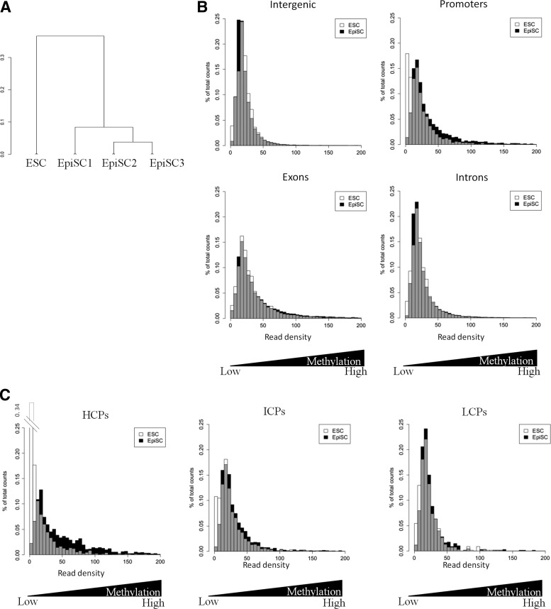
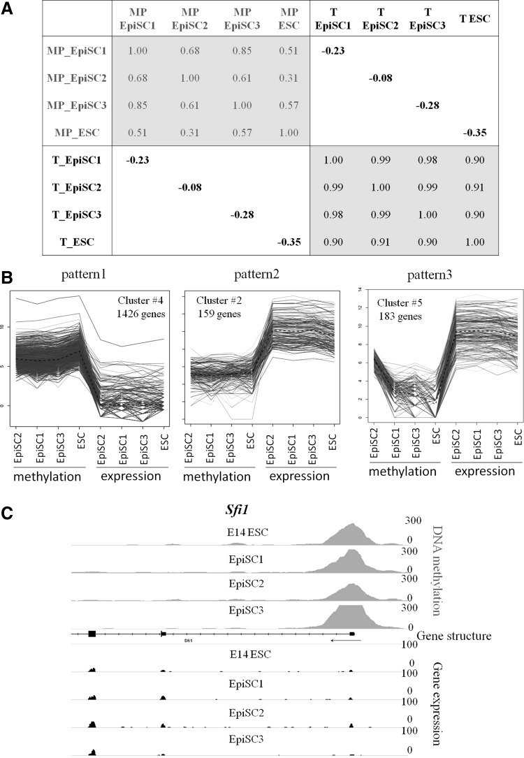
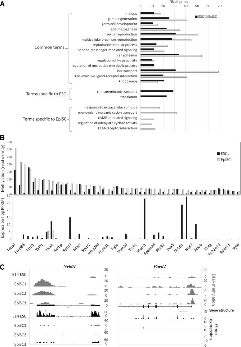
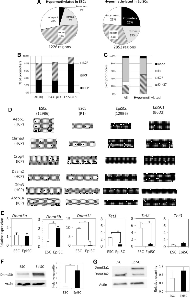
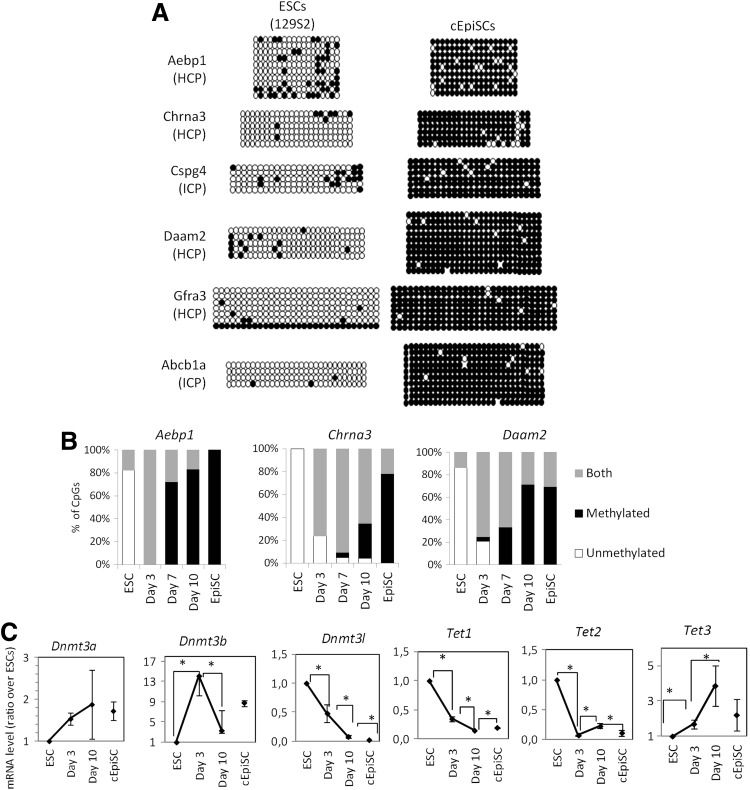
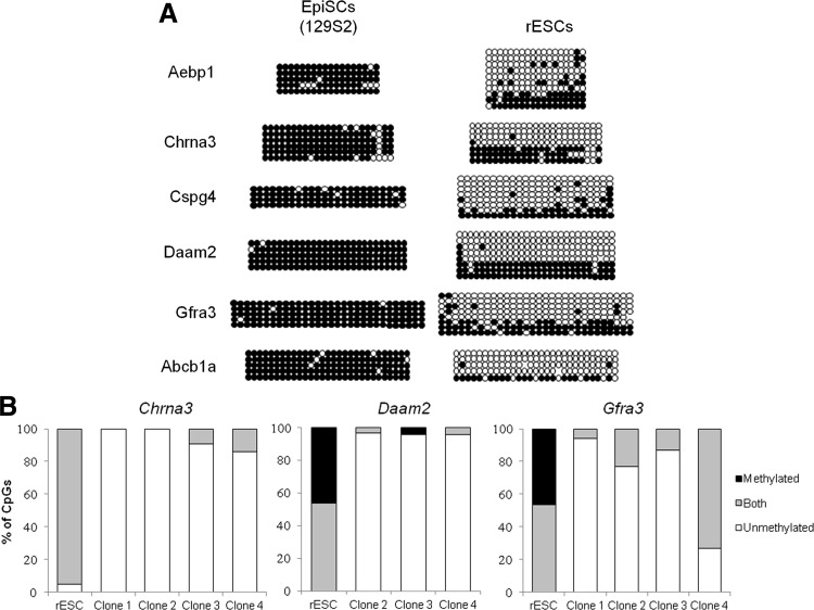
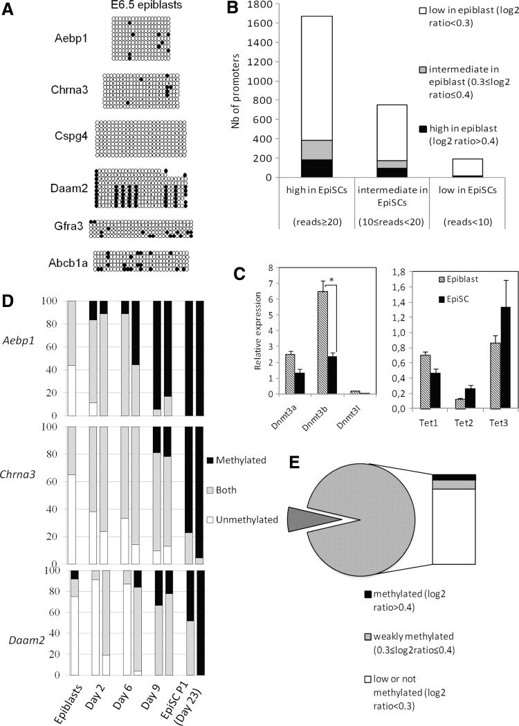
Embryonic Stem Cells (ESCs) and Epiblast Stem Cells (EpiSCs) are the in vitro representatives of naïve and primed pluripotency, respectively. It is currently unclear how their epigenomes underpin the phenotypic and molecular characteristics of these distinct pluripotent states. Here, we performed a genome-wide comparison of DNA methylation between ESCs and EpiSCs by MethylCap-Seq. We observe that promoters are preferential targets for methylation in EpiSC compared to ESCs, in particular high CpG island promoters. This is in line with upregulation of the de novo methyltransferases Dnmt3a1 and Dnmt3b in EpiSC, and downregulation of the demethylases Tet1 and Tet2. Remarkably, the observed DNA methylation signature is specific to EpiSCs and differs from that of their in vivo counterpart, the postimplantation epiblast. Using a subset of promoters that are differentially methylated, we show that DNA methylation is established within a few days during in vitro outgrowth of the epiblast, and also occurs when ESCs are converted to EpiSCs in vitro. Once established, this methylation is stable, as ES-like cells obtained by in vitro reversion of EpiSCs display an epigenetic memory that only extensive passaging and sub-cloning are able to almost completely erase.


Figures

<b>FIG. 1.</b>
FIG. 1.
Global analysis of DNA methylation in embryonic stem cell (ESC) and epiblast stem cells (EpiSC) lines. (A) Hierarchical clustering of methylation profiles, based on a Pearson's correlation distance matrix. (B, C) Distribution of read density for ESCs and EpiSCs (mean of the three cell lines) in each genomic category (B) and in promoters annotated as high (HCP), intermediate (ICP), or low (LCP) CpG content (C). Bars for EpiSC are in white, bars for ESC in black, the overlay in gray.
<b>FIG. 2.</b>
FIG. 2.
Relationships between promoter methylation and gene expression. (A) Correlation coefficients between DNA methylation on promoters (MP) and expression data (T). (B) Three main clusters (Qt-clust) illustrating the relation between promoter methylation and gene expression. (C) Example of a gene following pattern 1.
<b>FIG. 3.</b>
FIG. 3.
Functional annotation of methylated promoters. (A) GO terms associated with promoters methylated (read density >20) in EpiSCs and in ESCs, according to DAVID analysis (P-value≤1%). The *indicate KEGG pathways. (B) Plots comparing methylation and expression of the 49 genes associated with the union of “gamete generation” and “sexual reproduction” GO terms. (C) IGV browser view of two germline and ESC-specific genes, Nr0b1 and Piwil2, showing the de novo methylation and loss of gene expression in EpiSCs compared to ESCs.
<b>FIG. 4.</b>
FIG. 4.
EpiSCs tend to contain hypermethylated promoters. (A) Pie charts showing the classification of hypermethylated regions in ESCs (read density ratio ESC/EpiSC >3) and hypermethylated in EpiSCs (read density ratio EpiSC/ESC >3). The number of DMRs is indicated below the pies. ESC and EpiSC distribution differ significantly (Chi-squared test, P-value<10−53). (B) Classification of all methylated (read density ≠0) and differentially methylated promoters as HCP, ICP, and LCP. Hypermethylated promoters in EpiSCs are significantly enriched in HCP (Chi-squared test, P-value<10−76). (C) Classification of all promoters (read density in EpiSC ≠0) and differentially methylated promoters in EpiSC (ratio EpiSC/ESC >3) according to their association in ESC with H3K4me3 (gray), H3K27me3 (white), bivalent (H3K4me3+H3K27me3, hatched) or neither mark (black). Hypermethylated promoters are significantly different from all promoters (Chi-squared test, P-value<10−22). (D) Validation of differential methylation between EpiSCs and ESCs. The class of each promoter according to their CpG content is indicated. Circles represent CpG nucleotides either methylated (closed) or unmethylated (open). (E) Gene expression of DNA methylation modifying enzymes in ESCs and EpiSCs determined by RT-qPCR. Error bars represent SEM of three different cell lines. (F, G) Western blots showing the protein level of Dnmt3a and Dnmt3b in ESCs and EpiSCs. The average quantity relative to Actin is shown on the right. Error bars represent SEM of two (ESCs) to three (EpiSCs) different cell lines. *in E, F: P<0.05, Mann–Whitney U test.
<b>FIG. 5.</b>
FIG. 5.
Characterization of ESC conversion into cEpiSC. (A) Changes of methylation in the six promoters assessed by bisulfite-sequencing during conversion of ESCs into cEpiSCs. (B) Changes in the CpG level of methylation in the promoter of Aebp1, Chrna3, and Daam2 during the conversion process. Genomic DNA after bisulfite conversion was directly sequenced. Each CpG has been classified according to the presence of a C (methylated), a T (unmethylated), or a polymorphism meaning heterogeneity between the two forms in the cell population (both). (C) Changes in expression of DNA methylation modifying enzymes, during the conversion of ESCs determined by RT-qPCR. Bars represent SEM of three independent experiments. *P<0.05, Mann–Whitney U test.
<b>FIG. 6.</b>
FIG. 6.
Characterization of EpiSC reversion into rESC. (A) Changes of methylation in the six promoters assessed by bisulfite-sequencing during reversion of EpiSCs into rESCs. (B) CpG level of methylation in the promoter of Chrna3, Daam2, and Gfra3 after clonal expansion of rESCs. As in Fig. 5B, each CpG is classified as methylated, unmethylated, or polymorphic (both) in the cell population.
<b>FIG. 7.</b>
FIG. 7.
Differential methylation in embryo-derived pluripotent cells and in the epiblast. (A) Methylation status in the epiblast (E6.5). (B) Comparison of methylation in promoters common to Borgel et al. [12]) and our study. Promoters were classified according to their mean methylation status in EpiSCs and each category further separated according to their methylation values in E6.5 epiblasts. (C) Gene expression of DNA methylation modifying enzymes in E6.5 epiblast and EpiSCs determined by RT-qPCR. Bars represent SEM. *P<0.05, Mann–Whitney U test. (D) Changes in the CpG level of methylation in the promoter of Aebp1, Chrna3, and Daam2 during derivation of EpiSCs. Genomic DNA after bisulfite conversion was directly sequenced and the status of the CpGs was indicated as in Figs. 5 and 6. (E) Expression status in the epiblast of genes that contain methylated promoters in EpiSCs.

References

    1. Hutchins AP, Choo SH, Mistri TK, Rahmani M, Woon CT, Ng CK, Jauch R. and Robson P. (2013). Co-motif discovery identifies an Esrrb-Sox2-DNA ternary complex as a mediator of transcriptional differences between mouse embryonic and epiblast stem cells. Stem Cells 31:269–281 - PubMed
    1. Nichols J. and Smith A. (2012). Pluripotency in the embryo and in culture. Cold Spring Harb Perspect Biol 4:a008128. - PMC - PubMed
    1. Brons IGM, Smithers LE, Trotter MWB, Rugg-Gunn P, Sun B, Chuva de Sousa Lopes SM, Howlett SK, Clarkson A, Ahrlund-Richter L, Pedersen RA. and Vallier L. (2007). Derivation of pluripotent epiblast stem cells from mammalian embryos. Nature 448:191–195 - PubMed
    1. Chenoweth JG, McKay RDG. and Tesar PJ. (2010). Epiblast stem cells contribute new insight into pluripotency and gastrulation. Dev Growth Differ 52:293–301 - PubMed
    1. Nichols J. and Smith A. (2009). Naive and primed pluripotent states. Cell Stem Cell 4:487–492 - PubMed

Publication types