Review
. 2014 May;13(5):503-14.
doi: 10.1016/S1474-4422(14)70011-0.

Distinct neurological disorders with ATP1A3 mutations

Collaborators, Affiliations
Review

Distinct neurological disorders with ATP1A3 mutations

Erin L Heinzen et al. Lancet Neurol. 2014 May.

Abstract

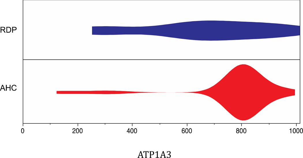
Genetic research has shown that mutations that modify the protein-coding sequence of ATP1A3, the gene encoding the α3 subunit of Na(+)/K(+)-ATPase, cause both rapid-onset dystonia parkinsonism and alternating hemiplegia of childhood. These discoveries link two clinically distinct neurological diseases to the same gene, however, ATP1A3 mutations are, with one exception, disease-specific. Although the exact mechanism of how these mutations lead to disease is still unknown, much knowledge has been gained about functional consequences of ATP1A3 mutations using a range of in-vitro and animal model systems, and the role of Na(+)/K(+)-ATPases in the brain. Researchers and clinicians are attempting to further characterise neurological manifestations associated with mutations in ATP1A3, and to build on the existing molecular knowledge to understand how specific mutations can lead to different diseases.


Figures

Figure 1
Figure 1
Structure of the Na, K-ATPase. A. Cartoon representation of the Na, K-ATPase in the potassium occluded state showing K+ (red spheres), the three protein subunits α (grey), β(purple) and FXYDγ (green), and the phosphorylation, which is mimicked by MgF42− (dark gray). Residues reported to be mutated in AHC and/or RDP (cf. Table 1) are indicated by spheres at the α carbon, yellow for one case of AHC, orange for more than one case of AHC, cyan for RDP, green for both AHC and RDP. Key ion binding residues are shown in stick. B. A 90° degree rotation in the membrane plane of the representation in A giving an extracellular view of the ion binding transmembrane part of the Na, K-ATPase with disease-causing mutations color coded as in A. Two of the ion binding residues (shown in sticks) have been found to be mutated both in RDP and in AHC patients. The figures were made from pdb code 2ZXE.
Figure 2
Figure 2
Schematic depicting the location of AHC-causing (red dots) and RDP-causing (blue dots) mutations in ATP1A3, mRNA and protein. The one mutation shared between disease phenotypes is located at D923N (blue dot with a red dot inside). Two rare polymorphisms identified in the general population are indicated by the green dots. Amino acid modifications are provided to the right of the dots.
Figure 3
Figure 3
Density plot showing the distribution of AHC and RDP mutations identified to date in 116 and 20 patients with ATP1A3 mutations, respectively. In general, RDP mutations appear to be more evenly distributed, whereas AHC mutations are heavily concentrated in particular sites in the protein.
Figure 4
Figure 4
Post-Albers model for the Na, K-ATPase reaction cycle, as reproduced from Toustrup-Jensen and co-workers.. E1 and E2 are major conformational states with preference for binding of Na+ and K+, respectively. Cytoplasmic and extracellular ions are indicated by subscripts c and e, respectively. Brackets indicate occlusion of the ions in a cavity in the protein. P indicates the bound phosphate.
Figure 5
Figure 5
Na, K-ATPase α3 genetic animal models. A. Atp1a3 mutant mice. The locations of three mutations in the mouse Atp1a3 genomic locus are depicted. Myshkin mice carry a T-to-A transversion in exon 18 that results in the substitution of asparagine for isoleucine at position 810 (I810N), Atp1a3tm1Ling mice carry a point mutation in intron 4 adjacent to the exon-intron splice site that results in aberrant splicing of the gene, adding 126 base pairs to the RNA transcript,,. Atp1a3tm2Kwk mice carry a STOP-polyA cassette that replaces exons 2–6 in Atp1a3. B. Atpα mutant Drosophila. AtpαCJ10 fruit flies carry a G to A transition that results in the substitution of glycine for serine at position 744 (G744S). G744S in the Drosophila α subunit is equivalent to mutation G755S in the human α3 subunit found in an AHC patient. C. Atp1a3a/b knockdown zebrafish. Knockdown of Atp1a3a or Atp1a3b RNA transcript by ~65% in 60 hpf embryos had similar phenotypic effects.

References

    1. Boycott KM, Vanstone MR, Bulman DE, MacKenzie AE. Rare-disease genetics in the era of next-generation sequencing: discovery to translation. Nat Rev Genet. 2013;14(10):681–691. - PubMed
    1. McGrail KM, Phillips JM, Sweadner KJ. Immunofluorescent localization of three Na, K-ATPase isozymes in the rat central nervous system: both neurons and glia can express more than one Na, K-ATPase. J Neurosci. 1991;11(2):381–391. - PMC - PubMed
    1. Bottger P, Tracz Z, Heuck A, Nissen P, Romero-Ramos M, Lykke-Hartmann K. Distribution of Na/K-ATPase alpha 3 isoform, a sodium-potassium P-type pump associated with rapid-onset of dystonia parkinsonism (RDP) in the adult mouse brain. J Comp Neurol. 2011;519(2):376–404. - PubMed
    1. Dobretsov M, Stimers JR. Neuronal function and alpha3 isoform of the Na/K-ATPase. Frontiers in bioscience : a journal and virtual library. 2005;10:2373–2396. - PubMed
    1. Dobretsov M, Hastings SL, Sims TJ, Stimers JR, Romanovsky D. Stretch receptor-associated expression of alpha 3 isoform of the Na+, K+-ATPase in rat peripheral nervous system. Neuroscience. 2003;116(4):1069–1080. - PubMed

Publication types

MeSH terms

Substances

Supplementary concepts