. 2014 May 30;289(22):15833-44.
doi: 10.1074/jbc.M113.530212. Epub 2014 Apr 16.

Binding of Streptococcus pneumoniae endopeptidase O (PepO) to complement component C1q modulates the complement attack and promotes host cell adherence

Affiliations

Binding of Streptococcus pneumoniae endopeptidase O (PepO) to complement component C1q modulates the complement attack and promotes host cell adherence

Vaibhav Agarwal et al. J Biol Chem. .

Abstract

The Gram-positive species Streptococcus pneumoniae is a human pathogen causing severe local and life-threatening invasive diseases associated with high mortality rates and death. We demonstrated recently that pneumococcal endopeptidase O (PepO) is a ubiquitously expressed, multifunctional plasminogen and fibronectin-binding protein facilitating host cell invasion and evasion of innate immunity. In this study, we found that PepO interacts directly with the complement C1q protein, thereby attenuating the classical complement pathway and facilitating pneumococcal complement escape. PepO binds both free C1q and C1 complex in a dose-dependent manner based on ionic interactions. Our results indicate that recombinant PepO specifically inhibits the classical pathway of complement activation in both hemolytic and complement deposition assays. This inhibition is due to direct interaction of PepO with C1q, leading to a strong activation of the classical complement pathway, and results in consumption of complement components. In addition, PepO binds the classical complement pathway inhibitor C4BP, thereby regulating downstream complement activation. Importantly, pneumococcal surface-exposed PepO-C1q interaction mediates bacterial adherence to host epithelial cells. Taken together, PepO facilitates C1q-mediated bacterial adherence, whereas its localized release consumes complement as a result of its activation following binding of C1q, thus representing an additional mechanism of human complement escape by this versatile pathogen.

Keywords: Cell Adhesion; Complement Activation; Complement System; Host Cell Adherence; Host-Pathogen Interaction; Innate Immunity; Pneumococci; Streptococcus; c1q.


Figures

FIGURE 1.
FIGURE 1.
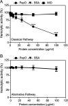
PepO inhibits the hemolytic activity of human serum. A, to measure the inhibitory effect of PepO on the classical pathway, antibody-coated erythrocytes were subjected to complement attack from NHS in the presence of increasing amounts of PepO. BSA and MID were used as negative controls. The degree of lysis was estimated by measurement of the release of hemoglobin. B, to study inhibition of the alternative pathway, rabbit erythrocytes were incubated with NHS with increasing concentrations of PepO. BSA was used as a negative control. Cell lysis was measured as in A. The absorbance obtained without an inhibitor present was set to 100%, and the graphs show the means ± S.D. of three independent experiments performed in duplicates. Statistical significance of differences was calculated using two-way analysis of variance and Bonferroni's post-test. *, p < 0.05; ****, p < 0.0001.
FIGURE 2.
FIGURE 2.
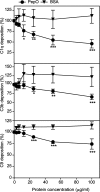
PepO inhibits the classical pathway of complement. PepO was preincubated with NHS in GVB++ buffer and added to microtiter plates coated with aggregated human IgG. BSA was used as a negative control. Deposition of C1q, C3b, and C9 was measured using polyclonal antibodies. The amount of deposited complement components in the absence of inhibitor was set as 100%. The data represent the means ± S.D. of three independent experiments performed in duplicates. Statistical significance of differences was calculated using two-way analysis of variance and Bonferroni's post-test. *, p < 0.05; **, p < 0.01; ***, p < 0.001.
FIGURE 3.
FIGURE 3.
PepO activates the complement cascade. A and B, the plates were coated with PepO, aggregated human IgG (hu-IgG) as positive control, or BSA as a negative control and incubated with the indicated concentrations of NHS in GVB++ buffer. Deposited components of the classical complement pathway C1q (A) and C3b (B) were detected with specific Abs. C, for lectin pathway, C1q-depleted serum in GVB++ buffer was added to mannan-coated plates, and the deposition of C3b was detected. D, for alternative pathway activation, NHS diluted in Mg2+EGTA buffer was added to zymosan-coated plates, and deposition of C3b was measured. Means ± S.D. of three independent experiments performed in duplicates are presented.
FIGURE 4.
FIGURE 4.
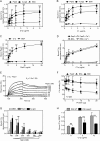
Binding of PepO to C1q. A and B, increasing concentrations of either plasma-purified C1q (A) or C1 complex (B) were added to microtiter plates coated with PepO, aggregated human IgG (hu-IgG), or BSA. C, plates were coated with C1q, and increasing concentrations of PepO were added. BSA was used as control. D, binding of C1q to PepO-coated wells was determined in the presence or absence of calcium. E, binding of PepO to immobilized C1q as analyzed by surface plasmon resonance. Increasing concentrations of PepO (22–1500 nm) were injected onto a C1q-coated CM5 sensor chip. The amount of PepO associating with the C1q was measured in response units (RU). Representative sensorgrams are presented. F, various concentrations of PepO were preincubated with 5 μg/ml C1q and added to the plates with immobilized aggregated human IgG. BSA and MID were used as controls. Bound C1q was detected with specific Abs. The amount of bound C1q in the absence of inhibitor was set as 100%. G, microtiter plates were coated with PepO, and the effect of different concentrations of NaCl on binding of plasma-purified C1q (5 μg/ml) was analyzed. Aggregated human IgG and BSA were used as positive and negative control, respectively. Bindings were detected with specific Abs. The data represent means ± S.D. of three independent experiments performed in duplicates. H, pneumococcal surface-exposed PepO functions as a C1q receptor. Pneumococcal wild-type strain D39 and its isogenic pepO mutant D39ΔpepO were incubated with 0, 5, and 10 μg/ml concentration of purified C1q for 60 min at 37 °C. Binding of C1q was analyzed by flow cytometry. Results were expressed as GMFI (means ± S.D.) from three independent experiments. Statistical significance was calculated using two-way ANOVA test. ns: not significant; *, p < 0.05; **, p < 0.01; ***, p < 0.001.
FIGURE 5.
FIGURE 5.
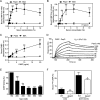
C4BP binding to PepO. A–C, PepO was immobilized, and increasing amounts of NHS (A), heat-inactivated serum (B), or plasma-purified C4BP (C) were added. BSA was used as control. Bound C4BP was detected using specific polyclonal Abs. Statistical significance was calculated using the two-way ANOVA test. ***, p < 0.001. D, binding of PepO to immobilized C4BP as analyzed by surface plasmon resonance. Increasing concentrations of PepO (22–1500 nm) were injected onto a C4BP-coated CM5 sensor chip. The amount of PepO associating with the C4BP was measured in response units. Representative sensorgrams are presented. E, the effect of different concentrations of NaCl on binding of plasma-purified C4BP (10 μg/ml) to PepO was analyzed. Specific polyclonal Abs detected bound C4BP. BSA was used as negative control. One-way ANOVA test was performed to calculate statistical differences of the groups as compared with the binding at 150 mm NaCl. ***, p < 0.001. F, Pneumococcal wild-type strain D39 and NCTC10319 and their respective isogenic pepO mutants were incubated with 100 kcpm 125I-C4BP for 1 h at 37 °C (where kcpm is kilo counts per minute). Binding was detected using a Wizard2 gamma counter. Statistical significance was calculated using Student's t test. *, p < 0.05; **, p < 0.01. Data are presented as means ± S.D. of three independent experiments done in duplicates.
FIGURE 6.
FIGURE 6.
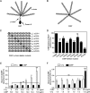
Mapping of binding site for PepO in C4BP. A–C, schematic representation of different variants of C4BP used: C4BP (A), rC4BP (B), and C4BP α-chain deletion mutants (C). The main isoform of C4BP contains seven identical α-chains and one unique β-chain. The β-chain-containing C4BP in circulation is bound to vitamin K-dependent anticoagulant Protein S (PS), forming a C4BP-PS complex. The rC4BP contains six α-chains and lacks the β-chain and the associated PS. C4BP α-chain deletion mutants lack single CCP domains (represented by circles with X in each α-chain). D, the binding of C4BP variants to immobilized PepO was analyzed. Bound C4BP was detected with polyclonal Abs. The graph represents data from two independent experiments done in duplicates ± S.D. E and F, simultaneous binding of C1q and C4BP to PepO immobilized on microtiter plates. A constant amount of C4BP (10 μg/ml) together with increasing amounts of C1q (E) or constant amount of C1q (1 μg/ml) with increasing amounts of C4BP (F) was added. Bound C4BP and C1q were detected using specific Abs. Data presented are from three independent duplicate experiments ± S.D. Statistical significance was calculated using one-way ANOVA and Dunnett's post-test. ns: not significant; *, p < 0.05; **, p < 0.01; ***, p < 0.001.
FIGURE 7.
FIGURE 7.
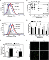
Blocking of C1q binding to pneumococci and C1q-mediated adherence to host cells by PepO. A–D, competitive inhibition experiments. Binding of plasma-purified C1q (A and B) to pneumococci (NCTC10319) or from NHS (0.5%) (C and D) was measured in the absence of exogenous added PepO proteins. BSA was used as negative control. Bound C1q was analyzed by flow cytometry using specific Abs. A representative flow cytometry histogram from three independent experiments is shown (A and C), and GMFI values (D) or normalized to the percentages (B) from three independent experiments performed in duplicates are presented. Statistical significance was calculated using two-way ANOVA test, **, p < 0.01; ***, p < 0.001. E and F, in cell culture blocking experiments, C1q-mediated adherence of pneumococci (NCTC10319) to A549 lung epithelial cells in the absence and presence of PepO (10 μg) was analyzed. E, adherence was determined by counting the cfu per well obtained from sample aliquots plated onto blood agar plates after 3 h of infection. F, representative immunofluorescence microscopy image of adherent pneumococci to A549 cells. Bar is equal to 10 μm. Two-way ANOVA test was performed to determine the statistical difference between the groups. Results present the means ± S.D. of at least three independent experiments. ns: not significant; **, p < 0.01 relative to infections carried out in the absence of C1q.
FIGURE 8.
FIGURE 8.
Effect of PepO on bacterial opsonization. A, deposition of C3b on pneumococci. Pneumococcal wild-type strain D39 and its isogenic pepO mutant D39ΔpepO were incubated with the indicated concentrations of NHS in GVB++ buffer for 15 min at 37 °C. The bacteria were thereafter washed and incubated with a FITC-conjugated anti-C3c Ab followed by flow cytometry analysis. Less C3b was deposited on the wild-type strain as compared with the mutant. B, in inhibition experiments, C3b deposition was investigated on the surface of mutant bacteria in the absence or the presence of PepO. Human serum albumin was used as a negative control. Results were expressed as GMFI (means ± S.D.) after subtracting the Ab background from two independent experiments performed in duplicates. C, PepO inhibits the bactericidal activity of human serum. E. coli DH5α (1000 cfu) were incubated for 30 min at 37 °C with 0.2% NHS pretreated with increasing concentrations of PepO or control protein A1AT, and the surviving bacteria were enumerated after overnight culture on LB agar plates. The survival was expressed as the percentage of inoculum. The data represent the means ± S.D. of three independent experiments performed in duplicates. Statistical significance was calculated using a two-way ANOVA test. ns: not significant; *, p < 0.05; ***, p < 0.001.
FIGURE 9.
FIGURE 9.
Schematic model demonstrating the different roles of PepO-C1q interaction in pneumococcal infections. The surface-presented PepO promotes C1q-mediated pneumococcal host cell colonization, and the secreted form consumes complement as a result of its activation following C1q interaction and thus helps in complement evasion. Furthermore, PepO binds the complement inhibitor C4BP, which when occurring at the bacterial surface further attenuates complement deposition. C4BP may also bind secreted PepO, but this does not compete out binding of C1q.

References

    1. Hackel M., Lascols C., Bouchillon S., Hilton B., Morgenstern D., Purdy J. (2013) Serotype prevalence and antibiotic resistance in Streptococcus pneumoniae clinical isolates among global populations. Vaccine 31, 4881–4887 - PubMed
    1. O'Brien K. L., Wolfson L. J., Watt J. P., Henkle E., Deloria-Knoll M., McCall N., Lee E., Mulholland K., Levine O. S., Cherian T. (2009) Burden of disease caused by Streptococcus pneumoniae in children younger than 5 years: global estimates. Lancet 374, 893–902 - PubMed
    1. Ricklin D., Hajishengallis G., Yang K., Lambris J. D. (2010) Complement: a key system for immune surveillance and homeostasis. Nat. Immunol. 11, 785–797 - PMC - PubMed
    1. Blom A. M., Hallström T., Riesbeck K. (2009) Complement evasion strategies of pathogens-acquisition of inhibitors and beyond. Mol. Immunol. 46, 2808–2817 - PubMed
    1. Lambris J. D., Ricklin D., Geisbrecht B. V. (2008) Complement evasion by human pathogens. Nat. Rev. Microbiol. 6, 132–142 - PMC - PubMed

Publication types

MeSH terms

LinkOut - more resources