. 2014 May 30;289(22):15666-79.
doi: 10.1074/jbc.M113.534222. Epub 2014 Apr 16.

Heat-induced irreversible denaturation of the camelid single domain VHH antibody is governed by chemical modifications

Affiliations

Heat-induced irreversible denaturation of the camelid single domain VHH antibody is governed by chemical modifications

Yoko Akazawa-Ogawa et al. J Biol Chem. .

Abstract

The variable domain of camelid heavy chain antibody (VHH) is highly heat-resistant and is therefore ideal for many applications. Although understanding the process of heat-induced irreversible denaturation is essential to improve the efficacy of VHH, its inactivation mechanism remains unclear. Here, we showed that chemical modifications predominantly governed the irreversible denaturation of VHH at high temperatures. After heat treatment, the activity of VHH was dependent only on the incubation time at 90 °C and was insensitive to the number of heating (90 °C)-cooling (20 °C) cycles, indicating a negligible role for folding/unfolding intermediates on permanent denaturation. The residual activity was independent of concentration; therefore, VHH lost its activity in a unimolecular manner, not by aggregation. A VHH mutant lacking Asn, which is susceptible to chemical modifications, had significantly higher heat resistance than did the wild-type protein, indicating the importance of chemical modifications to VHH denaturation.

Keywords: Antibody; Antibody Engineering; Protein Aggregation; Protein Chemical Modification; Protein Denaturation; Protein Folding; Protein Stability.


Figures

FIGURE 1.
FIGURE 1.
Experimental design and determination of residual antigen binding activity. A, the samples were subjected to heating-cooling cycles or continuous incubation as heat treatment. The high temperature (High-Temp) condition was typically 90 °C. All samples were allowed to cool to 20 °C, and their functional and structural properties were examined. B, typical SPR measurements obtained for standard curves. The broken line indicates the point at 350 s. C, SPR measurements of samples (1.3 μg/ml; 100 nm) treated with a continuous incubation at 90 °C for a given time. The broken line indicates the point at 350 s. D, standard curve constructed by plotting protein concentrations against SPR values at 350 s obtained in B. The fourth dimension polynomial fitting parameters were calculated and are indicated at the top left of the panel. E, fractions of residual activity at each time point were estimated from the SPR value at 350 s in C and the parameters obtained in D. F, to validate the analysis method for evaluation of the heat-induced inactivation of antibody using SPR spectra, the fractions of residual activities were estimated by using the SPR values at 150, 200, 250, 300, and 350 s. Error bars represent S.D. In B, C, D, E, and F, results of the histidine-tagged wild-type anti-hCG VHH purified from inclusion bodies are presented as a representative example. GPC, gel permeation chromatography; RU, resonance units.
FIGURE 2.
FIGURE 2.
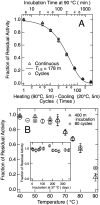
Heat-induced irreversible denaturation of anti-hCG VHH by repetitive heating-cooling cycles or continuous incubation at a high temperature and denaturation temperature dependence. A, anti-hCG VHH (1.3 μg/ml) was subjected to heating-cooling cycles (circles) or continuous incubation at 90 °C (triangles). The experiments were carried out at pH 7.4 in HBS-EP buffer. In heating-cooling cycles, a given number of reaction segments, which consisted of heating at 90 °C for 5 min and cooling at 20 °C for 5 min, were repeated. Residual activity at 20 °C was estimated using the strength of SPR signals compared with that of SPR signals from a series of diluted, untreated samples and was expressed as a fraction of the corresponding diluted, untreated sample. Because the time for unfolding was 5 min, one cycle corresponded to 5 min of incubation. The solid line represents a single exponential curve fitted to the VHH time-dependent denaturation where the first-order kinetic constant kden was 0.0039 min−1 and the time to reach half of the original activity was calculated to be 178 min. B, VHH (1.3 μg/ml) was subjected to 80 heating-cooling cycles (circles) or continuous incubation (400 min (m); triangles) where the heating temperature ranged from 40 to 90 °C in HBS-EP buffer. Inset, VHH (1.3 μg/ml) was incubated at 37 °C for the indicated time in HBS-EP buffer. In all panels, error bars represent S.D.
FIGURE 3.
FIGURE 3.
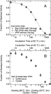
Heat resistance of anti-hCG VHH with different preparations and anti-β-lactamase VHH. A, anti-hCG VHH samples prepared from E. coli soluble (circles) and insoluble (squares) fractions were subjected to continuous incubation at 90 °C. Both samples were histidine-tagged at their C termini. B, anti-β-lactamase VHH was subjected to heating-cooling cycles (circles) or continuous incubation at 90 °C (triangles). In heating-cooling cycles, a given number of reaction segments, which consisted of heating at 90 °C for 5 min (m) and cooling at 20 °C for 5 min, were repeated. Both experiments were carried out at pH 7.4 in HBS-EP buffer at a protein concentration of 1.3 μg/ml. Error bars represent S.D. Sup, supernatant; Ppt, precipitate.
FIGURE 4.
FIGURE 4.
Effects of protein concentration on VHH denaturation and analysis of the molecular weight of heat-inactivated anti-hCG VHH by gel permeation chromatography and SDS-PAGE. A, VHH with different concentrations in HBS-EP buffer were subjected to 40 cycles of heating-cooling cycles (unfolding at 90 °C for 5 min and refolding at 20 °C for 5 min). Error bars represent S.D. B, room temperature gel permeation chromatography profiles of VHH without heat treatment, with 40 cycles of heating-cooling cycles, and with continuous incubation at 90 °C for 1,600 min (m) at a protein concentration of 10 μm. Arrowheads indicate the elution volumes of serum albumin (67 kDa), ribonuclease A (13.7 kDa), and trypsin inhibitor (6.5 kDa). C, VHH (10 μm) was treated with continuous incubation at 90 °C for 1,600 min and analyzed using a polyacrylamide gradient gel (5–20%) with or without reduction with 10 mm dithiothreitol. The numbers on the left of the panel indicate the molecular weights of marker proteins. mAU, milliabsorbance units.
FIGURE 5.
FIGURE 5.
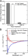
Effects of a denaturant on heat-inactivated anti-hCG VHH. A, the amount of residual activity after Gdn-HCl treatment was estimated and compared with that of VHH without Gdn-HCl treatment. Samples (40 μm) were unfolded using a final concentration of ∼6 m Gdn-HCl and then dialyzed against HBS-EP buffer without EDTA to remove the denaturant. Error bars represent S.D. B, comparison of CD spectra of heat-inactivated samples at 37 °C with or without ∼6 m Gdn-HCl treatment followed by refolding. m, minutes; deg, degrees.
FIGURE 6.
FIGURE 6.
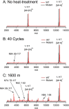
MALDI-TOF mass spectra of wild-type and N52S/N74S/N84T mutant anti-hCG VHH with and without heat treatment. A, MALDI-TOF mass spectra of wild-type (red) and N52S/N74S/N84T mutant (black) VHH (5 μm). B and C, samples were subjected to 40 heating-cooling cycles (B) or continuous 1,600-min (m) incubation (C) in HBS-EP buffer. The heating temperature was 90 °C. The spectra of wild-type VHH are presented with an offset of 200 m/z for clarity. The arrows indicate hypothetical fragments resulting from digestion occurring at the C terminus of Asn residues. In all panels, wild-type and N52S/N74S/N84T mutant VHH are abbreviated as WT and Mutant, respectively.
FIGURE 7.
FIGURE 7.
Effects of replacement of Asn residues in anti-hCG VHH. A, Asn residues 52, 74, and 84 in anti-hCG VHH-H14 were substituted by Ser, Ser, and Thr, respectively, resulting in the N52S/N74S/N84T mutant. B, N52S/N74S/N84T mutant (100 nm) was treated with heating (90 °C for 5 min)-cooling (20 °C for 5 min) cycles (black closed box) or continuous incubation at 90 °C followed by refolding at 20 °C for 5 min (black open box). The theoretical curve for N52S/N74S/N84T (solid line) was fitted using 0.0025 min−1 as a first-order kinetic constant kden. From this kden, the half-life of the N52S/N74S/N84T mutant was calculated to be 274 min (m). Data for wild-type VHH treated with continuous incubation and its theoretical curve are presented as red circles and broken line, respectively. The SPR signals for untreated wild-type (red broken line) and N52S/N74S/N84T (black solid line) VHHs (100 nm) are presented in the inset. In all panels, wild-type and N52S/N74S/N84T mutant VHHs are abbreviated as WT and Mutant, respectively. Error bars represent S.D. CDR, complementarity determining region.
FIGURE 8.
FIGURE 8.
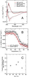
Effects of mutations of anti-hCG VHH on its biophysical characteristics. A, CD spectra of wild-type and N52S/N74S/N84T VHH (25 μm) at 37 and 90 °C in HBS-EP buffer without EDTA. B, equilibrium thermal unfolding curves of wild-type and N52S/N74S/N84T VHH were measured by changes in ellipticity at 235 nm at a protein concentration of 25 μm. Curves fitted by standard thermodynamic equations (63) are presented as solid lines, and Tm values of wild-type VHH and N52S/N74S/N84T mutant VHH were estimated to be 65 and 68 °C, respectively. C, Tm values and residual activities of wild-type VHH and VHH mutants (100 nm) after 40 heating-cooling cycles. The Tm values of disulfide mutants C22W/A49C/I70C/C96A (56 °C) and A49C/I70C (74 °C) are from Hagihara et al. (33). Wild-type and N52S/N74S/N84T mutant anti-hCG VHH are abbreviated as WT and Mutant, respectively. Error bars represent S.D. deg, degrees.
FIGURE 9.
FIGURE 9.
Structural changes in anti-hCG VHH induced by iterative DSC measurements. A, repetitive thermal unfolding and refolding in HBS-EP buffer induced changes in the heat absorption profiles of VHH at 54 μm. This experiment was carried out in a fixed cell without removing the samples from the DSC equipment. Heat absorption curves were measured while increasing the temperature at a rate of 1 °C/min. B, changes in Cp at 35 (open circles), 63 (closed triangles), and 85 °C (open squares) induced by repetitive DSC measurements.
FIGURE 10.
FIGURE 10.
Comparison of heat-induced structural changes and denaturation of anti-hCG VHH. A, VHH heat absorption curves. VHH samples (54 μm in HBS-EP buffer) were subjected to cycles of unfolding at 90 °C for 5 min and refolding at 20 °C for 5 min in a thermal cycler. The broken lines represent the extrapolated linear baseline of the unfolded state. The number of cycles is indicated on each curve. B, comparison between residual activities measured by SPR and the main peak of heat absorption curves corresponding to intact VHH after heat treatment. The main peaks were estimated by subtracting the baseline (unfolded state) from the peak heights for all samples, and the resulting heat-treated peak was then divided by the untreated sample peak. Error bars represent S.D. C, CD spectra of heat-treated VHH at 37 °C in HBS-EP buffer without EDTA. Samples at a protein concentration of 25 μm were subjected to cycles of unfolding at 90 °C for 5 min and refolding at 20 °C for 5 min. For comparison, CD spectra of thermally unfolded intact and heat-denatured VHH at 90 °C are also shown. D, equilibrium thermal unfolding curves of heat-treated VHH measured by changes in ellipticity at 235 nm. Samples were the same as in C. deg, degrees.
FIGURE 11.
FIGURE 11.
Heat-induced irreversible denaturation of anti-hCG antibody fragments. A–C, scFv (A), Fab (B), and IgG (C) samples (1 μg/ml) were subjected to heating (90 °C for 5 min)-cooling (20 °C for 5 min) cycles at pH 7.4. In addition, dependence of scFv denaturation on incubation time at 90 °C is also shown (A). Residual activity at 20 °C was estimated using the strength of the SPR signal compared with that of SPR signals from a series of diluted untreated samples and is presented as the fraction of the corresponding untreated diluted sample. D, concentration-dependent denaturation of scFv was examined. ScFv samples with the given concentrations were subjected to five cycles of unfolding at 90 °C for 5 min and refolding at 20 °C for 5 min. Inset, scFv (1 μg/ml) was subjected to seven heating-cooling cycles where the heating temperatures ranged from 40 to 90 °C in HBS-EP buffer. Error bars represent S.D. in all panels.
FIGURE 12.
FIGURE 12.
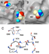
Atomic interaction of Asn residues in the crystal structure of anti-hCG VHH. A and B, solvent-excluded surface (gray) of anti-hCG VHH molecule around Asn residues, which are indicated by Corey-Pauling-Koltun model. Surface was generated by CueMol2 using Protein Data Bank code 1HCV data (39) with the probe radius of 4 Å. C, hydrogen bond network related to the Asn residues. Both the side chain and main chain atoms of Asn residues form hydrogen bonds with proximal atoms of other amino acid residues (dashed line). Hydrogen bonds were detected, and the graphic was drawn by using UCSF Chimera (64).

References

    1. Manning M. C., Chou D. K., Murphy B. M., Payne R. W., Katayama D. S. (2010) Stability of protein pharmaceuticals: an update. Pharm. Res. 27, 544–575 - PubMed
    1. Saerens D., Huang L., Bonroy K., Muyldermans S. (2008) Antibody fragments as probe in biosensor development. Sensors 8, 4669–4686 - PMC - PubMed
    1. Hamers-Casterman C., Atarhouch T., Muyldermans S., Robinson G., Hamers C., Songa E. B., Bendahman N., Hamers R. (1993) Naturally occurring antibodies devoid of light chains. Nature 363, 446–448 - PubMed
    1. Muyldermans S. (2013) Nanobodies: natural single-domain antibodies. Annu. Rev. Biochem. 82, 775–797 - PubMed
    1. van der Linden R. H., Frenken L. G., de Geus B., Harmsen M. M., Ruuls R. C., Stok W., de Ron L., Wilson S., Davis P., Verrips C. T. (1999) Comparison of physical chemical properties of llama VHH antibody fragments and mouse monoclonal antibodies. Biochim. Biophys. Acta 1431, 37–46 - PubMed

Publication types

MeSH terms

Substances

LinkOut - more resources