. 2014 May;14(5):359-70.
doi: 10.1038/nrc3711. Epub 2014 Apr 17.

Unravelling mechanisms of p53-mediated tumour suppression

Affiliations

Unravelling mechanisms of p53-mediated tumour suppression

Kathryn T Bieging et al. Nat Rev Cancer. 2014 May.

Abstract

p53 is a crucial tumour suppressor that responds to diverse stress signals by orchestrating specific cellular responses, including transient cell cycle arrest, cellular senescence and apoptosis, which are all processes associated with tumour suppression. However, recent studies have challenged the relative importance of these canonical cellular responses for p53-mediated tumour suppression and have highlighted roles for p53 in modulating other cellular processes, including metabolism, stem cell maintenance, invasion and metastasis, as well as communication within the tumour microenvironment. In this Opinion article, we discuss the roles of classical p53 functions, as well as emerging p53-regulated processes, in tumour suppression.


Figures

Figure 1
Figure 1. The classical view of p53 activation and response
The most well-elaborated molecular models for p53 activation are those in response to acute DNA damage signals and hyperproliferative signals. p53 induction by acute DNA damage begins when DNA double-strand breaks trigger activation of ataxia-telangiectasia mutated (ATM) — a kinase that phosphorylates the CHK2 kinase — or when stalled or collapsed DNA replication forks recruit ataxia telangiectasia and RAD3-related (ATR), which phosphorylates CHK1 (REF. 157). p53 is a substrate for both the ATM and ATR kinases, as well as for CHK1 and CHK2, which coordinately phosphorylate (P) p53 to promote its stabilization. Phosphorylation of p53 occurs at several sites, particularly at the amino-terminus, such as at serines 15 and 20 (REFS 158–160). These phosphorylation events are important for p53 stabilization, as some of the modifications disrupt the interaction between p53 and its negative regulators MDM2 and MDM4 (REFS 159,–163). MDM2 and MDM4 bind to the transcriptional activation domains of p53, thereby inhibiting p53 transactivation function, and MDM2 has additional activity as an E3 ubiquitin ligase that causes proteasome-mediated degradation of p53 (REFS 167,168). Phosphorylation also allows the interaction of p53 with transcriptional cofactors, which is ultimately important for activation of target genes and for responses such as cell cycle arrest, DNA repair, apoptosis and senescence. Hyperproliferative signals similarly activate p53 through perturbation of the MDM2–p53 interaction. These signals can function by liberating the E2F transcription factor, which can stimulate transcription of the ARF tumour suppressor,. ARF in turn inhibits MDM2 by antagonizing MDM2 ubiquitin ligase activity, and/or sequestering MDM2 to nucleoli. As a consequence, ARF activation enhances p53 stability and activity, promoting p53 responses such as apoptosis or cellular senescence.
Figure 2
Figure 2. A revised view of p53-activating signals and responses that are important for tumour suppression
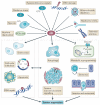
A host of different stresses can activate p53 in the context of tumour initiation or progression, including nutrient deprivation, hypoxia, oxidative stress, hyperproliferative signals (which could also promote chronic DNA damage or oxidative stress), DNA damage (which might most typically be chronic DNA damage triggered by replicative stress, telomere attrition, or oxidative stress), and ribonucleotide depletion. p53 activation by these signals, or potentially even ‘basal’ p53 action in some contexts, can consequently promote diverse responses that lead to tumour suppression. This view expands the set of stress signals that can activate p53 to promote responses of cell cycle arrest, senescence, apoptosis and DNA repair, which could potentially occur through pathways that are distinct from those used upon acute DNA damage,. The revised view also suggests that, in addition to the ability of p53 to fully block cell cycle progression in response to a stress signal, basal p53 levels may also simply dampen the rate of progression through the cell cycle. Beyond triggering classical responses, p53 that is activated by various stress signals can modulate several additional cellular processes that are relevant to suppressing tumour development, including opposing oncogenic metabolic reprogramming and limiting the accumulation of reactive oxygen species (ROS), activating autophagy, promoting communication within the tumour microenvironment, inhibiting stem cell self-renewal and reprogramming of differentiated cells into stem cells, and restraining invasion and metastasis. Regulation of these processes by p53 may directly promote tumour suppression or may impinge on the canonical functions, such as apoptosis or senescence. For example, the inhibition of metabolic reprogramming by p53 may impede tumorigenesis by limiting proliferation or activating apoptosis, and the induction of autophagy may also suppress cancer by facilitating apoptosis. Similarly, classical responses may affect novel functions. For example, p53-induced senescence precipitates signalling to the tumour microenvironment that ultimately provokes tumour suppression,.
Figure 3
Figure 3. p53 suppresses cancer through transcriptional activation, by regulating diverse biological processes through transactivation of target genes
a ∣ The p53 protein contains two amino-terminal transcriptional activation domains (TADs), a proline-rich domain (PRD), a DNA-binding domain (DBD), a tetramerization domain (TET) and a carboxy-terminal region that is rich in basic residues (Basic). Inactivation of p53 in human tumours typically occurs through missense mutations in the DBD of the p53 protein. Six common p53 ‘hot-spot’ mutations are categorized as either structural or contact p53 mutants, both of which disrupt the protein–DNA interaction and the transactivation of p53 target genes (see also BOX 1). b ∣ Lists of key p53-induced target genes involved in processes that are important for tumour suppression, including the canonical p53-associated responses — apoptosis, cell cycle arrest, senescence and DNA repair (purple) — as well as processes that have recently been associated with p53-dependent tumour suppression, such as metabolism control, autophagy, tumour microenvironment crosstalk, invasion and metastasis, and stem cell biology,,,,– (beige). The evidence for p53-dependent regulation of the genes on this list comes from mouse and/or human cells. Most of the genes are regulated by p53 in both human and mouse cells, but a few of the genes have currently only been identified and/or shown to be regulated by p53 in one of these species. Adora2b, adenosine A2b receptor; Aldh4, aldehyde dehydrogenase 9 family, member A1; Apaf1, apoptotic peptidase activating factor 1; Atg, autophagy related; Bai1, brain-specific angiogenesis inhibitor 1; Bax, BCL-2-associated X protein; Btg2, B cell translocation gene 2, anti-proliferative; Cdkn1a, cyclin-dependent kinase inhibitor 1A; Ctsd, cathepsin D; Cx3cl1, chemokine (C-X3-C motif) ligand 1; Ddb2, damage-specific DNA binding protein 2; Ddit4, DNA-damage-inducible transcript 4; Dram1, DNA-damage regulated autophagy modulator 1; Ercc5, excision repair cross-complementing rodent repair deficiency, complementation group 5; Fancc, Fanconi anaemia, complementation group C; Foxo3, forkhead box O3; Gadd45a, growth arrest and DNA-damage-inducible 45α; Gamt, guanidinoacetate N-methyltransferase; Gls2, glutaminase 2; Gpx1, glutathione peroxidase 1; Icam1, intercellular adhesion molecule 1; Irf, interferon regulatory factor; Laptm4a, lysosomal protein transmembrane 4α; Lkb1, liver kinase B1 (also known as Stk11); Lpin1, lipin 1; Mcp1, monocyte chemoattractant protein 1 (also known as Ccl2); Mgmt, O-6-methylguanine-DNA methyltransferase; Ncf2, neutrophil cytosolic factor 2; Pai1, plasminogen activator inhibitor; Perp, p53 apoptosis effector; Pig3, p53 inducible protein 3 (also known as Tp53i3); Pidd, p53-induced death domain protein; Pik3r3, phosphoinositide-3-kinase, regulatory subunit 3; Pml, promyelocytic leukaemia; pol, polymerase; Polk, DNA polymerase-κ; Prka, protein kinase, AMP-activated; Prkag2, protein kinase, AMP-activated, γ2 non-catalytic subunit; Ptprv, protein tyrosine phosphatase, receptor type, V; Sesn, sestrin; Tigar, TP53-induced glycolysis and apoptosis regulator; Tlr, Toll-like receptor; TP53AIP1, tumour protein p53 regulated apoptosis inducing protein 1; Tp53inp1, tumour protein p53 inducible nuclear protein 1; Tpp1, tripeptidyl peptidase I; Tsc2, tuberous sclerosis 2; Tsp1, thrombospondin 1; Ulbp, UL16 binding protein; Ulk, UNC-51 like autophagy activating kinase; Uvrag, UV radiation resistance associated; Vamp4, vesicle-associated membrane protein 4; Vmp1, vacuole membrane protein 1; Xpc, xeroderma pigmentosum, complementation group C.

Similar articles

Cited by

References

    1. Vousden KH, Prives C. Blinded by the light: the growing complexity of p53. Cell. 2009;137:413–431. - PubMed
    1. Brady CA, Attardi LD. p53 at a glance. J. Cell Sci. 2010;123:2527–2532. - PMC - PubMed
    1. Lang GA, et al. Gain of function of a p53 hot spot mutation in a mouse model of Li-Fraumeni syndrome. Cell. 2004;119:861–872. - PubMed
    1. Song H, Hollstein M, Xu Y. p53 gain-of-function cancer mutants induce genetic instability by inactivating ATM. Nature Cell Biol. 2007;9:573–580. - PubMed
    1. Fearon ER, Vogelstein B. A genetic model for colorectal tumorigenesis. Cell. 1990;61:759–767. - PubMed

Substances