. 2014 Apr 16;5(2):54.
doi: 10.1186/scrt443.

Umbilical cord tissue-derived mesenchymal stem cells induce apoptosis in PC-3 prostate cancer cells through activation of JNK and downregulation of PI3K/AKT signaling

Umbilical cord tissue-derived mesenchymal stem cells induce apoptosis in PC-3 prostate cancer cells through activation of JNK and downregulation of PI3K/AKT signaling

Ihn Han et al. Stem Cell Res Ther. .

Retraction in

Abstract

Introduction: Although mesenchymal stem cells (MSCs) have antitumor potential in hepatocellular carcinoma and breast cancer cells, the antitumor mechanism of human umbilical cord mesenchymal stem cells (hUCMSCs) in prostate cancer cells still remains unclear. Thus, in the present study, we elucidated the antitumor activity of hUCMSCs in PC-3 prostate cancer cells in vitro and in vivo.

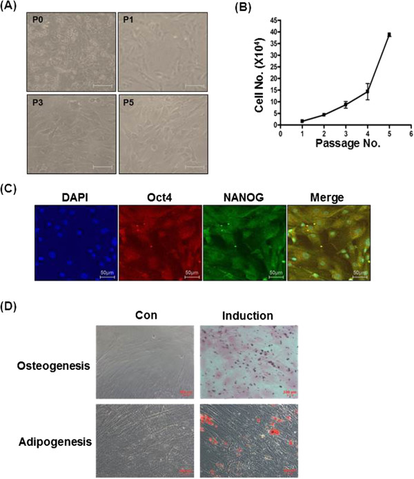
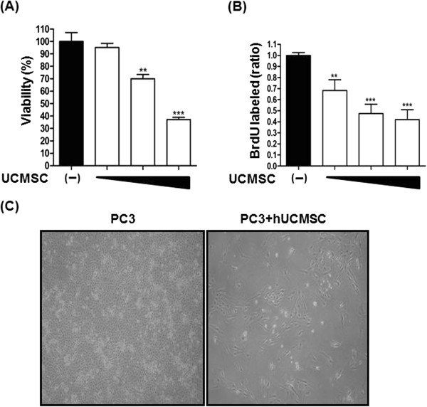
Methods: hUCMSCs were isolated from Wharton jelly of umbilical cord and characterized via induction of differentiations, osteogenesis, and adipogenesis. Antitumor effects of UCMSCs on tumor growth were evaluated in a co-culture condition with PC-3 prostate cancer cells. PC-3 cells were subcutaneously (sc) injected into the left flank of nude mice, and UCMSCs were sc injected into the right flank of the same mouse.

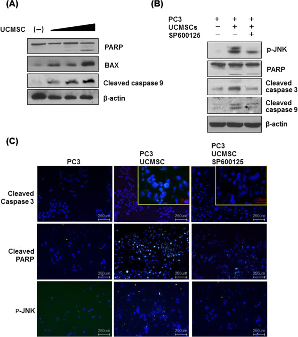
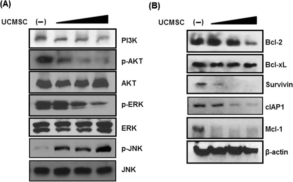
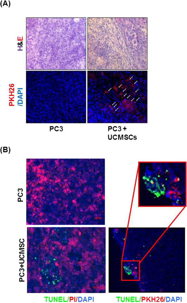
Results: We found that hUCMSCs inhibited the proliferation of PC-3 cells in the co-culture condition. Furthermore, co-culture of hUCMSCs induced the cleavage of caspase 9/3 and PARP, activated c-jun NH2-terminal kinase (JNK), and Bax, and attenuated the phosphorylation of phosphatidylinositol 3-kinase (PI3K)/ AKT, extracellular signal-regulated kinase (ERK), and the expression of survival genes such as Bcl-2, Bcl-xL, Survivin, Mcl-1, and cIAP-1 in PC-3 cells in Western blotting assay. Conversely, we found that treatment of specific JNK inhibitor SP600125 suppressed the cleavages of caspase 9/3 and PARP induced by hUCMSCs in PC-3 cells by Western blotting and immunofluorescence assay. The homing of hUCMSCs to, and TUNEL-positive cells on, the K562 xenograft tumor region were detected in Nu/nu-BALB/c mouse.

Conclusions: These results suggest that UCMSCs inhibit tumor growth and have the antitumor potential for PC-3 prostate cancer treatment.


Figures

Figure 1
Figure 1
Characterization of mesenchymal stem cells. (A) Beta-galactosidase staining was used to check SA-β-gal activity in early passages (0, 1, 3, and 5) of hUCMSCs. Cell morphology was observed by phase-contrast microscopy. Scale bar, 50 μm. (B) Growth kinetics of the hUCMSCs according to the passages of hUCMSCs. (C) Characterization of isolated hUCMSCs was performed by identification of MSC markers, OCT4 (green) and NANOG (red). Nuclei were stained with DAPI (blue). Scale bar, 50 μm. (D) Characterization of isolated hUCMSCs cultured in adipogenic and osteogenic differentiation media for 14 days was performed by identification of the adipogenic and osteogenic differentiation assays, as described in Materials and Methods. The cells were stained with Oil Red-O and Alizarin Red dye staining, respectively. Scale bar, 100 μm.
Figure 2
Figure 2
The inhibitory effect of hUCMSCs on prostate cancer cell growth in direct or indirect coculture condition. PC-3 cells (5 × 104) were cocultured for 24 hours with or without hUCMSCs at ratio of 1:10, 1:5, and 1:3 (hUCMSCs:PC-3s). PC-3 cells were placed in the lower Transwell chamber, and different numbers of hUCMSCs (hUCMSCs:PC-3s, 1:10, 1:5, 1:3) were seeded in the upper chamber. (A) Effect of hUCMSCs on the viability of PC-3 cells by MTT assay. (B) Effect of hUCMSCs on the proliferation of PC-3 cells by BrdU assay. (C) Analysis of morphology of PC-3 cells cocultured with hUCMSCs by phase-contrast microscopy. **P < 0.01, ***P < 0.001 versus untreated control.
Figure 3
Figure 3
Effect of JNK SP60015 on PARP, cleaved caspase 3, cleaved caspase 9, and p-JNK induced by hUCMSCs in PC-3 cells. (A) Effect of hUCMSCs on PARP, Bax, and cleaved caspase 9 in PC-3 cells. (B) Effect of JNK SP60015 on PARP, cleaved caspase 3, cleaved caspase 9, and p-JNK induced by hUCMSCs in PC-3 cells was detected in immunoblotting assay. (C) In the same condition as in (B), expression levels of cleaved caspase, cleaved PARP, and p-JNK were analyzed by immunofluorescence assay. Each primary antibody was diluted 1/300. Light green indicates cleaved caspase 3, cleaved PARP, and p-JNK. DAPI (Blue).
Figure 4
Figure 4
Effect of hUCMSCs on survival genes and JNK in PC-3 cells. PC-3 cells were cultured for 24 hours alone or in the presence of hUCMSCs at the ratio (hUCMSCs:PC-3, 1:3). Cells lysates were immunoblotted with PI3K, p-AKT, AKT, p-ERK, ERK, p-JNK, JNK and PARP, Bax, cleaved caspase 9, and β-actin antibodies. (A) Effect of hUCMSCs on PI3K, AKT, ERK, and JNK in PC-3 cells. (B) Effect of hUCMSCs on Bcl-2, Bcl-xL, survivin, cIAP-1, and β-actin in PC-3 cells.
Figure 5
Figure 5
Homing of hUCMSCs to PC-3 tumor site in Balb-c/nu-nude mice and their effect on TUNEL-positive cells in PC-3 tumor section. (A) Paraffin sections for H&E and IHC staining with PKH26 dye (red). The PKH26-labeled cells tracking toward the PC-3 tumor region from the opposite-side flank. White arrows indicate the labeled PKH26 (red). (B) Representative photographs of TUNEL/PI staining. Red fluorescence (PKH26) marks transplanted hUCMSCs, and green indicates TUNEL-positive cells. DAPI (blue).

References

    1. Jootar S, Pornprasertsud N, Petvises S, Rerkamnuaychoke B, Disthabanchong S, Pakakasama S, Ungkanont A, Hongeng S. Bone marrow derived mesenchymal stem cells from chronic myeloid leukemia t(9;22) patients are devoid of Philadelphia chromosome and support cord blood stem cell expansion. Leukoc Res. 2006;30:1493–1498. doi: 10.1016/j.leukres.2006.04.013. - DOI - PubMed
    1. Iwamoto S, Mihara K, Downing JR, Pui CH, Campana D. Mesenchymal cells regulate the response of acute lymphoblastic leukemia cells to asparaginase. J Clin Invest. 2007;117:1049–1057. doi: 10.1172/JCI30235. - DOI - PMC - PubMed
    1. Lescaudron L, Naveilhan P, Neveu I. The use of stem cells in regenerative medicine for Parkinson’s and Huntington’s diseases. Curr Med Chem. 2012;19:6018–6035. doi: 10.2174/092986712804485881. - DOI - PubMed
    1. Kim YJ, Park HJ, Lee G, Bang OY, Ahn YH, Joe E, Kim HO, Lee PH. Neuroprotective effects of human mesenchymal stem cells on dopaminergic neurons through anti-inflammatory action. Glia. 2009;57:13–23. doi: 10.1002/glia.20731. - DOI - PubMed
    1. Anzalone R, Lo Iacono M, Loria T, Di Stefano A, Giannuzzi P, Farina F, La Rocca G. Wharton’s jelly mesenchymal stem cells as candidates for beta cells regeneration: extending the differentiative and immunomodulatory benefits of adult mesenchymal stem cells for the treatment of type 1 diabetes. Stem Cell Rev. 2010;7:342–363. - PubMed

Publication types

MeSH terms

Substances