. 2014 May 8;509(7499):230-4.
doi: 10.1038/nature13168. Epub 2014 Apr 13.

Listeria monocytogenes exploits efferocytosis to promote cell-to-cell spread

Affiliations

Listeria monocytogenes exploits efferocytosis to promote cell-to-cell spread

Mark A Czuczman et al. Nature. .

Abstract

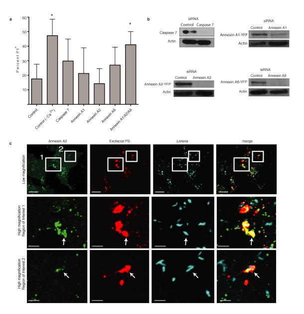
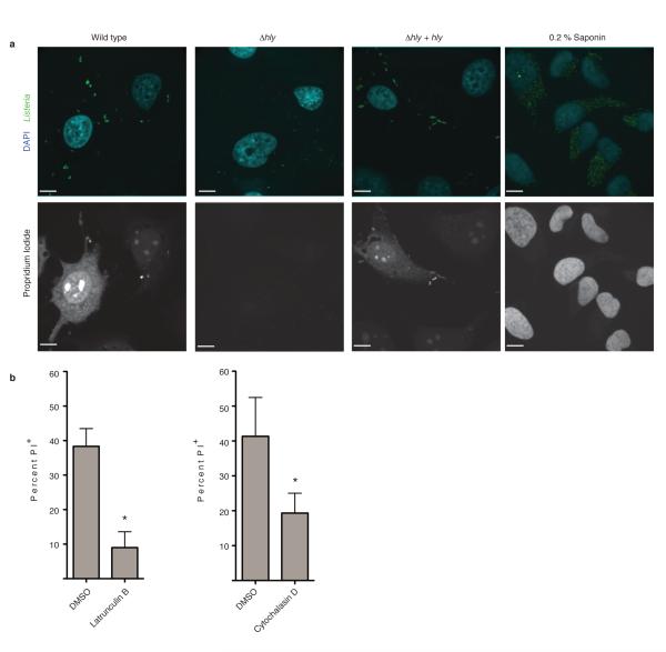
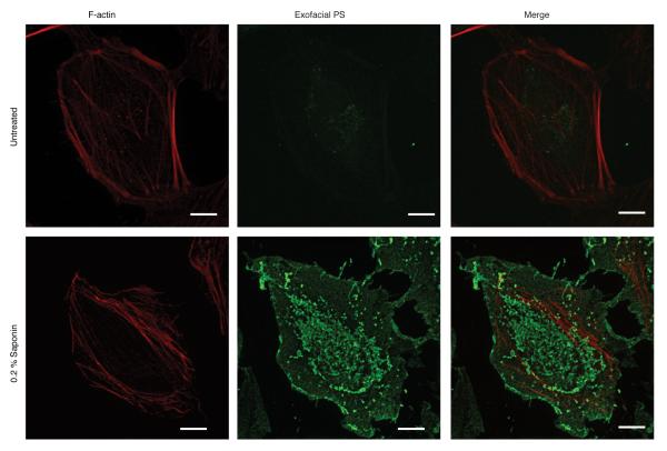
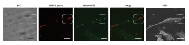
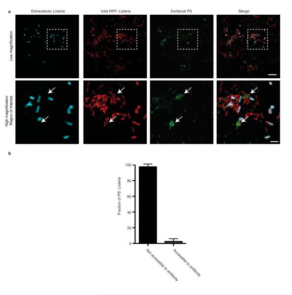
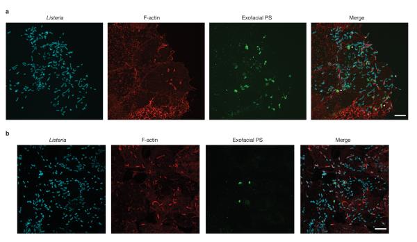
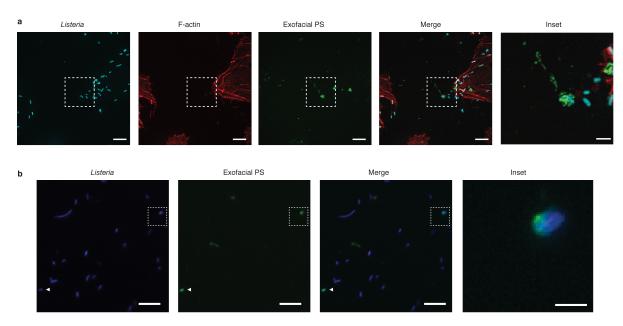
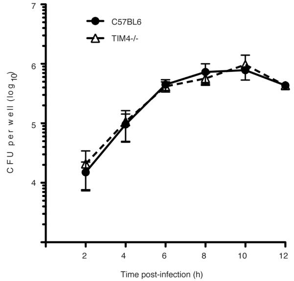
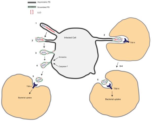
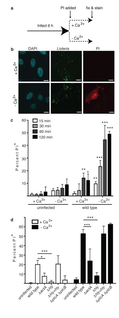
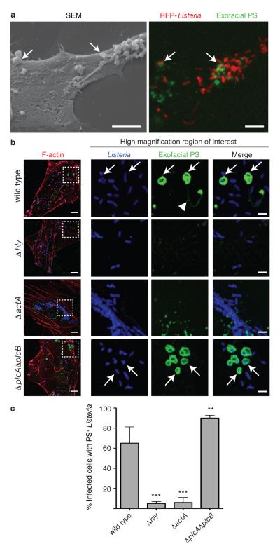
Efferocytosis, the process by which dying or dead cells are removed by phagocytosis, has an important role in development, tissue homeostasis and innate immunity. Efferocytosis is mediated, in part, by receptors that bind to exofacial phosphatidylserine (PS) on cells or cellular debris after loss of plasma membrane asymmetry. Here we show that a bacterial pathogen, Listeria monocytogenes, can exploit efferocytosis to promote cell-to-cell spread during infection. These bacteria can escape the phagosome in host cells by using the pore-forming toxin listeriolysin O (LLO) and two phospholipase C enzymes. Expression of the cell surface protein ActA allows L. monocytogenes to activate host actin regulatory factors and undergo actin-based motility in the cytosol, eventually leading to formation of actin-rich protrusions at the cell surface. Here we show that protrusion formation is associated with plasma membrane damage due to LLO's pore-forming activity. LLO also promotes the release of bacteria-containing protrusions from the host cell, generating membrane-derived vesicles with exofacial PS. The PS-binding receptor TIM-4 (encoded by the Timd4 gene) contributes to efficient cell-to-cell spread by L. monocytogenes in macrophages in vitro and growth of these bacteria is impaired in Timd4(-/-) mice. Thus, L. monocytogenes promotes its dissemination in a host by exploiting efferocytosis. Our results indicate that PS-targeted therapeutics may be useful in the fight against infections by L. monocytogenes and other bacteria that use similar strategies of cell-to-cell spread during infection.


Figures

Extended Data Figure 1
Extended Data Figure 1. Annexins promote membrane repair during Lm infection
A. HeLa cells were treated with the indicated siRNA for 48 h and then infected with wild type Lm at an MOI of 100. At 6 h p.i., medium was switched to Tyrodes buffer containing 0.5 mg/mL propidium iodide (PI) with or without calcium. Cells were fixed at 60 min after PI addition and then stained for bacteria and DNA (DAPI). PI+ cells were enumerated by microscopic analysis. Averages +/− s.d. for 3 independent experiments are shown. P values calculated using one-Way ANOVA. *P <0.05. B. Knockdown of gene expression by siRNA was confirmed by western blotting. Images representative of 2 independent experiments. C. Recruitment of Annexin A2 to PS+ structures containing bacteria. Boxes in low magnification image indicate areas enlarged in lower panels. Arrows indicate PS+ structures that colocalize with Annexin A2. Images representative of 3 independent experiments. Scale bars, 10 μm for low magnification images, 2 μm for enlarged regions of interest.
Extended Data Figure 2
Extended Data Figure 2. Actin-based motility promotes LLO-mediated membrane damage during Lm infection
A. HeLa cells were infected with the indicated Lm strain. At 6 h p.i., medium was switched to Tyrodes buffer containing 0.5 mg/mL propidium iodide (PI) with or without calcium. Cells were fixed at 60 min after PI addition and then stained for bacteria and DNA (DAPI). Confocal images representative of 3 independent experiments are shown. PI+ cells were enumerated and results are shown in Figure 1D. Where indicated, uninfected cells were treated with Saponin to permeabilize membranes and allow PI entry, serving as a positive control. Scale bars, 10 μm. B. HeLa cells were infected with wild type Lm and subjected to membrane damage assay as in A in the presence of either DMSO or the actin cytoskeleton inhibitors Latrunculin B or Cytochalasin D. Averages +/− s.d. for 3 independent experiments are shown. P values were calculated using two-tailed Student’s t test. *P <0.05.
Extended Data Figure 3
Extended Data Figure 3. Annexin V-Alexa 488 as a probe to label PS
Live HeLa cells were cooled on ice and stained with a fluorescent probe (Annexin V-Alexa 488) for 10 min to label exofacial PS. Cells were then fixed and stained with phalloidin Alexa 568 to visualize F-actin. In uninfected control experiments, low amounts of exofacial PS was detected in the membranes of cells, due to asymmetry of PS distribution in the plasma membrane. In contrast, treatment of cells with the pore-forming surfactant saponin led to robust staining of cells with Annexin V-Alexa 488. Images representative of 3 independent experiments. Scale bars, 10 μm.
Extended Data Figure 4
Extended Data Figure 4. Formation of PS+ structures during Lm infection
Low magnification images used to generate images shown in Figure 2a. HeLa cells were infected with wild type Lm expressing RFP for 6 h and then cooled on ice and stained with a fluorescent probe (Annexin V-Alexa 488) for 10 min to label exofacial PS. Cells were then fixed and analyzed by fluorescence microscopy to identify PS+ structures and bacteria. Scanning electron microscopy (SEM) of the same cell revealed that PS+ structures were associated with the dorsal surface of infected cells. Differential interference contrast (DIC) microscopy of cells was also performed to help identify cells for correlative imaging analysis. Images representative of 2 independent experiments. Scale bars, 20 μm.
Extended Data Figure 5
Extended Data Figure 5. PS+ bacteria are present with a host-derived membrane structure
A. HeLa cells were infected with wild type Lm expressing RFP for 8 h and then labeled with a probe for exofacial PS (Annexin V-Alexa 488). Cells were then rapidly stained with anti-Listeria antibodies (5 min) to label extracellular bacteria. Cells were then fixed and analyzed by fluorescence microscopy to identify PS+ structures and bacteria. Bacteria that colocalize with exofacial PS but are not labeled with anti-Listeria antibodies in the extracellular medium are indicated with arrows. Extracellular bacteria do not label with Annexin V-Alexa 488, indicating this probe does not bind non-specifically to bacteria. Box in low magnification image indicates area enlarged in lower panels. Images representative of 3 independent experiments. Scale bars, 10 μm for low magnification, 2 μm for high magnification. B. Cells were infected and stained as in A and analyzed by fluorescence microscopy. Bacteria that colocalize with exofacial PS were scored for their accessibility to anti-Listeria antibodies present in the extracellular medium. Data shows that the majority of PS+ bacteria are not accessible to anti-Listeria antibodies. Averages +/− s.d. for two independent experiments are shown.
Extended Data Figure 6
Extended Data Figure 6. Formation of PS+ structures during Lm infection of epithelial cells and macrophages
A. Henle-407 human intestinal epithelial cells were infected with wild type Lm for 6 h and then incubated with a probe for exofacial PS (Annexin V-Alexa 488; green). Cells were then fixed and stained with phalloidin to visualize F-actin (red) and for bacteria (blue). Cells were analyzed by fluorescence microscopy to identify PS+ structures and bacteria. Images representative of 3 independent experiments. B. Mouse bone marrow-derived macrophages (BMDM) from C57BL/6 mice were infected and stained as in A. Scale bars, 10 μm. Images representative of 3 independent experiments.
Extended Data Figure 7
Extended Data Figure 7. Release of PS+ structures containing Lm from infected cells
A. HeLa cells were infected with wild type Lm for 6 h and then incubated with a probe for exofacial PS (Annexin V-Alexa 488; green). Cells were then fixed and stained with phalloidin to visualize F-actin (red) and for bacteria (blue). Cells were analyzed by fluorescence microscopy to identify PS+ structures and bacteria. Inset shows PS+ bacteria that are not cell associated. Images representative of 3 independent experiments. B. HeLa cells were infected with ΔplcAΔplcB mutant bacteria for 6 h. The supernatant from the infected cultures was then removed and centrifuged onto poly-L-Lysine coated coverslips. Bacteria associated with coverslips were then stained with a probe for exofacial PS (Annexin V-Alexa 488; green). Cells were then fixed and stained for bacteria (blue). Coverslips were analyzed by fluorescence microscopy to identify PS+ bacteria. Inset shows PS+ bacteria. Scale bars, 10 μm for low magnification images, 2 μm for insets. Images representative of 3 independent experiments.
Extended Data Figure 8
Extended Data Figure 8. Growth of Lm in the cytosol of TIM-4−/− macrophages is not impaired
Gentamicin protection assay to measure intracellular bacterial growth. Bone marrow-derived macrophages were harvested from C57BL/6 or TIM-4−/− mice and seeded at a density of 3 ×10⁵ cells/well. Cells were then infected with wild type Lm in the presence of extracellular Gentamicin. At the indicated times, cell lysates were plated and intracellular bacterial numbers (CFU) were determined. Averages +/− s.d. for two independent experiments are shown.
Extended Data Figure 9
Extended Data Figure 9. Cytokine measurements
A. Measurement of cytokines following in vitro infection of bone marrow-derived macrophages from C57BL/6 or TIM4−/− mice with wild type Lm for the indicated time. Data from one of two independent experiments is shown. B. Measurement of basal cytokines in tissues of C57BL/6 or TIM4−/− mice without infection. Averages +/− s.d. for three independent experiments are shown. P values were calculated using one-Way ANOVA.
Extended Data Figure 10
Extended Data Figure 10. Lm exploits efferocytosis to promote cell-to-cell spread during infection
Model shows the steps that promote cell-to-cell spread by L. monocytogenes. 1. Protrusion formation via actin-based motility. 2. LLO-mediated damage to the plasma membrane leads to loss of membrane asymmetry and exofacial phosphatidylserine (PS) on protrusions. The exofacial exposure of PS promotes protrusion association with neighboring cells (right side). 3. Loss of membrane asymmetry and PS exposure extends along length of protrusions. 4. Calcium entry activates membrane repair pathways that promote scission of the protrusion. Bacteria are released from the cell in PS+ vesicles. 5. Macrophages mediate uptake of PS+ vesicles containing bacteria via the PS-binding receptor TIM-4. PS+ vesicles may be engulfed by neighboring cells either near the infected cell surface (left side) or within enclosed spaces that form as a result of protrusion penetration into the neighboring cell (right side). TIM-4 may also promote Lm infection indirectly, through its ability to suppress basal levels of pro-inflammatory cytokines as part of its homeostatic function in the immune system.
Figure 1
Figure 1. Actin-based motility promotes LLO-mediated membrane damage
A. Experimental design for membrane damage assay. B. Confocal images of HeLa cells infected as in A with wild type Lm at an MOI of 100. Scale bars,10 μm. Images representative of 3 independent experiments. C. Cells were infected as in A for the indicated time and PI+ cells were enumerated. Averages +/− s.d. for 3 independent experiments. Asterisks indicate significantly different from uninfected cells. P values calculated using two-tailed Student’s t test. D. Cells were infected with the indicated strain for 60 min. PI+ cells were enumerated. Averages +/− s.d. for 3 independent experiments. P values calculated using one-Way ANOVA. *P <0.05 **P <0.01 ***P <0.001.
Figure 2
Figure 2. Formation of PS+ structures during Lm infection
A. HeLa cells were infected at an MOI of 100 with wild type Lm expressing RFP (red) for 6 h and then incubated with a probe for exofacial PS (Annexin V-Alexa 488; green). Cells were then fixed and analyzed by fluorescence microscopy to identify PS+ structures and Lm (right panel). Scanning electron microscopy of the same cell shows PS+ structures (left panel). Images representative of 2 independent experiments. Scale bars, 5 μm. B. Confocal images of cells infected with the indicated strain for 6 h. Boxed areas in left hand low mag image (a 3 color merge, includes phalloidin stain for F-actin) are enlarged to right. Arrows indicate Lm that colocalize with PS and arrowhead indicates PS+ filaments. Images representative of 3 independent experiments. Scale bars,10 μm for low mag merge, and 3 μm for enlarged panels. C. Cells containing PS+ bacteria enumerated for B. Averages +/− s.d. for 3 independent experiments. P values calculated using one-Way ANOVA. **P <0.01 ***P <0.001.
Figure 3
Figure 3. Protrusions give rise to PS+ vesicles containing Lm
A. PS+ bacteria and ezrin+ protrusions were enumerated in HeLa cells infected at an MOI of 100 with wild type Lm for the indicated time. Averages +/− s.d. for 3 independent experiments. B. Cells were transfected with LifeAct-RFP (F-actin probe) and then infected with wild type Lm expressing GFP for 6 h. Live infected cells were analyzed by spinning disk confocal microscopy with Annexin V-Alexa 647 in the medium to label exofacial PS (green). Successive frames are shown. Arrows indicates protrusion that acquires PS. Images representative of 4 independent experiments. Scale bar, 10 μm.
Figure 4
Figure 4. TIM-4 promotes Lm cell-to-cell spread in macrophages and growth in mice
A. Infection focus assay for measuring cell-to-cell spread. BMDM were infected with Lm at an MOI of 0.01. Images representative of 3 independent experiments. Dotted lines delineate infection foci. Scale bars,100 μm. B. Infection foci from A were enumerated by fluorescence microscopy. A total of 100 infection foci were analyzed for each genotype/strain. Where indicated, cells were treated with anti-PS, anti-TIM-4 or non-specific rat IgG (control) during infection. Averages +/− s.d. for 3 independent experiments. P values calculated using two-way ANOVA with Bonferroni post test. C. Monolayers of control or TIM-4−/− BMDM were infected at an MOI of 0.01 with Lm expressing RFP and examined by live cell imaging. Images representative of 2 independent experiments. Dotted lines delineate infection foci. Scale bars,100 μm. D. The number of infected cells/focus for C. Averages +/− s.d. for 2 independent experiments. P values calculated using two-tailed Student’s t test. E. Cell-to-cell spread assay. Averages +/− s.d. for 3 independent experiments. P values calculated using two-tailed Student’s t test. F. Images from E. Arrows indicate actin+ protrusions associated with secondary cells. Images representative of 4 independent experiments. Scale bars,10 μm. G. Actin+ protrusions associated with secondary cells were enumerated. Averages +/− s.d. for 4 independent experiments. H, I. Mice were infected with 5 × 10⁴ CFU wild type (H) or 1 × 10⁷ CFU ΔactA mutant (I) Lm by tail vein injection. Mice were sacrificed and livers and spleen were harvested at 72 h p.i. for quantification of bacterial load (CFU/gram tissue). Data are expressed as Mean ± s.e.m.. P values calculated using one-tailed Mann Whitney test.

Comment in

References

    1. Ravichandran KS. Beginnings of a good apoptotic meal: the find-me and eat-me signaling pathways. Immunity. 2011;35:445–455. - PMC - PubMed
    1. Mostowy S, Cossart P. Virulence factors that modulate the cell biology of Listeria infection and the host response. Adv Immunol. 2012;113:19–32. - PubMed
    1. Robbins JR, et al. Listeria monocytogenes exploits normal host cell processes to spread from cell to cell. J Cell Biol. 1999;146:1333–1350. - PMC - PubMed
    1. Alberti-Segui C, Goeden KR, Higgins DE. Differential function of Listeria monocytogenes listeriolysin O and phospholipases C in vacuolar dissolution following cell-to-cell spread. Cell Microbiol. 2007;9:179–195. - PubMed
    1. Gedde MM, Higgins DE, Tilney LG, Portnoy DA. Role of listeriolysin O in cell-to-cell spread of Listeria monocytogenes. Infect Immun. 2000;68:999–1003. - PMC - PubMed

Methods References

    1. Bishop DK, Hinrichs DJ. Adoptive transfer of immunity to Listeria monocytogenes. The influence of in vitro stimulation on lymphocyte subset requirements. J Immunol. 1987;139:2005–2009. - PubMed
    1. Jones S, Portnoy DA. Characterization of Listeria monocytogenes pathogenesis in a strain expressing perfringolysin O in place of listeriolysin O. Infection and immunity. 1994;62:5608–5613. - PMC - PubMed
    1. Lauer P, Chow MY, Loessner MJ, Portnoy DA, Calendar R. Construction, characterization, and use of two Listeria monocytogenes site-specific phage integration vectors. Journal of bacteriology. 2002;184:4177–4186. - PMC - PubMed
    1. Skoble J, Portnoy DA, Welch MD. Three regions within ActA promote Arp2/3 complex-mediated actin nucleation and Listeria monocytogenes motility. J Cell Biol. 2000;150:527–538. - PMC - PubMed
    1. Smith GA, et al. The two distinct phospholipases C of Listeria monocytogenes have overlapping roles in escape from a vacuole and cell-to-cell spread. Infect Immun. 1995;63:4231–4237. - PMC - PubMed

Publication types

MeSH terms