. 2014 Jun 5;510(7503):152-6.
doi: 10.1038/nature13279. Epub 2014 Apr 13.

Focused specificity of intestinal TH17 cells towards commensal bacterial antigens

Affiliations

Focused specificity of intestinal TH17 cells towards commensal bacterial antigens

Yi Yang et al. Nature. .

Abstract

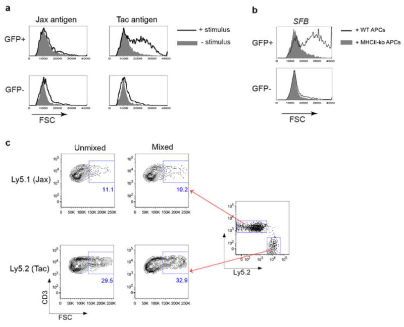
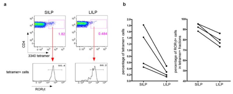
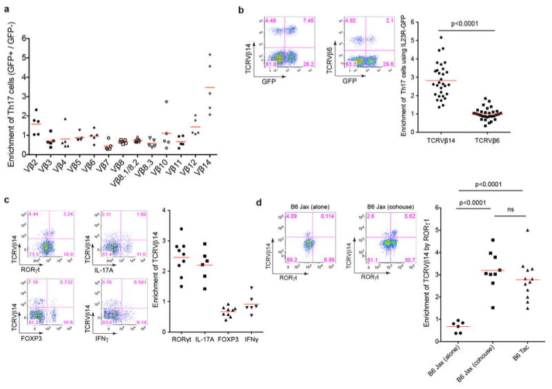
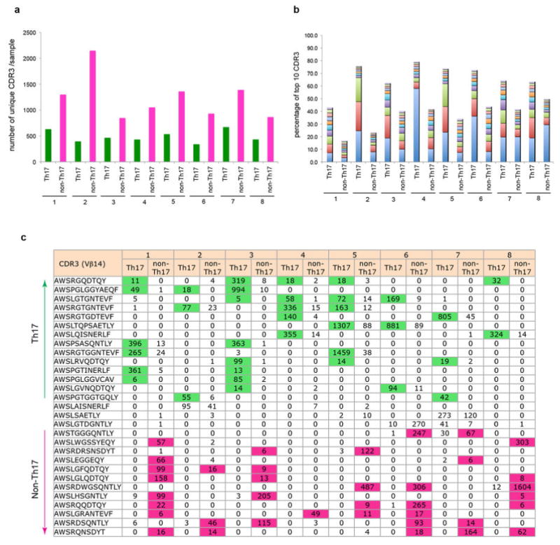
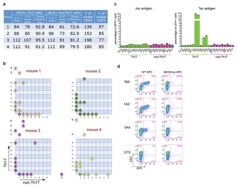
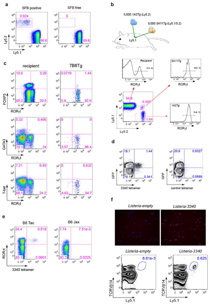
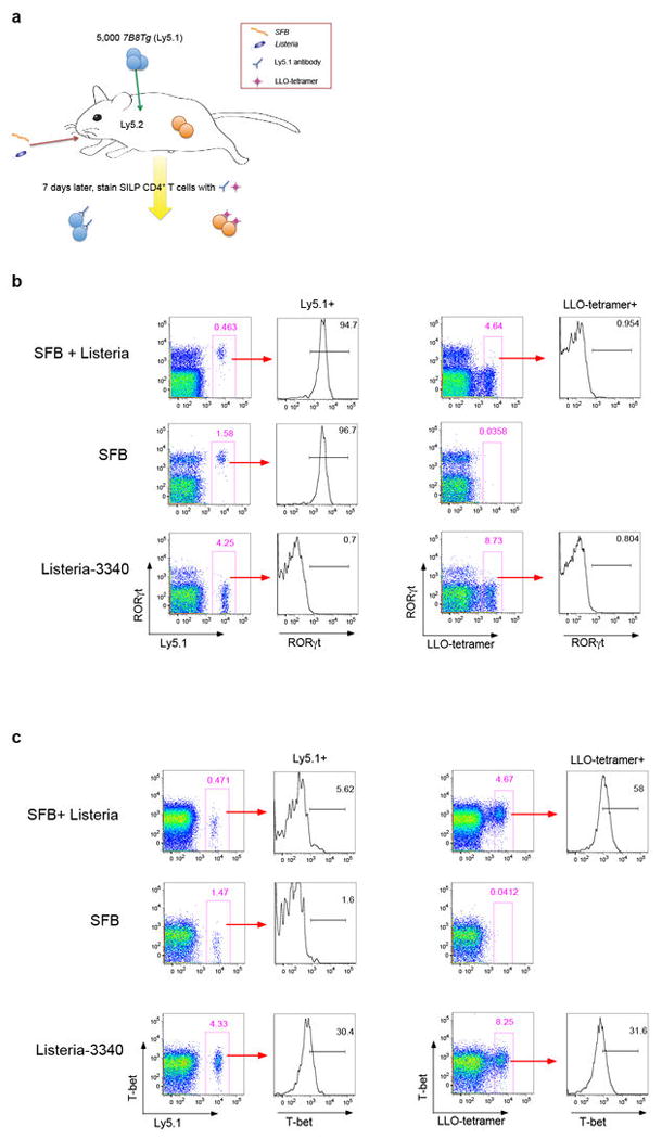
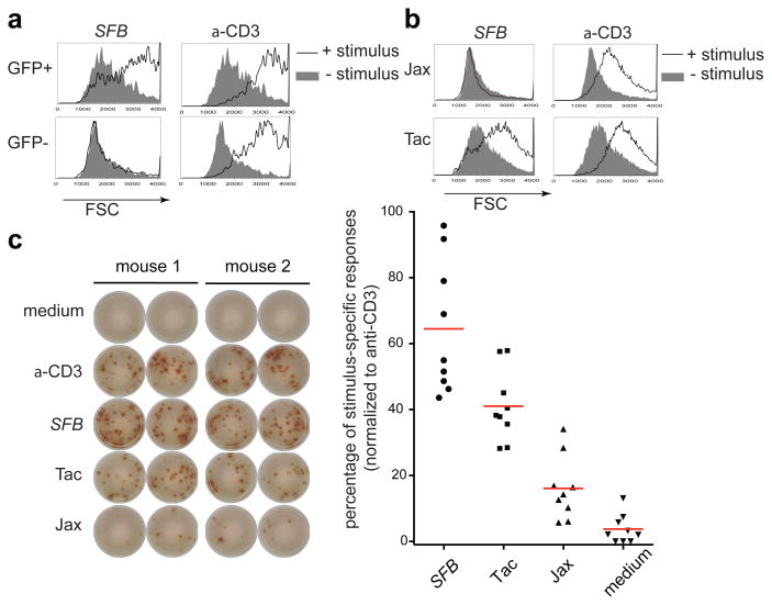
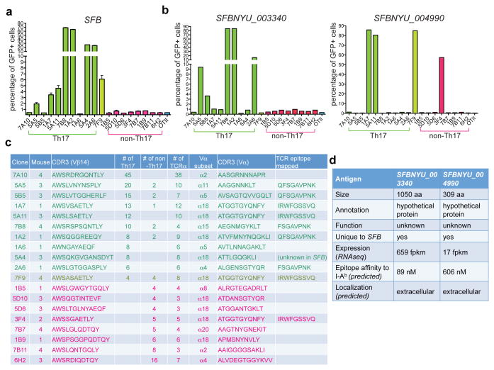
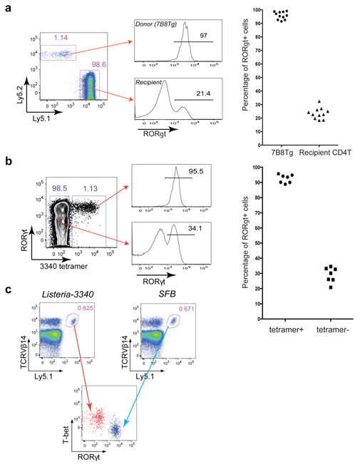
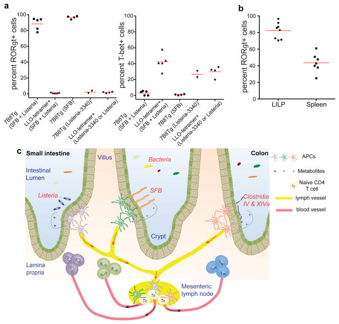
T-helper-17 (TH17) cells have critical roles in mucosal defence and in autoimmune disease pathogenesis. They are most abundant in the small intestine lamina propria, where their presence requires colonization of mice with microbiota. Segmented filamentous bacteria (SFB) are sufficient to induce TH17 cells and to promote TH17-dependent autoimmune disease in animal models. However, the specificity of TH17 cells, the mechanism of their induction by distinct bacteria, and the means by which they foster tissue-specific inflammation remain unknown. Here we show that the T-cell antigen receptor (TCR) repertoire of intestinal TH17 cells in SFB-colonized mice has minimal overlap with that of other intestinal CD4(+) T cells and that most TH17 cells, but not other T cells, recognize antigens encoded by SFB. T cells with antigen receptors specific for SFB-encoded peptides differentiated into RORγt-expressing TH17 cells, even if SFB-colonized mice also harboured a strong TH1 cell inducer, Listeria monocytogenes, in their intestine. The match of T-cell effector function with antigen specificity is thus determined by the type of bacteria that produce the antigen. These findings have significant implications for understanding how commensal microbiota contribute to organ-specific autoimmunity and for developing novel mucosal vaccines.


Figures

Extended Data Fig. 1
Extended Data Fig. 1. Stimulation of SILP Th17 cells requires intestinal microbiota antigen presentation
(a) Intestinal GFP+ CD4+ T cells from Il-23rGFP/+ mice stimulated with fecal material from Jax and Tac mice in the presence of syngeneic splenic APCs. Forward scatter was evaluated after 2 days. (b) Th17 cell activation by fecal material from SFB-monoassociated mice in the presence of APCs sufficient (WT) or deficient (KO) for MHC class II. (c) Evaluation of potential activation of bystander CD4+ T cells upon stimulation with SFB antigen. SILP CD4+ T cells from mice with Jax flora (Ly5.1) and Taconic flora (Ly5.2) were co-cultured or stimulated separately with APCs and SFB-monoassociated fecal material, and FSC was evaluated.
Extended Data Fig. 2
Extended Data Fig. 2. Microbiota-dependent TCR usage bias among SILP Th17 cells
(a) SILP CD4+ T cells from Il-23rGFP/+ mice were analyzed for utilization of Vβ's in Th17 cells versus non-Th17 cells. Ratios of the percentage of each TCR Vβ in GFP+ vs. GFP- cells are shown. Each symbol represents one mouse. (b) Relative expression of Vβ14 and Vβ6 TCRs by SILP Th17 versus non-Th17 CD4+ T cells from Il-23rGFP/+ mice. Left: Representative FACS plots; Right: Analysis of multiple animals. (c) Specific enrichment of Vβ14 TCRs in CD4+ T cells expressing RORγt and IL-17A, but not FOXP3 or IFNγ. Left: Representative FACS plots. Right: Analysis of multiple animals. Each symbol represents one mouse. (d) Correlation of Vβ14 enrichment in Th17 cells with the presence of specific commensal microbiota. B6 Jax mice were housed alone or cohoused with B6 Tac mice for two weeks. Left: Representative FACS analyses. Right: Analysis of multiple animals.
Extended Data Fig. 3
Extended Data Fig. 3. Th17 TCR repertoire analysis by pyrosequencing
(a) Numbers of unique Vβ14 CDR3 sequences of individual SILP Th17 and non-Th17 samples. The sequences were normalized for numbers of cells and total reads. (b) Preferential expansion of Vβ14+ clones in the Th17 compartment in the SILP. The proportions of the 10 most abundant Vβ14 CDR3 sequences from Th17 and non-Th17 cells from 8 mice are shown. (c) Th17-non Th17 bias of unique Vβ14 CDR3 sequences in the SILP of multiple mice.
Extended Data Fig. 4
Extended Data Fig. 4. Single-cell TCR cloning and TCR hybridoma screen
(a) Efficiency of single-cell Vβ14 cloning from SILP Th17 and non-Th17 cells of multiple mice. (b) Distributions of unique Vβ14 sequences in Th17 and non-Th17 cells within the SILP. Each plot represents one mouse shown in (a). y and x axes represent numbers of Th17 cells and non-Th17 cells for each unique Vβ14 sequence. Numbers of unique sequences are shown in colored circles. (c) Responses of Th17 and non-Th17 TCR hybridomas to small intestinal luminal contents from B6 Tac and B6 Jax mice. (d) Stimulation of Th17 TCR hybridomas by SFB-monoassociated antigens in the presence of APCs sufficient (WT) or deficient (KO) for MHC class II.
Extended Data Fig. 5
Extended Data Fig. 5. Identification of SFBNYU_003340 epitopes recognized by a subset of the Th17 TCR hybridomas
(a) Schematic representation of the antigen screen using a whole-genome shotgun SFB library. (b) Stimulation of the 7B8 hybridoma by bacterial pool 3F12. (c) Reactivity of 7B8 and four other TCR hybridomas with bacterial clone 3F12-E8. (d) Diversity of the CDR3 sequences of TCRs specific for 3F12-E8. Note that they belong to different Vα subsets and have distinct Vβ14 CDR3 sequences. (e) Responses of the 3F12-E8-specific TCR hybridomas to core epitopes encoded by minigenes expressed in E.coli.
Extended Data Fig. 6
Extended Data Fig. 6. Identification of SFBNYU_004990 epitopes recognized by related TCRs
(a) Top: The distribution in Th17 and non-Th17 cells of four TCRs that share an identical TCRα chain. Bottom: Amino acid alignment of the Vβ14 CDR3 sequences. The green box highlights the sequence differences. (b) Stimulation of the 5A11 hybridoma by bacterial pool 2D10 in the SFB antigen screen. (c) Responses of 4 TCR hybridomas, including a non-Th17 hybridoma, to bacterial clone 2D10-A10. (d) Responses of the 2D10-A10-specific TCR hybridomas to core epitopes encoded by minigenes expressed in E.coli. (e) TCR hybridoma responses to titrated synthetic peptide (IRWFGSSVQKV) in the presence of APCs.
Extended Data Fig. 7
Extended Data Fig. 7. SFB epitopes recognized by diverse Th17 cell TCRs
(a) The epitopes recognized by the Vβ14+ TCR hybridomas stimulate only Vβ14+ Th17 cells from the SILP. Th17 cells sorted from Il-23rGFP/+ mice were stimulated with indicated peptides (listed in (d)) in the presence of APCs. Left: Representative IL-17A ELISPOT assay with triplicates. Right: Normalized peptide-specific Th17 responses. Each dot represents one mouse. (b) Polyclonal responses of Vβ14+ and Vβ14- SILP Th17 cells to SFB antigens. Representative FACS plots from five experiments are shown. (c) Bioinformatics filtering approach to select candidate SFB epitopes. (d) Summary of newly-selected and the known A6 and A15 SFB peptides. (e) IL-17A ELISPOT screen for indicated peptides using SILP Th17 cells sorted from SFB-colonized Il-23rGFP/+ mice. The A6 peptide from SFBNYU_003340 and anti-CD3 served as positive controls. (f) Vβ14 usage in Th17 cells specific for peptide N5. Left: Representative IL-17A ELISPOT assay with triplicates for peptide N5, using Vβ14+ and Vβ14- SILP Th17 cells sorted from Il-23rGFP/+ mice. Right: Normalized N5-specific Th17 responses. Each dot represents one mouse.
Extended Data Fig. 8
Extended Data Fig. 8. SFB-specific T cells become Th17 cells in SFB-colonized mice
(a) SFB-dependent 7B8Tg T cell accumulation in the SILP. 2×10⁴ naive 7B8Tg T cells were transferred into congenic Ly5.1 recipient mice that were SFB-colonized or SFB-free. CD4+ T cells in the SILP were examined for donor and recipient isotype markers after 13 days. (b) Top: Strategy for co-transfer of congenic 1A2Tg and 5A11Tg T cells into SFB-colonized recipient mice. Bottom: FACS analysis of RORγt expression in host- and donor-derived CD4+ T cells in the SILP at 7 days after transfer. (c) FACS analysis of transcription factors in host- and donor- derived SILP CD4+ T cells after transfer of naïve 7B8Tg T cells as in (a). (d) FACS analysis of SILP T cells from Il-23rGFP/+ mice, stained with I-Ab/3340-A6 tetramer and control tetramer (2W). (e) FACS analysis of SILP T cells of B6 mice from colonies with different microbiota, stained with I-Ab/3340-A6 tetramer and intracellular RORγt antibody. (f) Expansion of 7B8Tg T cells in mice colonized with Listeria monocytogenes expressing SFBNYU_003340. Top: Immunofluorescence microscopic visualization of the expression of SFB protein by L. monocytogenes. Listeria-3340 and Listeria-empty were stained with anti-3340 rabbit polyclonal antibody. Red: anti-3340 antibody staining. Blue: DAPI staining. Bottom: Naive Ly5.1+ 7B8Tg cells were transferred into congenic mice infected with Listeria-3340 or Listeria-empty. Seven days after transfer, donor derived CD4+ T cells in the SILP were analyzed by FACS.
Extended Data Fig. 9
Extended Data Fig. 9. Transcription factor expression in SFB-specific and Listeria-specific T cells in co-infected mice (representative of data plotted in Fig. 4b)
(a) Experimental design for tracking both SFB- and Listeria- specific CD4+ T cells following intestinal colonization with both bacteria. Ly5.2 B6 mice were colonized with Listeria monocytogenes, SFB, or both bacteria, and 7B8Tg T cells from Ly5.1 mice were injected IV. Expression of Th1 and Th17 transcription factors in the SFB-specific 7B8Tg cells and LLO tetramer-specific recipient T cells was evaluated. (b) Intracellular stain for RORγt. (c) Intracellular stain for T-bet.
Extended Data Fig. 10
Extended Data Fig. 10. SFB-specific Th17 cells are present in both SILP and LILP of SFB-colonized mice
T cells were stained with I-Ab/3340-A6 tetramer and antibody to intracellular RORγt. (a) Representative FACS plots (gated on CD4+ T cells). (b) Analysis of multiple animals. Left: percent of tetramer-positive cells among total CD4+ T cells in each region of the intestine. Right: percent of RORγt+ cells among the tetramer-positive cells. Each symbol represents cells from a separate animal.
Fig. 1
Fig. 1. Intestinal Th17 cells are specific for SFB- and other microbiota-derived antigens
(a) Selective activation of intestinal GFP+ CD4+ T cells from Il-23rGFP/+ mice by fecal extract from SFB-monoassociated mice. Forward scatter (FSC) was evaluated after 2 days. (b) Activation of SILP CD4+ T cells from B6 Tac mice and B6 Jax mice with fecal extract from SFB-monoassociated mice. (c) IL17A ELISPOT assay of intestinal GFP+ CD4+ T cells from SFB-colonized Il-23rGFP/+ mice treated with indicated stimuli. Left: Representative ELISPOT images. Right: Compilation of results from multiple animals. Each symbol represents cells from a separate animal.
Fig. 2
Fig. 2. Most Th17 TCR hybridomas recognize SFB-unique proteins
(a) Responses of the TCR hybridomas, prepared from Th17 and non-Th17 intestinal CD4+ T cells, to fecal material from SFB-monoassociated mice. (b) Responses of the TCR hybridomas to E.coli clones expressing full-length SFBNYU_003340 and SFBNYU_004990. Note that a non-Th17 TCR hybridoma also responded to the clone expressing SFBNYU_004990. (c) Summary of the nineteen dominant clonotypic TCR clones. Ten Th17-biased clones are highlighted in green, and eight non-Th17-biased clones are highlighted in red. (d) Features of the two antigenic proteins of SFB.
Fig. 3
Fig. 3. SFB-specific T cells become Th17 cells in the SILP
(a) 7B8Tg cells (Ly5.2) were transferred into SFB-colonized mice (Ly5.1), and SILP T cells were analyzed after 8-15 days. Left: Representative FACS plots. Right: Analysis of multiple animals (one symbol/animal). (b) I-Ab/3340-A6 tetramer stain of SILP T cells from SFB-colonized B6 mice. Left: Representative FACS plots. Right: Analysis of multiple animals (one symbol/animal). (c) 7B8Tg cells (Ly5.1) were transferred into Ly5.2 congenic hosts orally colonized with Listeria-3340 or SFB. Seven days after transfer, donor-derived cells in the SILP were analyzed. The results are representative of three experiments.
Fig. 4
Fig. 4. TCR specificity for distinct luminal bacteria underlies divergent T helper cell differentiation in the SILP
(a) Th17 (RORγt) versus Th1 (T-bet) differentiation of SFB- (7B8Tg) and Listeria (LLO-tetramer)-specific CD4+ T cells in mice colonized with either or both bacteria. Each symbol represents cells from one animal. (b) Proportions of donor-derived 7B8Tg T cells that express RORγt in the colon and spleen of SFB-colonized mice. (c) Model for intestinal niches that promote diverse microbiota-dependent CD4+ effector T cell programs. Microbial signals, may induce polarizing cytokines or preformed niche-specific antigen presenting cells may interact with different T cell-inducing bacteria.

References

    1. Bettelli E, Korn T, Oukka M, Kuchroo VK. Induction and effector functions of T(H)17 cells. Nature. 2008;453:1051–1057. doi: 10.1038/nature07036. - DOI - PMC - PubMed
    1. McGeachy MJ, Cua DJ. The link between IL-23 and Th17 cell-mediated immune pathologies. Seminars in immunology. 2007;19:372–376. doi: 10.1016/j.smim.2007.10.012. - DOI - PubMed
    1. Littman DR, Rudensky AY. Th17 and regulatory T cells in mediating and restraining inflammation. Cell. 2010;140:845–858. doi: 10.1016/j.cell.2010.02.021. - DOI - PubMed
    1. Ivanov II, et al. The orphan nuclear receptor RORgammat directs the differentiation program of proinflammatory IL-17+ T helper cells. Cell. 2006;126:1121–1133. doi: 10.1016/j.cell.2006.07.035. - DOI - PubMed
    1. Ivanov II, et al. Specific microbiota direct the differentiation of IL-17-producing T-helper cells in the mucosa of the small intestine. Cell host & microbe. 2008;4:337–349. doi: 10.1016/j.chom.2008.09.009. - DOI - PMC - PubMed

Publication types

MeSH terms

Substances