. 2014 Apr 16;9(4):e95432.
doi: 10.1371/journal.pone.0095432. eCollection 2014.

Myeloid-specific Rictor deletion induces M1 macrophage polarization and potentiates in vivo pro-inflammatory response to lipopolysaccharide

Affiliations

Myeloid-specific Rictor deletion induces M1 macrophage polarization and potentiates in vivo pro-inflammatory response to lipopolysaccharide

William T Festuccia et al. PLoS One. .

Abstract

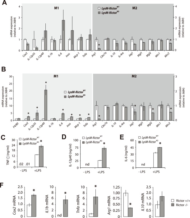
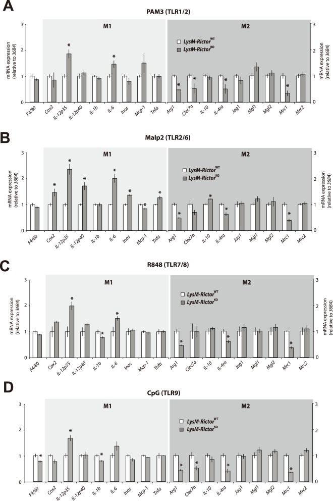
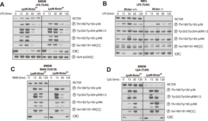
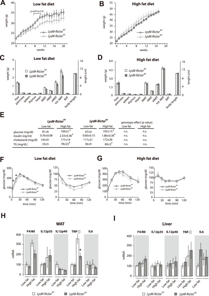
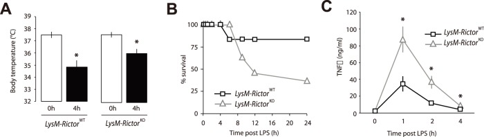
The phosphoinositide-3-kinase (PI3K)/protein kinase B (Akt) axis plays a central role in attenuating inflammation upon macrophage stimulation with toll-like receptor (TLR) ligands. The mechanistic target of rapamycin complex 2 (mTORC2) relays signal from PI3K to Akt but its role in modulating inflammation in vivo has never been investigated. To evaluate the role of mTORC2 in the regulation of inflammation in vivo, we have generated a mouse model lacking Rictor, an essential mTORC2 component, in myeloid cells. Primary macrophages isolated from myeloid-specific Rictor null mice exhibited an exaggerated response to TLRs ligands, and expressed high levels of M1 genes and lower levels of M2 markers. To determine whether the loss of Rictor similarly affected inflammation in vivo, mice were either fed a high fat diet, a situation promoting chronic but low-grade inflammation, or were injected with lipopolysaccharide (LPS), which mimics an acute, severe septic inflammatory condition. Although high fat feeding contributed to promote obesity, inflammation, macrophage infiltration in adipose tissue and systemic insulin resistance, we did not observe a significant impact of Rictor loss on these parameters. However, mice lacking Rictor exhibited a higher sensitivity to septic shock when injected with LPS. Altogether, these results indicate that mTORC2 is a key negative regulator of macrophages TLR signalling and that its role in modulating inflammation is particularly important in the context of severe inflammatory challenges. These observations suggest that approaches aimed at modulating mTORC2 activity may represent a possible therapeutic approach for diseases linked to excessive inflammation.


Conflict of interest statement

Competing Interests: The authors have declared that no competing interests exist.

Figures

Figure 1
Figure 1. TLR4 activation with LPS induces the phosphorylation of AKT.
(A) Response of Raw264.7 macrophages to LPS. Raw264.7 macrophages were plated and incubated overnight. Cells were then washed twice with PBS and incubated for 16 hours in DMEM 0.1% FBS. Then, cells were treated with LPS (500ng/ml) for the indicated time. Proteins were extracted and lysates were analysed by immunoblotting for indicated proteins. (B) Impact of LPS on cell signalling in Rictor +/+ and Rictor −/− MEFs. MEFs were plated 24 hours before the experimentation. The next day, cells were washed twice with PBS and incubated in DMEM 0.1% FBS for 2 hours. Cells were then treated with LPS (5ug/ml) for the indicate time. Proteins were extracted and lysates were analysed by immunoblotting for indicated proteins.
Figure 2
Figure 2. Production of a macrophage-specific Rictor knock-out mouse model.
(A) PCR strategy for the genotyping and identification of wild-type and KO mice. On the left panel is presented the PCR reaction showing the presence or the absence of the Lox allele in the Rictor gene. PCR was performed from a piece of tail collected from mice. The right part of the panel shows the efficiency of the recombination of the Rictor allele in BMDM isolated from the Rictor wt/wt, Rictor Lox/wt, Rictor Lox/Lox expressing or not the LysMcre. The presence of the Δ allele confirms the recombination of the Lox sites and the deletion of the targeted exon. (B to D) Confirmation of the specific loss of RICTOR in (B) BMDM and (C) TEM but not in (D) other mouse tissues. BMDM and TEM were isolated from mice and cultured as described in the methods sections. Proteins were extracted from cells and tissues and lysates were analysed by immunoblotting for indicated proteins. In B, C, and D, each line represents one mouse. (E–F) The loss of Rictor does not affect cell morphology and proliferation of (E) BMDM or (F) TEM.
Figure 3
Figure 3. TLR activation promotes AKT phosphorylation through the activation of mTORC2.
BMDM were isolated from LysM-Rictor WT or LysM-Rictor KO mice and were differentiated for 6 days in vitro. Cells were then plated and incubated 24 hours. Cells were wash twice with PBS, incubated 2 hours in RPMI 0.1% FBS and then treated with (A) LPS (250ng/ml), (B) R848 (0.1uM), or (C) CpG (0.5uM) for the indicated times. Proteins were extracted and lysates were analysed by immunoblotting for indicated proteins.
Figure 4
Figure 4. mTORC2 loss exacerbates the pro-inflammatory profile of BMDM in response to LPS.
(A) Basal gene expression in macrophages isolated from LysM-Rictor WT or LysM-Rictor KO mice. BMDM were isolated and differentiated for 6 days as described in the methods sections. For each genotype, BMDM isolated from 3 mice were pooled together to get enough biological material for all the studies. Cells were then allowed to rest for 48 hours. mRNA expression of markers of the classically activated (M1) and alternatively activated (M2) macrophages were measured by qRT-PCR and normalized to 36B4 mRNA levels. Data are expressed as the mean ± SEM for n = 3 per condition. *p<0.05 versus control. (B) Impact of LPS on gene expression in macrophages isolated from LysM-Rictor WT or LysM-Rictor KO mice. Cells isolated and differentiated as in A were treated with LPS (250ng/ml) for 10 hours. RNA expression was measured as described in A. Data are expressed as the mean ± SEM for n = 3 per condition. *p<0.05 versus control. This experiment was performed 3 times with similar outcome. (C to E) Secretion of pro-inflammatory cytokines by BMDM isolated from LysM-Rictor WT or LysM-Rictor KO mice in response to LPS. Cell culture media in which BMDM were incubated was collected and (C) TNF-α, (D) IL-12P40 and (E) IL-6 secretion levels were measured by ELISA. This measurement was repeated in another independent experiment and similar results were observed. (F) Impact of LPS on gene expression in Rictor +/+ and Rictor −/− MEFs. MEFs were plated 24 hours before the experimentation. The next day, cells were then treated with LPS (5ug/ml) for 10 hours. mRNA expression of several genes was measured by qRT-PCR and normalized to 36B4 mRNA levels. Data are expressed as the mean ± SEM for n = 4 per condition. *p<0.05 versus control. This experiment was repeated once with similar outcome.
Figure 5
Figure 5. mTORC2 loss exacerbates the pro-inflammatory profile of BMDM in response to several TLR agonists.
(A to D) Impact of several TLR agonists on gene expression in macrophages isolated from LysM-Rictor WT or LysM-Rictor KO mice. BMDM were isolated from LysM-Rictor WT or LysM-Rictor KO mice and were differentiated for 6 days in vitro. Cells were then plated and incubated 24 hours. Cells were then treated with (A) PAM3 (1ug/ml), (B) MALP2 (0.1ug/ml), (C) R848 (0.1uM), or CpG (0.5uM) for 8 hours. mRNA expression of markers of the classically activated (M1) and alternatively activated (M2) macrophages were measured by qRT-PCR and normalized to 36B4 mRNA levels. Data are expressed as the mean ± SEM for n = 4 per condition. *p<0.05 versus control. This experiment was repeated twice and similar results were observed.
Figure 6
Figure 6. The pro-inflammatory profile induced by Rictor loss is not associated with consistent modulations in MAP kinase signalling and IKKα/β activation.
(A) Impact of LPS on cell signalling in BMDM isolated from LysM-Rictor WT or LysM-Rictor KO mice. BMDM were isolated from LysM-Rictor WT or LysM-Rictor KO mice and were differentiated for 6 days in vitro. Cells were then plated, incubated 24 hours and then treated with LPS (250ng/ml) for the indicated times. Proteins were extracted and lysates were analysed by immunoblotting for indicated proteins. (B) Impact of LPS on cell signalling in Rictor +/+ and Rictor −/− MEFs. MEFs were plated 24 hours before the experimentation. The next day, cells were treated with LPS (5ug/ml) for the indicate time. Proteins were extracted and lysates were analysed by immunoblotting for indicated proteins. (C–D) Impact of other TLR agonists on cell signalling in BMDM isolated from LysM-Rictor WT or LysM-Rictor KO mice. Cells were isolated as described in A and were treated with either (C) R848 or (D) CpG for the indicated times. Protein were extracted and treated as described in A.
Figure 7
Figure 7. Myeloid-specific deletion of Rictor does not affect metabolic homeostasis in mice fed a low or a high-fat diet.
(A–D) Body and tissue weight of LysM-Rictor WT or LysM-Rictor KO mice fed a (A and C) low or (B a D) high fat diet for 21 and 23 weeks respectively. (E) Blood parameters of LysM-Rictor WT or LysM-Rictor KO mice fed either a low or a high fat diet. Mice were fasted for 6 hours before blood collection. Ŧ denotes a significant difference between low fat fed and high fat fed mice (p<0.05). The p value for the genotype effect is indicated on the right side of the table. (F–G) GTT and ITT of LysM-Rictor WT or LysM-Rictor KO mice fed with (F) low or (G) high fat diet. For the panels A to G, n = 6–13 mice per group. (H–I) Gene expression in (H) WAT and (I) liver of LysM-Rictor WT or LysM-Rictor KO mice fed a low or a high fat diet. mRNA expression was measured by qRT-PCR and normalized to 36B4 mRNA levels. Data are expressed as the mean ± SEM for n = 6–10 per condition. Ŧ denotes a significant difference between low fat fed and high fat fed mice (p<0.05). * denotes a significant difference between LysM-Rictor WT or LysM-Rictor KO mice (p<0.05).
Figure 8
Figure 8. LysM-Rictor KO mice exhibit a higher susceptibility to LPS-induced septic shock.
LysM-Rictor WT and LysM-Rictor KOmice were injected intraperitoneally with LPS (2.5 mg/kg of body weight) and body temperature was measured 4 hours post-injection. * denotes a significant difference between LysM-Rictor WT or LysM-Rictor KO mice (p<0.05). Data are expressed as the mean ± SEM for n = 6–10 per condition. (B) Survival rate of LysM-Rictor WT or LysM-Rictor KO mice following the injection of LPS. Mice having a core body temperature dropping down to 33.0+/−0.2°C or below following LPS injection were killed. (C) The sensitivity of LysM-Rictor KO mice to LPS-induced hypothermia is linked to an elevation in circulating levels of TNF-α. Plasma was collected from the mice used in the experimentation described in A before LPS injection and 1,2, and 4 hours post injection. TNF-α levels were measured by ELISA. Data are expressed as the mean ± SEM. The graph presented in B is based on the plasma measurements of TNF-α of all LysM-Rictor WT mice (n = 6) vs. the LysM-Rictor KO mice that died over the experiment (n = 7). *p<0.05 versus control.

Similar articles

Cited by

References

    1. Murray PJ, Wynn TA (2011) Protective and pathogenic functions of macrophage subsets. Nat Rev Immunol 11: 723–737. - PMC - PubMed
    1. Gordon S (2003) Alternative activation of macrophages. Nat Rev Immunol 3: 23–35. - PubMed
    1. Kawai T, Akira S (2010) The role of pattern-recognition receptors in innate immunity: update on Toll-like receptors. Nat Immunol 11: 373–384. - PubMed
    1. O’Neill LA, Bryant CE, Doyle SL (2009) Therapeutic targeting of Toll-like receptors for infectious and inflammatory diseases and cancer. Pharmacol Rev 61: 177–197. - PMC - PubMed
    1. Olefsky JM, Glass CK (2010) Macrophages, inflammation, and insulin resistance. Annu Rev Physiol 72: 219–246. - PubMed

MeSH terms

Substances