Review
. 2014 Apr 17;508(7496):331-9.
doi: 10.1038/nature13001.

The ensemble nature of allostery

Affiliations
Review

The ensemble nature of allostery

Hesam N Motlagh et al. Nature. .

Abstract

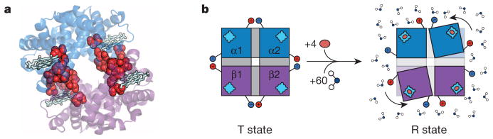
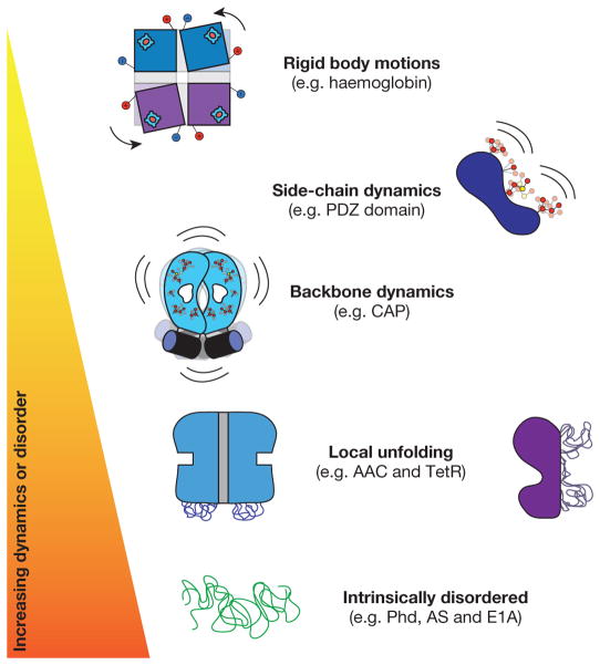
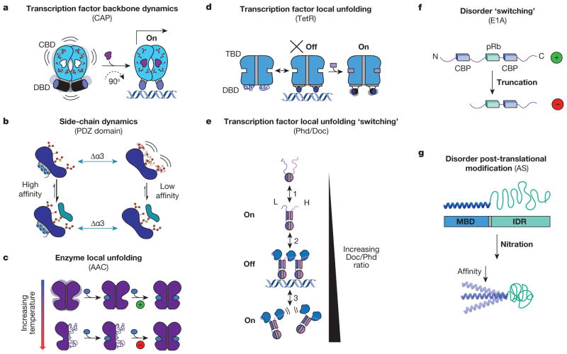
Allostery is the process by which biological macromolecules (mostly proteins) transmit the effect of binding at one site to another, often distal, functional site, allowing for regulation of activity. Recent experimental observations demonstrating that allostery can be facilitated by dynamic and intrinsically disordered proteins have resulted in a new paradigm for understanding allosteric mechanisms, which focuses on the conformational ensemble and the statistical nature of the interactions responsible for the transmission of information. Analysis of allosteric ensembles reveals a rich spectrum of regulatory strategies, as well as a framework to unify the description of allosteric mechanisms from different systems.


Conflict of interest statement

The authors declare no competing financial interests.

Figures

Figure 1
Figure 1. Structure-based views of allostery
a, Ribbon diagram representation of tetrameric haemoglobin (PDB accession 1GZX) rendered in PyMol (Schrödinger). The proposed pathway responsible for the cooperative transition from tensed (T) to relaxed (R) is highlighted with red spheres and the haem groups are represented as light blue sticks. b, Allosteric transition of tetrameric haemoglobin, as proposed by Perutz,. Tetrameric haemoglobin in the T state is depicted on the left with the two α-subunits (blue) and the two β-subunits (purple) each with their own haem group (light blue). Salt bridges, depicted as the red positive and blue negative charges, hold the molecule in the T conformation, and these salt bridges are released upon binding of oxygen (orange oval) in the transition to the R conformation (on the right) accompanied by a 15° turn of the subunits relative to each another. Also contributing to the equilibrium are 60 additional water molecules preferentially binding the R state.
Figure 2
Figure 2. The dynamic continuum of allosteric phenomena
Schematic representation of allosteric systems with increasing dynamics, disorder or fluctuations on the vertical axis.
Figure 3
Figure 3. Allosteric systems from the dynamic continuum
a, CAP homodimer with a cyclic nucleotide binding domain (CBD, blue domains with side chains) and a DNA binding domain (DBD, blue cylinders). Binding energetics of cAMP (purple ligand) quench dynamics in the bound-state ensemble and induce a 90° change in the conformation of the DBD, allowing it to bind DNA and turn on transcription. b, Side-chain dynamics modulate the binding affinity of a canonical PDZ domain to its native ligand with and without α-helix 3 (Δα3, green arrows). c, AAC homodimer with each monomer represented as one purple domain. Binding of the allosteric effector acetyl-CoA (blue oval) is positively cooperative at low temperatures (green ‘+’), and negatively cooperative at high temperatures (red ‘−’). d, TetR homodimer depicted as a two-domain protein with a tetracycline binding domain (TBD, blue region, top) and a DBD (blue region, bottom). e, Doc/Phd toxin–antitoxin system equilibrium. Phd is depicted as a homodimer (top, blue and purple monomers) and Doc is depicted as the blue ligand. f, Representation of variants of E1A shown from N to C terminus with binding sites for ligands in the two variants represented by blue rectangles (CBP) and green rectangles (pRb). g, Schematic representation of α-synuclein (AS) with its N-terminal membrane-binding domain (MBD) coupled to its C-terminal IDR. Upon oxidative stress (nitration) the affinity of the MBD decreases (bottom).

Similar articles

Cited by

References

    1. Monod J, Jacob F. Teleonomic mechanisms in cellular metabolism, growth, and differentiation. Cold Spring Harb Symp Quant Biol. 1961;26:389–401. - PubMed
    1. Changeux JP. The feedback control mechanisms of biosynthetic L-threonine deaminase by L-isoleucine. Cold Spring Harb Symp Quant Biol. 1961;26:313–318. - PubMed
    1. Monod J, Wyman J, Changeux JP. On the nature of allosteric transitions: a plausible model. J Mol Biol. 1965;12:88–118. - PubMed
    1. Freiburger LA, et al. Competing allosteric mechanisms modulate substrate binding in a dimeric enzyme. Nature Struct Mol Biol. 2011;18:288–294. - PMC - PubMed
    1. Nussinov R, Tsai CJ, Ma B. The (still) underappreciated role of allostery in the cellular network. Annu Rev Biophys. 2013;42:169–189. - PMC - PubMed

Publication types