

Gel formation in protein amyloid aggregation: a physical mechanism for cytotoxicity
- PMID: 24740416
- PMCID: PMC3989237
- DOI: 10.1371/journal.pone.0094789
Gel formation in protein amyloid aggregation: a physical mechanism for cytotoxicity
Erratum in
- PLoS One. 2014;9(7):e104152. Cole, Lisa [corrected to Burnett, Lisa Cole]
Abstract
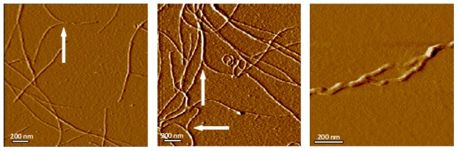
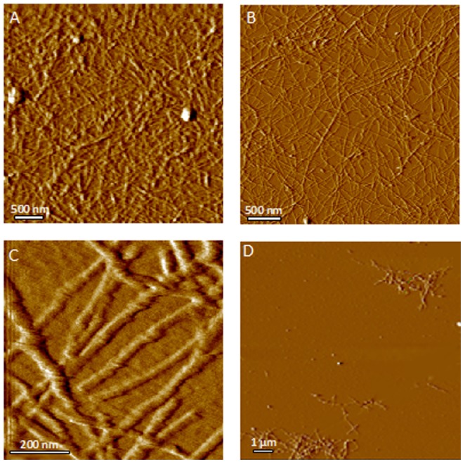
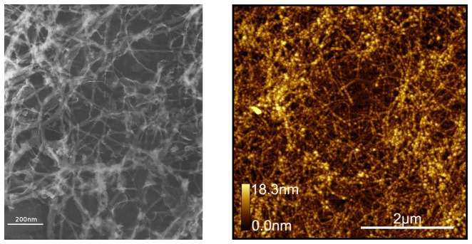
Amyloid fibers are associated with disease but have little chemical reactivity. We investigated the formation and structure of amyloids to identify potential mechanisms for their pathogenic effects. We incubated lysozyme 20 mg/ml at 55C and pH 2.5 in a glycine-HCl buffer and prepared slides on mica substrates for examination by atomic force microscopy. Structures observed early in the aggregation process included monomers, small colloidal aggregates, and amyloid fibers. Amyloid fibers were observed to further self-assemble by two mechanisms. Two or more fibers may merge together laterally to form a single fiber bundle, usually in the form of a helix. Alternatively, fibers may become bound at points where they cross, ultimately forming an apparently irreversible macromolecular network. As the fibers assemble into a continuous network, the colloidal suspension undergoes a transition from a Newtonian fluid into a viscoelastic gel. Addition of salt did not affect fiber formation but inhibits transition of fibers from linear to helical conformation, and accelerates gel formation. Based on our observations, we considered the effects of gel formation on biological transport. Analysis of network geometry indicates that amyloid gels will have negligible effects on diffusion of small molecules, but they prevent movement of colloidal-sized structures. Consequently gel formation within neurons could completely block movement of transport vesicles in neuronal processes. Forced convection of extracellular fluid is essential for the transport of nutrients and metabolic wastes in the brain. Amyloid gel in the extracellular space can essentially halt this convection because of its low permeability. These effects may provide a physical mechanism for the cytotoxicity of chemically inactive amyloid fibers in neurodegenerative disease.
Conflict of interest statement
Figures
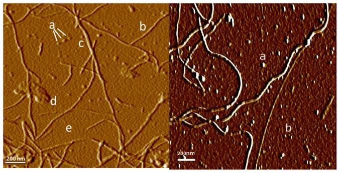
 nm (n = 20), probably representing individual lysozyme monomers.
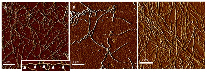
 m. Fibers forming in buffer with 150 mM NaCl (right) demonstrate virtually no helical structure even after 31 days.
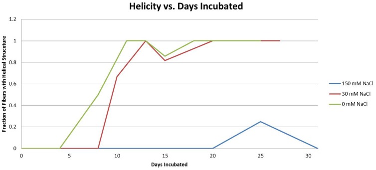
References
-
- Aggeli A, Boden N (2006) Self-assembling peptide gels. In: Molecular Gels: Materials With Selfassembled Fibrillar Networks, Springer, New York. pp. 721–742.
-
- Estroff LA, Hamilton AD (2006) Cryo-tem, x-ray diffraction and modeling of an organic hydrogel. In: Molecular Gels, Springer. pp. 721–742.
-
- Astbury W, Beighton E, Parker K (1959) The cross-β configuration in supercontracted proteins. Biochimica et biophysica acta 35: 17–25. - PubMed
-
- Xu S (2009) Cross-beta-sheet structure in amyloid fiber formation. J Phys Chem B 113: 12447–12455. - PubMed
Publication types
MeSH terms
Substances
LinkOut - more resources
Full Text Sources
Other Literature Sources
Research Materials
