. 2014 May 15;192(10):4813-20.
doi: 10.4049/jimmunol.1303369. Epub 2014 Apr 16.

Protection from intestinal inflammation by bacterial exopolysaccharides

Affiliations

Protection from intestinal inflammation by bacterial exopolysaccharides

Sara E Jones et al. J Immunol. .

Abstract

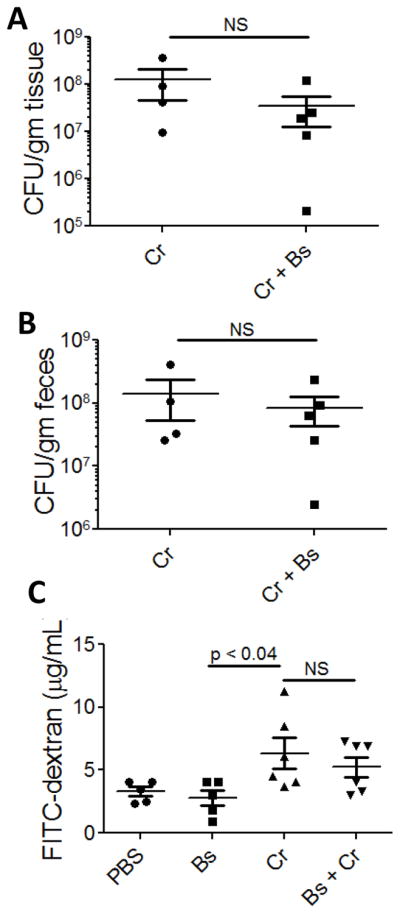
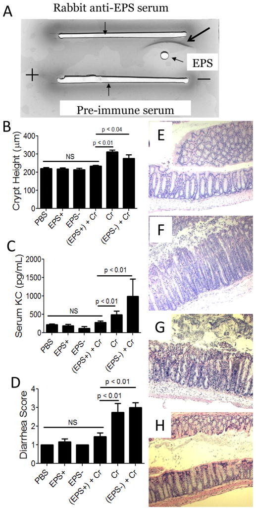
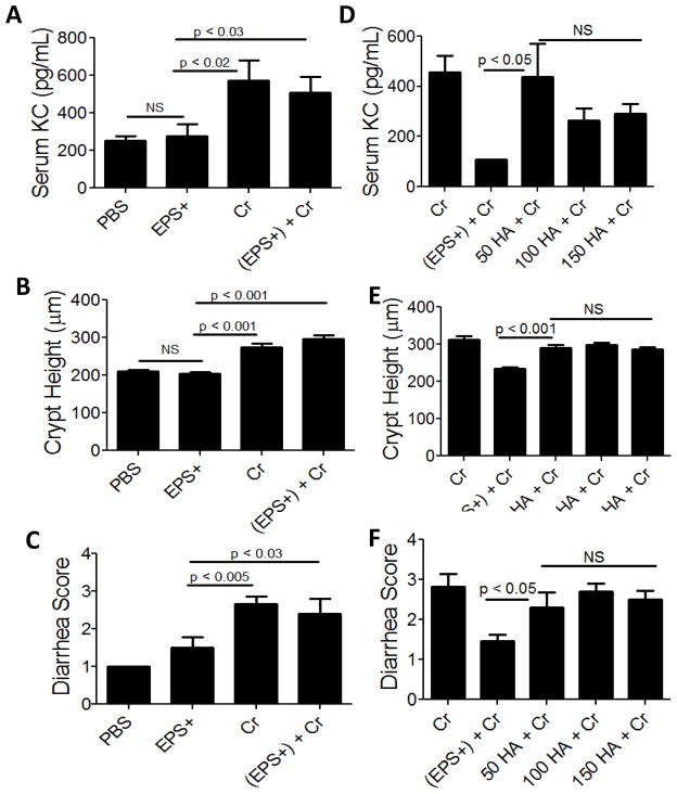
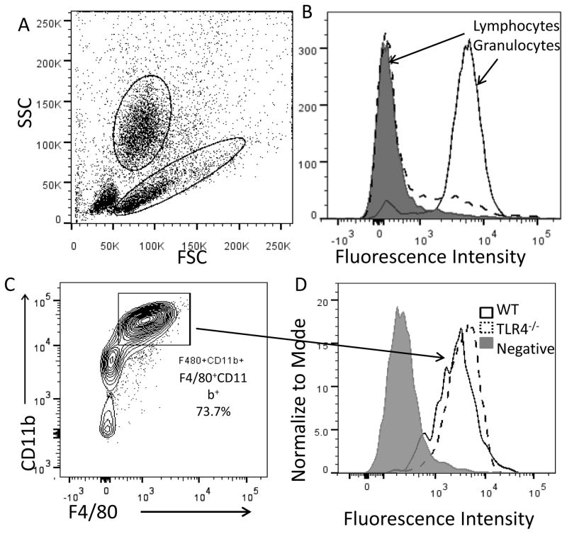
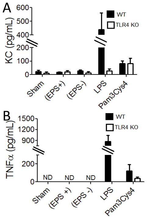
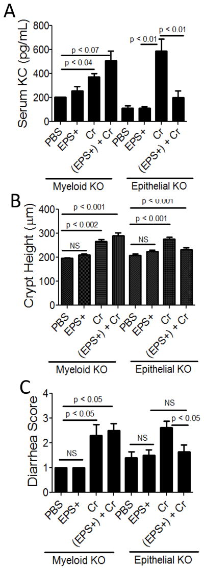
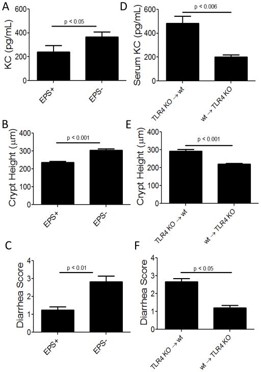
Host inflammatory responses against pathogenic organisms can be abrogated by commensals; however, the molecular mechanisms by which pathogenesis is prevented are still poorly understood. Previous studies demonstrated that administration of a single dose of Bacillus subtilis prevented disease and inflammation by the enteric mouse pathogen Citrobacter rodentium, which causes disease similar to the human pathogen enteropathogenic Escherichia coli. No protection was observed when an exopolysaccharide (EPS)-deficient mutant of B. subtilis was used, suggesting that EPS are the protective factor. In this study, we isolated and characterized EPS and showed that they also prevent C. rodentium-associated intestinal disease after a single injection. Protection is TLR4 dependent because EPS-treated TLR4 knockout mice developed disease. Furthermore, protection could be conveyed to wild-type mice by adoptive transfer of macrophage-rich peritoneal cells from EPS-treated mice. We found that EPS specifically bind peritoneal macrophages, and because mice lacking MyD88 signaling in myeloid cells were not protected by EPS, we conclude that bacterial EPS prevent colitis in a TLR4-dependent manner that requires myeloid cells. These studies provide a simple means of preventing intestinal inflammation caused by enteric pathogens.


Conflict of interest statement

Conflicts of interest: The authors have no conflicts of interest.

Figures

Figure 1
Figure 1. Colonization and gut-induced leakiness in C. rodentium-infected mice after treatment with B. subtilis
Mucosal (A) and lumenal (B) colonization of C. rodentium 11 dpi. FITC-dextran in serum of mice 11 dpi with lux+ C. rodentium (C). Cr, C. rodentium-infected mice; Cr + Bs, mice treated with B. subtilis 24 hr prior to C. rodentium; PBS, phosphate-buffered saline. The results are averages from 2 independent experiments and a total of 5–6 mice were assessed for each group. No statistical difference was found between C. rodentium-infected mice and those that received B. subtilis prior to infection.
Figure 2
Figure 2. Assessment of the B. subtilis exopolysaccharides on C. rodentium-associated disease 10 days post-infection dpi of wt mice
A) Immunoelectrophoresis analysis of purified EPS (arrow points to precipitation arc). B) Average colonic crypt heights from each treatment group. Serum KC levels (C), and evidence of diarrhea (D) were also used as disease markers. Results are averages from at least three independent experiments; a total of 5–12 mice were assessed for each group. PBS, phosphate-buffered saline; EPS+, exopolysaccharide from B. subtilis strain DS991; EPS−, material from B. subtilis strain DS5187; Cr, C. rodentium. Representative images of H&E stained colons from wildtype mice (100X). Images are representative of mice that received EPS from DS991 prior to C. rodentium infection (E), or material from the non-EPS producing strain DS5187 prior to pathogen infection (F). Representative images from myeloid MyD88 KO mice (G) and epithelial MyD88 KO mice (H) treated with EPS prior to infection with C. rodentium.
Figure 3
Figure 3. Assessment of C. rodentium-associated disease in EPS or TLR4 agonist-treated TLR4 KO or wt mice
Quantification by ELISA of pro-inflammatory KC in serum of TLR4 KO mice infected with C. rodentium (Cr) with or without EPS (EPS+); PBS and EPS+ are negative controls (A). Summary of colonic crypt heights from each treatment group (B). Diarrhea (C) also served as a disease marker. Results are averages from at least three independent experiments; a total of 5–10 mice were assessed for each group. (D–F): Wt mice were treated with 50, 100, or 150 μg of the TLR4 agonist hyaluronic acid (HA) prior to infection and then assessed for disease 10 dpi. Serum KC was measured by ELISA (D), colonic crypt heights from each treatment group were measured (E), and diarrhea (F) also served as a disease marker. Results are averages from at two independent experiments; a total of 4–5 mice were assessed for each group.
Figure 4
Figure 4. Flow cytometric analysis of EPS-binding to peritoneal cells from wt and TLR4 KO mice
FSC vs. SSC (A); granulocyte and lymphocyte binding to EPS (B) - gray peak is negative isotype control; staining of wt or TLR4 KO F4/80+CD11b+ gated cells with EPS (C and D). Fluorescence intensity represents EPS binding. Data shown are from one of three independent experiments.
Figure 5
Figure 5. ELISA analysis of cytokines induced by in vitro culture of EPS with peritoneal cells from wt and TLR4 KO mice
Peritoneal cells were incubated with EPS (EPS+) (30 μg/ml), material from the non-EPS producing strain (EPS−) (30 μg/mL), LPS (100 ng/mL), Pam3Cys4 (100 ng/mL), or without addition (sham). Results are averages from three independent experiments. ND, not detectable
Figure 6
Figure 6. Assessment of C. rodentium-associated disease in EPS-treated mice lacking MyD88 in myeloid or epithelial cells
Myeloid MyD88 KO and epithelial MyD88 KO mice were treated with EPS (EPS+) (i.p.) 1 day prior to infection with C. rodentium (Cr) and disease was assessed 10 dpi. Injection with PBS and EPS+ alone served as negative controls. Serum KC levels (A), colonic crypt height (B), and diarrhea (C) were used as disease markers. Results are averages from at least two independent experiments and a total of 2–5 mice were assessed for each group.
Figure 7
Figure 7. Assessment of disease after transfer of peritoneal cells from EPS-treated wt and TLR4 KO mice to C. rodentium-infected wt mice
Donor wt mice were treated with EPS+ or EPS− material (i.p.) 2–3 days before peritoneal cells (6 × 10⁴) were transferred i.p. to naïve recipient wt (A–C) mice 1 day prior to, 1 dpi and 3 dpi with C. rodentium. Peritoneal cells from wt or TLR4 KO mice similarly treated with EPS+ were transferred i.p. into naïve wt or TLR4 KO mice (D–F). Disease was assessed for all mice 10 dpi; serum KC (A and D), crypt hyperplasia (B and E), and diarrhea (C and F) were used as disease markers. KC quantification, crypt height and diarrhea scores can be compared to uninfected (PBS-treated) mice shown in Figure 2B–D. Results are averages from at least three independent experiments and a total of 6–10 mice were assessed for each group.

Similar articles

Cited by

References

    1. Hayashi A, Sato T, Kamada N, Mikami Y, Matsuoka K, Hisamatsu T, Hibi T, Roers A, Yagita H, Ohteki T, Yoshimura A, Kanai T. A single strain of Clostridium butyricum induces intestinal IL-10-producing macrophages to suppress acute experimental colitis in mice. Cell Host Microbe. 2013;13:711–722. - PubMed
    1. Round JL, Lee SM, Li J, Tran G, Jabri B, Chatila TA, Mazmanian SK. The Toll-like receptor 2 pathway establishes colonization by a commensal of the human microbiota. Science. 2011;332:974–977. - PMC - PubMed
    1. Gais P, Reim D, Jusek G, Rossmann-Bloeck T, Weighardt H, Pfeffer K, Altmayr F, Janssen KP, Holzmann B. Cutting edge: Divergent cell-specific functions of MyD88 for inflammatory responses and organ injury in septic peritonitis. J Immunol. 2012;188:5833–5837. - PubMed
    1. D’Arienzo R, Maurano F, Mazzarella G, Luongo D, Stefanile R, Ricca E, Rossi M. Bacillus subtilis spores reduce susceptibility to Citrobacter rodentium-mediated enteropathy in a mouse model. Res Microbiol. 2006;157:891–897. - PubMed
    1. Wiles S, Pickard KM, Peng K, MacDonald TT, Frankel G. In vivo bioluminescence imaging of the murine pathogen Citrobacter rodentium. Infect Immun. 2006;74:5391–5396. - PMC - PubMed

Publication types

MeSH terms