Review
. 2014 Jun 1;306(11):C987-96.
doi: 10.1152/ajpcell.00321.2013. Epub 2014 Apr 16.

Cellular mechanisms of tissue fibrosis. 7. New insights into the cellular mechanisms of pulmonary fibrosis

Affiliations
Review

Cellular mechanisms of tissue fibrosis. 7. New insights into the cellular mechanisms of pulmonary fibrosis

Christina E Barkauskas et al. Am J Physiol Cell Physiol. .

Abstract

Idiopathic pulmonary fibrosis (IPF) is a devastating disease characterized by severe and progressive scar formation in the gas-exchange regions of the lung. Despite years of research, therapeutic treatments remain elusive and there is a pressing need for deeper mechanistic insights into the pathogenesis of the disease. In this article, we review our current knowledge of the triggers and/or perpetuators of pulmonary fibrosis with special emphasis on the alveolar epithelium and the underlying mesenchyme. In doing so, we raise a number of questions highlighting critical voids and limitations in our current understanding and study of this disease.

Keywords: epithelium; lung fibrosis; myofibroblast.


Figures

Fig. 1.
Fig. 1.
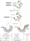
Proposed model for the development of pulmonary fibrosis in human lung. The distal human lung consists of distal bronchioles (<3 mm in diameter) containing basal cells, Scgb1a1+ secretory cells, ciliated cells, and some mucus-producing cells [commonly MUC5B+, occasionally MUC5AC+ (71)]. The distal bronchioles lead to alveolar ducts that are lined by poorly defined cuboidal epithelium. The alveolar ducts ultimately open to the alveoli that contain Sftpc+ type 1 and 2 alveolar epithelial cells (AEC1s and AEC2s) in close association with a number of different mesenchymal cell types (top). At steady state when tissue turnover is low, AEC2s will occasionally self-renew and differentiate into AEC1s. Note the close approximation of AEC2s and the niche-comprising resident fibroblasts in the alveolar space. Following injury or “wear and tear” to the alveolar epithelium in otherwise normal lung, dead alveolar epithelial cells are replaced by descendants of AEC2 stem cells that self-renew and differentiate into AEC1s. We also hypothesize that Scgb1a1+ cells and/or basal cells serve as a source of AEC2s following injury. This hypothesis is based on murine data revealing that bronchiolar epithelial cells in the mouse differentiate into both AEC2s and AEC1s following injury (68, 81, 92, 93). Note that in mouse bronchioles basal cells are not normally present. These repair processes effectively cover denuded basal lamina, and fibrosis does not develop. If injury occurs to alveolar epithelium that is already stressed or defective (as is hypothesized to be the case in IPF), the AEC2 stem cells cannot differentiate to effectively cover denuded basal lamina and profibrotic signals are likely released (middle). Based on the detailed description of human IPF histology, we hypothesize that the persistently denuded basal lamina is eventually covered via one of 2 proposed mechanisms (bottom). Both of these mechanisms result in the destruction of delicate alveolar architecture and deposition of significant amounts of extracellular matrix. The first ineffective repair model (bottom left) stems from human IPF histology data revealing the unusual presence of pseudostratified epithelium (with many mucus-producing cells) in distal airways and lining “honeycomb cysts” of IPF patients. At least 2, nonexclusive, hypotheses can explain this finding. First, conditions may be such that Scgb1a1+ cells in the distal bronchioles, differentiate predominantly into mucus producing goblet cells rather than AEC2 cells. Second, alveolar duct cells may change their phenotype and resemble bronchiolar epithelium, including basal cells. In the second ineffective repair model (bottom right), we hypothesize that stressed AEC2s are unable to differentiate into AEC1s but they retain some capacity to self-renew and cover the denuded basal lamina with a hyperplastic Sftpc+ cuboidal epithelium that lines the airspace. This epithelial type is associated with fibroblast foci. This model is speculative but based on extensive lineage-tracing of epithelial cells in the mouse (5, 68, 81, 93) and on detailed analysis of human IPF histology (71).
Fig. 2.
Fig. 2.
Potential pathways involved in the development of pulmonary fibrosis. The biology underlying the pathogenesis of pulmonary fibrosis is complex. This figure illustrates known and hypothesized relationships between two components of the alveolar region of the lung, the epithelium and the mesenchyme. We hypothesize that dysregulated cross talk between these two components leads to development of the disease. As depicted here, while there may be an inciting factor or event that predisposes an individual to developing the disease (e.g., chronic cellular stress or epithelial injury), a host of other abnormal responses (e.g., abnormal proliferation of fibroblasts and excessive stiffening of the mesenchyme) are likely to be integral in the progression of fibrosis. Concepts written in italics within the concept boxes in the figure are hypothesized and yet to be experimentally proven in the precise context presented. ER, endoplasmic reticulum; SNP, single-nucleotide polymorphisms; TGF-β1, transforming growth factor-β1.

References

    1. Alder JK, Chen JJ, Lancaster L, Danoff S, Su SC, Cogan JD, Vulto I, Xie M, Qi X, Tuder RM, Phillips JA, 3rd, Lansdorp PM, Loyd JE, Armanios MY. Short telomeres are a risk factor for idiopathic pulmonary fibrosis. Proc Natl Acad Sci USA 105: 13051–13056, 2008. - PMC - PubMed
    1. Armanios M, Chen JL, Chang YP, Brodsky RA, Hawkins A, Griffin CA, Eshleman JR, Cohen AR, Chakravarti A, Hamosh A, Greider CW. Haploinsufficiency of telomerase reverse transcriptase leads to anticipation in autosomal dominant dyskeratosis congenita. Proc Natl Acad Sci USA 102: 15960–15964, 2005. - PMC - PubMed
    1. Armanios MY, Chen JJ, Cogan JD, Alder JK, Ingersoll RG, Markin C, Lawson WE, Xie M, Vulto I, Phillips JA, 3rd, Lansdorp PM, Greider CW, Loyd JE. Telomerase mutations in families with idiopathic pulmonary fibrosis. N Engl J Med 356: 1317–1326, 2007. - PubMed
    1. Azuma A, Nukiwa T, Tsuboi E, Suga M, Abe S, Nakata K, Taguchi Y, Nagai S, Itoh H, Ohi M, Sato A, Kudoh S. Double-blind, placebo-controlled trial of pirfenidone in patients with idiopathic pulmonary fibrosis. Am J Respir Crit Care Med 171: 1040–1047, 2005. - PubMed
    1. Barkauskas CE, Cronce MJ, Rackley CR, Bowie EJ, Keene DR, Stripp BR, Randell SH, Noble PW, Hogan BL. Type 2 alveolar cells are stem cells in adult lung. J Clin Invest 123: 3025–3036, 2013. - PMC - PubMed