. 2014 Jun 15;306(12):R925-33.
doi: 10.1152/ajpregu.00027.2014. Epub 2014 Apr 16.

Overexpression of TRB3 in muscle alters muscle fiber type and improves exercise capacity in mice

Affiliations

Overexpression of TRB3 in muscle alters muscle fiber type and improves exercise capacity in mice

Ding An et al. Am J Physiol Regul Integr Comp Physiol. .

Abstract

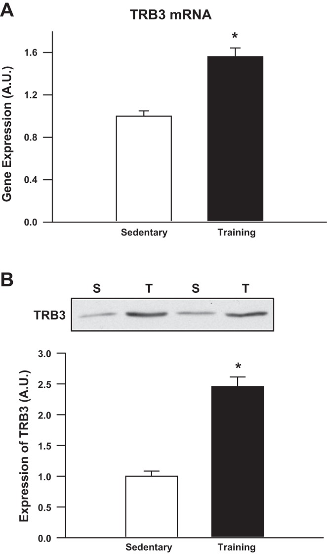
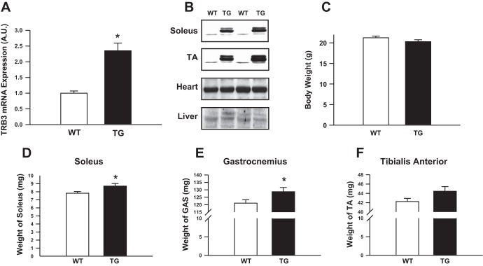
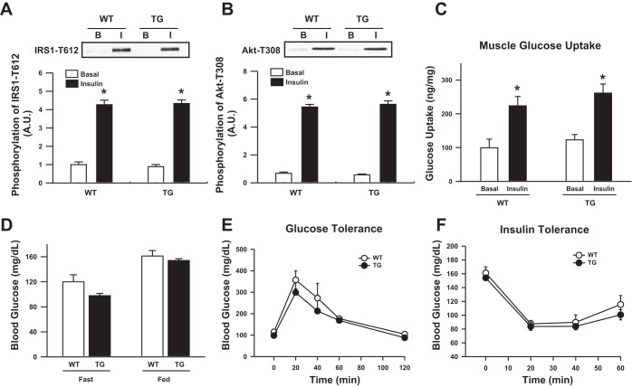
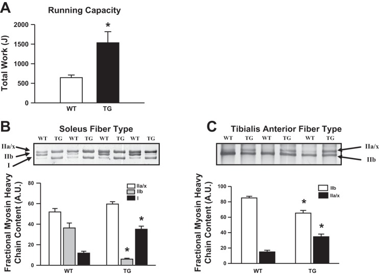
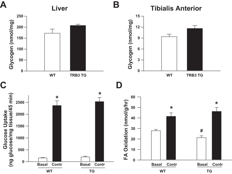
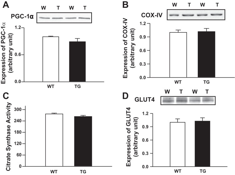
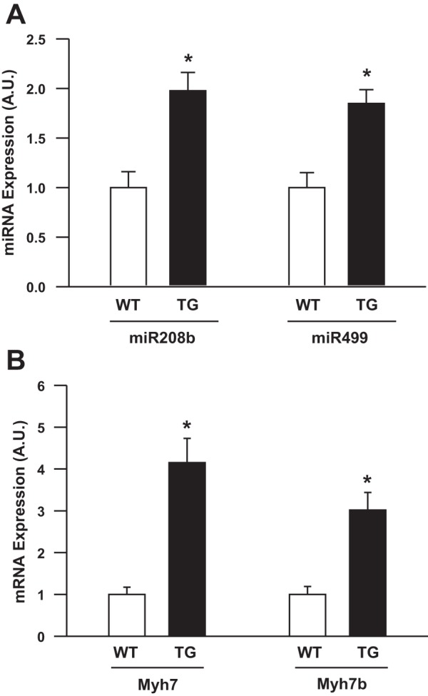
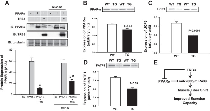
Increasing evidence suggests that TRB3, a mammalian homolog of Drosophila tribbles, plays an important role in cell growth, differentiation, and metabolism. In the liver, TRB3 binds and inhibits Akt activity, whereas in adipocytes, TRB3 upregulates fatty acid oxidation. In cultured muscle cells, TRB3 has been identified as a potential regulator of insulin signaling. However, little is known about the function and regulation of TRB3 in skeletal muscle in vivo. In the current study, we found that 4 wk of voluntary wheel running (6.6 ± 0.4 km/day) increased TRB3 mRNA by 1.6-fold and protein by 2.5-fold in the triceps muscle. Consistent with this finding, muscle-specific transgenic mice that overexpress TRB3 (TG) had a pronounced increase in exercise capacity compared with wild-type (WT) littermates (TG: 1,535 ± 283; WT: 644 ± 67 joules). The increase in exercise capacity in TRB3 TG mice was not associated with changes in glucose uptake or glycogen levels; however, these mice displayed a dramatic shift toward a more oxidative/fatigue-resistant (type I/IIA) muscle fiber type, including threefold more type I fibers in soleus muscles. Skeletal muscle from TRB3 TG mice had significantly decreased PPARα expression, twofold higher levels of miR208b and miR499, and corresponding increases in the myosin heavy chain isoforms Myh7 and Myb7b, which encode these microRNAs. These findings suggest that TRB3 regulates muscle fiber type via a peroxisome proliferator-activated receptor-α (PPAR-α)-regulated miR499/miR208b pathway, revealing a novel function for TRB3 in the regulation of skeletal muscle fiber type and exercise capacity.

Keywords: PPAR-α; TRB3; exercise capacity; microRNAs; muscle.


Figures

Fig. 1.
Fig. 1.
Exercise training increases tribbles homolog 3 (TRB3) mRNA and protein expression. C57BL/6 mice (Charles River Laboratories, 10 wk old) were housed in wheel cages for 4 wk (Training; T), and sedentary (S) mice were housed in separate cages without a wheel. Mice were euthanized and triceps muscles were snap frozen in liquid nitrogen. RNA and protein were extracted and TRB3 mRNA (A) and protein (B) were determined. Data are means ± SE, n = 9 mice/group. *P < 0.05 compared with sedentary mice.
Fig. 2.
Fig. 2.
Characterization of muscle-specific TRB3 transgenic (TG) mice. Wild-type (WT) and TRB3 TG mice (8 wk old) were euthanized and tissues were harvested. TRB3 mRNA expression was determined in tibialis anterior (TA) muscle (A) and TRB3 protein expression determined in the soleus and TA muscles, heart, and liver (B). TRB3 TG mice had similar body weights (C) compared with WT littermates. Muscle weights of the soleus (D) gastrocnemius (GAS) (E), and TA (F) were measured. Data are means ± SE, n = 8 mice/group. *P < 0.05 compared with WT mice.
Fig. 3.
Fig. 3.
Effect of TRB3 overexpression on insulin signaling and glucose uptake in TA muscle. After treatment with insulin, basal (B) and insulin-stimulated (I) phosphorylation of IRS1-T612 (A) and Akt-T308 (B) were measured by Western blotting. In vivo skeletal muscle 2-[3H]deoxyglucose uptake in response to a glucose bolus was similar between TRB3 TG mice and WT littermates (C). Whole body glucose homeostasis was measured by fasting blood glucose (D) and by performing glucose- and insulin-tolerance tests (E and F). *P < 0.05 vs. basal conditions.
Fig. 4.
Fig. 4.
TRB3 TG mice have increased exercise capacity, and muscles from TRB3 TG mice exhibit a transformation toward oxidative fiber type. Maximal exercise capacity was determined by running female WT and TRB3 TG mice on a treadmill until the mice reached exhaustion (A). WT and TRB3 TG mice (8 wk old) were euthanized, and soleus and TA muscle were harvested. Protein lysates were prepared from muscles and myosin heavy chain isoforms were separated. Gels were silver-stained with the SilverSNAP Stain Kit II (Pierce, 24612) and myosin heavy chain bands from the soleus (B) and TA (C) muscles were quantified from images of scanned gels using the 1D Multifunction of AlphaEase FC software. Data are means ± SE, n = 8 mice/group. *P < 0.05 compared with WT mice.
Fig. 5.
Fig. 5.
Overexpression of TRB3 does not alter glycogen or contraction-stimulated substrate metabolism. Liver (A) and muscle (B) glycogen concentrations were slightly but not significantly elevated in TRB3 TG mice in the basal state. Overexpression of TRB3 did not alter contraction-stimulated glucose transport in vivo (C) or fatty acid oxidation in vitro (D), although TRB3 TG muscle had decreased basal rates of fatty acid oxidation. Data are means ± SE, n = 6–8 mice/group. *P < 0.05 compared with sedentary (Basal) group. #P < 0.05 compared with WT mice.
Fig. 6.
Fig. 6.
Expression of PGC1α and COX IV, citrate synthase activity (e.g., mitochondrial markers), and glucose transporter type 4 (GLUT4) in muscle of TRB3 TG and WT animals. PGC1α expression (A), COX IV expression (B), citrate synthase activity (C), and GLUT4 (D) were determined in TA muscle of 8-wk-old female TRB3 TG and WT animals. TRB3 overexpression was not associated with an increase in PGC1α expression or an increase in COX IV and GLUT4 expression and citrate synthase activity. n = 8 mice/group.
Fig. 7.
Fig. 7.
Muscles from TRB3 TG mice have increased expression of microRNAs miR208b and miR499 and their associated mRNAs Myh7 and Myh7b. RNA was isolated from gastrocnemius muscles from WT and TRB3 TG mice. cDNA was synthesized and expression of miR208b and miR499 (A) and their associated mRNAs Myh7 and Myh7b (B) were determined. Data are means ± SE, n = 6–8 mice/group. *P < 0.05 compared with WT mice.
Fig. 8.
Fig. 8.
Overexpression of TRB3 decreases peroxisome proliferator-activated receptor-α (PPAR-α), fatty acid transport protein (FATP), and uncoupling protein 3 (UCP3). To determine whether TRB3 regulates PPAR-α, TRB3 and PPAR-α were overexpressed in HEK293 cells. In some experiments, transfected cells were incubated with proteasome inhibitor MG132 overnight before harvesting. A: expression of TRB3 and PPAR-α were determined by Western blotting. Protein was isolated from gastrocnemius muscles from WT and TRB3 TG mice and Western blots were performed to determine protein expression of PPA-α (B), UCP3 (C), and FATP1 (D). E: proposed mechanisms by which TRB3 regulates muscle fiber type and exercise capacity. Overexpression of TRB3 decreased PPAR-α activity and increased the expression of miR208b and miR499, inducing a transformation toward a more oxidative fiber type, which in turn, contributes to increased exercise capacity in TRB3 TG mice (E). Data are means ± SE, n = 8 mice/group for A and n = 3 separate experiments for A. *P < 0.05, PPAR-α compared with PPAR-α +TRB3, #P < 0.05 PPAR-α +TRB3 untreated compared with PPAR-α +TRB3 treated with MG132.

References

    1. Anuppalle M, Maddirevula S, Huh TL, Rhee M. Trb3 regulates LR axis formation in zebrafish embryos. Mol Cells 36: 542–547, 2013 - PMC - PubMed
    1. Avery J, Etzion S, DeBosch BJ, Jin X, Lupu TS, Beitinjaneh B, Grand J, Kovacs A, Sambandam N, Muslin AJ. TRB3 function in cardiac endoplasmic reticulum stress. Circ Res 106: 1516–1523, 2010 - PMC - PubMed
    1. Avery J, Etzion S, DeBosch BJ, Jin X, Lupu TS, Beitinjaneh B, Grand J, Kovacs A, Sambandam N, Muslin AJ. TRB3 function in cardiac endoplasmic reticulum stress. Circ Res 106: 1516–1523, 2010 - PMC - PubMed
    1. Cline GW, Vidal-Puig AJ, Dufour S, Cadman KS, Lowell BB, Shulman GI. In vivo effects of uncoupling protein-3 gene disruption on mitochondrial energy metabolism. J Biol Chem 276: 20240–20244, 2001 - PubMed
    1. Conley DL, Krahenbuhl GS. Running economy and distance running performance of highly trained athletes. Med Sci Sports Exerc 12: 357–360, 1980 - PubMed

Publication types

LinkOut - more resources