. 2014 Jun 15;306(12):F1400-9.
doi: 10.1152/ajprenal.00624.2013. Epub 2014 Apr 16.

Mesangial cell αvβ8-integrin regulates glomerular capillary integrity and repair

Affiliations

Mesangial cell αvβ8-integrin regulates glomerular capillary integrity and repair

Sujata Lakhe-Reddy et al. Am J Physiol Renal Physiol. .

Abstract

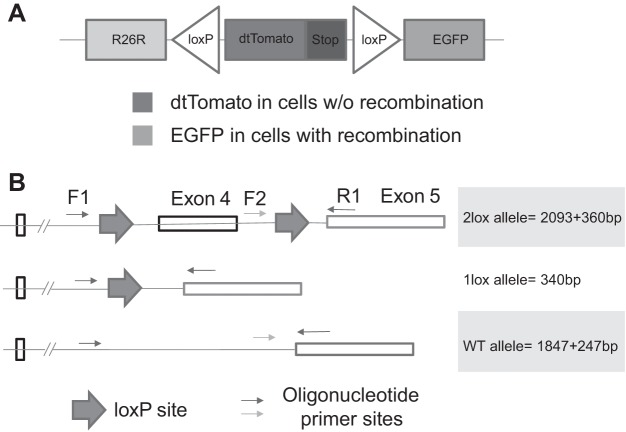
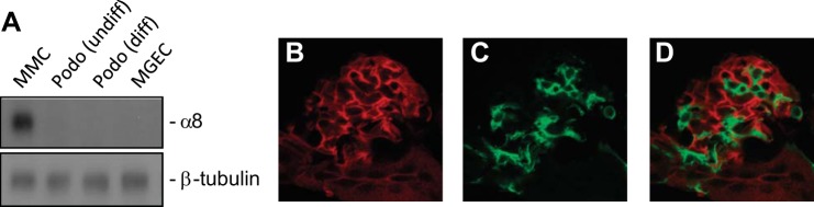
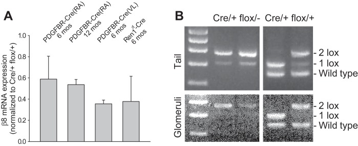
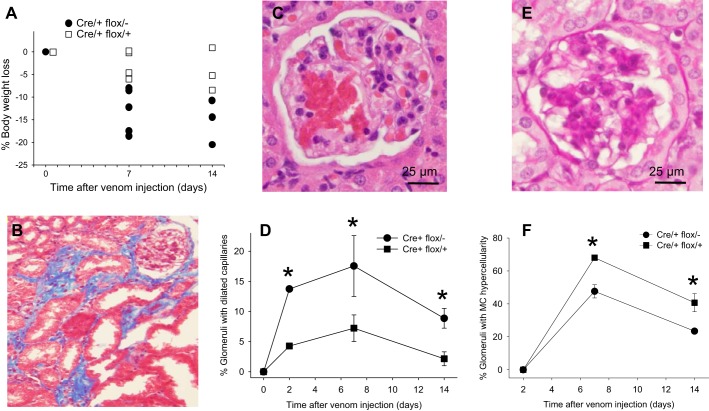
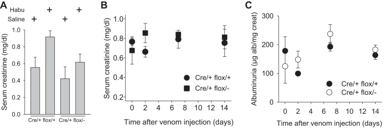
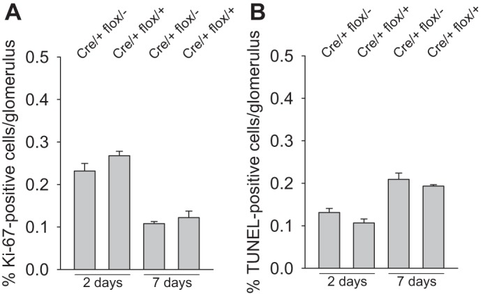
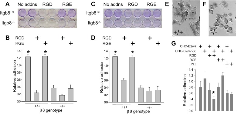
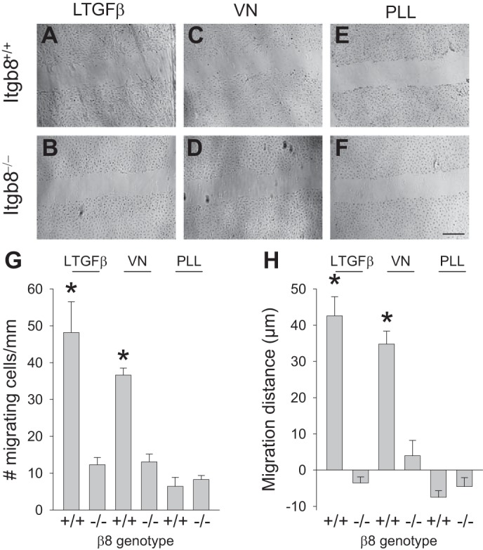
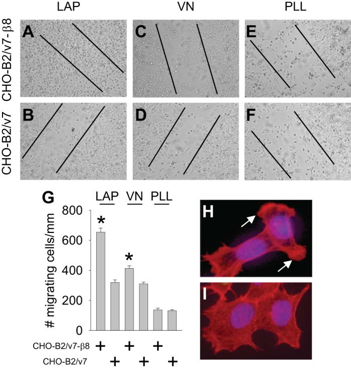
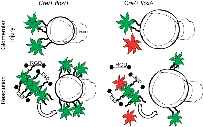
αvβ8-Integrin is most abundantly expressed in the kidney, brain, and female reproductive organs, and its cognate ligand is latent transforming growth factor (LTGF)-β. Kidney αvβ8-integrin localizes to mesangial cells, and global β8-integrin gene (Itgb8) deletion results in embryonic lethality due to impaired placentation and cerebral hemorrhage. To circumvent the lethality and better define kidney αvβ8-integrin function, Cre-lox technology was used to generate mesangial-specific Itgb8-null mice. Platelet-derived growth factor-β receptor (PDGFBR)-Cre mice crossed with a reporter strain revealed functional Cre recombinase activity in a predicted mesangial pattern. However, mating between two different PDGFBR-Cre or Ren1(d)-Cre strains with Itgb8 (flox/-) mice consistently resulted in incomplete recombination, with no renal phenotype in mosaic offspring. Induction of a renal phenotype with Habu snake venom, a reversible mesangiolytic agent, caused exaggerated glomerular capillary microaneurysms and delayed recovery in Cre(+/-) PDGFRB (flox/-) mice compared with Cre(+/-) PDGFRB (flox/+) control mice. To establish the mechanism, in vitro experiments were conducted in Itgb8-null versus Itgb8-expressing mesangial cells and fibroblasts, which revealed β8-integrin-regulated adhesion to Arg-Gly-Asp (RGD) peptides within a mesangial-conditioned matrix as well as β8-integrin-dependent migration on RGD-containing LTGF-β or vitronectin matrices. We speculate that kidney αvβ8-integrin indirectly controls glomerular capillary integrity through mechanical tension generated by binding RGD peptides in the mesangial matrix, and healing after glomerular injury may be facilitated by mesangial cell migration, which is guided by transient β8-integrin interactions with RGD ligands.

Keywords: cell migration; extracellular matrix; glomerular endothelial cell; glomerular injury.


Figures

Fig. 1.
Fig. 1.
Cre-lox recombination efficiency assays. A: reporter gene map. B: multiplex PCR strategy to identify one lox, two lox, and wild-type (WT) β8-integrin alleles. EGFP, enhanced green fluorescent protein.
Fig. 2.
Fig. 2.
Platelet-derived growth factor-β receptor (PDGFBR)-Cre activity in a mesangial pattern. A: β8-integrin immunoblot of cell culture lysates from mouse mesangial cells (MMC), undifferentiated and differentiated mouse podocytes (Podo), and mouse glomerular endothelial cells (MGEC). PDGFBR-Cre mice were crossed with floxed reporter mice. B: the red tag marks nonrecombined cells. C: the green tag marks cells where recombination has occurred. D: merged image.
Fig. 3.
Fig. 3.
Floxed β8-integrin is incompletely deleted by Cre recombinase. A: β8-integrin mRNA expression was determined by quantitative RT-PCR in isolated glomeruli derived from crosses between floxed β8-integrin and three different Cre mouse strains. B: to investigate incomplete recombination as a possible mechanism, a PCR-based strategy was used, whereby complete recombination collapses the target gene to one loxP site (1 lox), whereas the unrecombined target gene retains both loxP sites (2 lox). Results from two mice per genotype revealed incomplete recombination in PDGFRB(VL)-Cre/+ flox/− mice and variable, but sometimes complete, recombination in PDGFRB(VL)-Cre/+ flox/+ mice.
Fig. 4.
Fig. 4.
Phenotypes in Habu venom-injected mice. A: body weights in Cre/+ flox/+ and Cre/+ flox/− mice after Habu injection. B: a Masson's trichrome-stained Cre/+ flox/− kidney revealed interstitial fibrosis 14 days after Habu injection. C: glomerular capillary dilation in a Cre/+ flox/− kidney 7 days after Habu injection. D and E: glomerular capillary dilation was quantitated in Cre/+ flox/+ and Cre/+ flox/− mice at the indicated time points. E: mesangial hypercellularity in a Cre/+ flox/− kidney 7 days after Habu injection. F: mesangial hypercellularity was quantitated in Cre/+ flox/+ and Cre/+ flox/− mice at the indicated time points. Results are expressed as means ± SE. *P < 0.05 compared with Cre/+ flox/+ kidneys at the same time point.
Fig. 5.
Fig. 5.
Renal function in Habu-treated mice. Renal function was determined by serum creatinine (A and B) or by the urine albumin-to-creatinine ratio (C). Data in A were generated 2 days after Habu venom or saline control injection in Cre/+ flox/+ and Cre/+ flox/− mice. Data in B and C were generated at the indicated time points after Habu toxin or saline injection.
Fig. 6.
Fig. 6.
β8-Integrin effects on apoptosis and proliferation. Mice were evaluated for mesangial cell proliferation by Ki-67 labeling (A) or apoptosis by TUNEL (B) 2 and 7 days after Habu injection.
Fig. 7.
Fig. 7.
β8-Integrin regulates adhesion to the mesangial matrix. A–F: mesangial cells derived from Itgb8+/+ and Itgb8−/− kidneys were seeded in 24-well plates coated with an extracellular matrix deposited by Itgb8+/+ (A, B, E, and F) or Itgb8−/− (C and D) mesangial cells with or without GRGDSP [Arg-Gly-Asp (RDG)] or GRGESP [Arg-Gly-Glu (RGE)] peptides. Adherent cells were fixed in paraformaldehyde, and nuclei were stained with crystal violet, which was eluted and quantitated, as described in materials and methods. A and C: representative plates with samples in duplicate. B and D: quantification of adhesion. E and F: representative phase-contrast microscopy fields for Itgb8+/+ (+/+; E) and Itgb8−/− (−/−; F) mouse mesangial cells at ×40 magnification. G: CHO-B2/v7 and CHO-B2/v7-β8 cells were maintained in serum-free media overnight, washed in PBS, and then seeded in 96-well plates with or without RGD or RGE peptides on a matrix deposited by wild-type mesangial cells or poly-l-lysine (PLL). Adhesion was assayed as described in A–D. * P < 0.05 compared with cells on a mesangial matrix without RGD peptide.
Fig. 8.
Fig. 8.
Mesangial cell β8-integrin regulates migration on a RGD-containing matrix. A–F: scratch migration assays for wild-type (Itgb8+/+; A, C, and E) and Itgb−/− (B, D, and F) mesangial cells plated on latent transforming growth factor (LTGF)-β (A and B), vitronectin (VN; C and D), or PLL (E and F), all at 5 μg/ml, for 15 h. G and H: quantification of the scratch migration data. Results are representative of 5 scratches/well. *P < 0.05 compared with the similarly treated Itgb8−/− group. Bar = 300 μm.
Fig. 9.
Fig. 9.
β8-Integrin regulates migration on a RGD-containing matrix. A–F: scratch migration assays for fibroblasts expressing β8-integrin (CHO-B2/v7-β8; A, C, and E) and devoid of β8-integrin (CHO-B2/v7; B, D, and F) plated on latency-associated peptide-β1 (LAP; A and B), VN (C and D), or PLL (E and F), all at 5 μg/ml, for 14 h. Lines represent the width of the scratch at time 0. G: quantification of the scratch migration data. Results are representative of 3 scratches/well. *P < 0.05 compared with CHO-B2/v7 cells. H and I: morphology of CHO-B2/v7-β8 cells on LAP (5 μg/ml, 2 h; H) or glass coverslips (I). Actin was stained in red with phalloidin. Arrows mark lamellipodia.
Fig. 10.
Fig. 10.
Schematic diagram of the β8-integrin influence on glomerular capillary injury and repair. The reduced Cre-lox recombination in Cre/+ flox/− mice, resulting in mosaicism, is shown by the greater number of mesangial cells, which do not express β8-integrin (shown in red). The reduced number of β8-integrin-expressing mesangial cells (shown in green), causes enhanced capillary dilation with injury and delayed mesangial cell migration during the recovery phase in Cre/+ flox/− glomeruli. GEC, glomerular endothelial cell.

References

    1. Abu Jawdeh BG, Khan S, Deschênes I, Hoshi M, Goel M, Lock JT, Shinlapawittayatorn K, Babcock G, Lakhe-Reddy S, DeCaro G, Yadav SP, Mohan ML, Naga Prasad SV, Schilling WP, Ficker E, Schelling JR. Phosphoinositide binding differentially regulates NHE1 Na+/H+ exchanger-dependent proximal tubule cell survival. J Biol Chem 286: 42435–42445, 2011 - PMC - PubMed
    1. Akis N, Madaio MP. Isolation, culture, and characterization of endothelial cells from mouse glomeruli. Kidney Int 65: 2223–2227, 2004 - PubMed
    1. Barnes JL, Abboud HE. Temporal expression of autocrine growth factors corresponds to morphological features of mesangial proliferation in Habu snake venom-induced glomerulonephritis. Am J Pathol 143: 1366–1376, 1993 - PMC - PubMed
    1. Barnes JL, Hevey KA, Hastings RR, Bocanegra RA. Mesangial cell migration precedes proliferation in Habu snake venom-induced glomerular injury. Lab Invest 70: 460–467, 1994 - PubMed
    1. Betz UA, Vosshenrich CA, Rajewsky K, Muller W. Bypass of lethality with mosaic mice generated by Cre-loxP-mediated recombination. Curr Biol 6: 1307–1316, 1996 - PubMed

Publication types

MeSH terms

LinkOut - more resources