. 2014 Jun 19;123(25):3979-87.
doi: 10.1182/blood-2014-01-551812. Epub 2014 Apr 16.

TFPI cofactor function of protein S: essential role of the protein S SHBG-like domain

Affiliations

TFPI cofactor function of protein S: essential role of the protein S SHBG-like domain

Natalia Reglińska-Matveyev et al. Blood. .

Abstract

Protein S is a cofactor for tissue factor pathway inhibitor (TFPI), accelerating the inhibition of activated factor X (FXa). TFPI Kunitz domain 3 residue Glu226 is essential for enhancement of TFPI by protein S. To investigate the complementary functional interaction site on protein S, we screened 44 protein S point, composite or domain swap variants spanning the whole protein S molecule for their TFPI cofactor function using a thrombin generation assay. Of these variants, two protein S/growth arrest-specific 6 chimeras, with either the whole sex hormone-binding globulin (SHBG)-like domain (Val243-Ser635; chimera III) or the SHBG laminin G-type 1 subunit (Ser283-Val459; chimera I), respectively, substituted by the corresponding domain in growth arrest-specific 6, were unable to enhance TFPI. The importance of the protein S SHBG-like domain (and its laminin G-type 1 subunit) for binding and enhancement of TFPI was confirmed in FXa inhibition assays and using surface plasmon resonance. In addition, protein S bound to C4b binding protein showed greatly reduced enhancement of TFPI-mediated inhibition of FXa compared with free protein S. We show that binding of TFPI to the protein S SHBG-like domain enables TFPI to interact optimally with FXa on a phospholipid membrane.


Figures

Figure 1
Figure 1
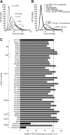
Protein S enhancement of the TFPI-mediated inhibition of thrombin generation in protein S–depleted plasma. Thrombin generation was measured in protein Sdepleted plasma supplemented with 50 µM phospholipids and 1 pM TF in the presence or absence of 0 to 2 nM (A) or 1 nM TFPI (B-C) and in the presence or absence of 50 nM WT PS (B-C). (A) A dose-dependent decrease in thrombin generation could be seen after the addition of increasing concentrations of TFPI. (B) Inhibitory antibodies against TFPI (a mix of 120 nM anti-TFPI Kunitz domain 1, 120 nM anti-TFPI Kunitz domain 2, and 120 nM anti-TFPI C-terminus) did not affect thrombin generation in protein S–depleted plasma, whereas they successfully repressed all inhibition of thrombin generation by TFPI in the presence or absence of protein S. In addition, inhibitory antibodies against protein S (2.8 µM; Dako) inhibited the enhancement of TFPI seen in the presence of protein S but had no effect on the absence of protein S. (C) The TFPI cofactor function of WT protein S and protein S variants was evaluated at 1 nM TFPI and 50 nM protein S. Pure WT protein S and WT protein S in concentrated conditioned media gave identical results. Protein S variants with amino acid substitutions in the Gla-TSR-EGF1-EGF2-EGF3-EGF4 domains (see supplemental Table 1 for full details of amino acid substitutions in the protein S variants used in this study) were screened in concentrated conditioned media. Protein S/Gas6 chimeras were purified before all experiments. All protein S variants were compared with WT protein S in concentrated conditioned media or were purified, as suitable. The relative protein S enhancement was determined as the decrease in peak height compared with WT protein S. The decrease in peak height in the presence of WT protein S compared with TFPI alone was set as 100% (n = 2). Results are expressed as mean ± standard deviation (SD). (A-B) Representative experiments are shown (n = 3). PS, protein S.
Figure 2
Figure 2
Enhancement of the TFPI-mediated inhibition of thrombin generation by WT protein S and protein S/Gas6 chimeras I to III. Thrombin generation was measured in protein S–depleted plasma supplemented with 50 μM phospholipids and 1 pM TF, in the presence or absence of 1 nM TFPI, and in the presence or absence of titrated (0-100 nM) WT protein S (A), chimera I (B), chimera II (C), or chimera III (D). Representative experiments are shown (n = 3). (E) Peak thrombin was plotted against protein S concentrations. The peak height in the presence of TFPI alone represents 100%. Results are expressed as mean ± SD (n = 3). PS, protein S.
Figure 3
Figure 3
Enhancement of TFPI in the inhibition of FXa by WT protein S and protein S/Gas6 chimeras I to III. FXa activity (0.5 nM) was followed in real time through cleavage of S-2765 (200 µM) at 405 mm in the presence of 25 μM phospholipids, the presence or absence of TFPI (2 nM), and in increasing concentrations (0-160 nM) of WT protein S (A), chimera I (B), chimera II (C), or chimera III (D). Results from representative experiments are shown (n = 3). (E) The initial velocity (V0) was calculated and plotted against protein S concentration. Results are given as mean ± SD and are expressed as percentage of the V0 in the presence of TFPI alone (n = 3). PS, protein S.
Figure 4
Figure 4
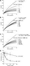
Enhancement of TFPI inhibition of FXa by protein S–, C4BP-, and C4BP-bound protein S. FXa activity (0.5 nM) was followed in real time through cleavage of S-2765 (200 µM) at 405 mm in the presence of 25 μM phospholipids, the presence or absence of TFPI (2 nM), and in increasing concentrations (0-160 nM) of protein S (A), 0 to 320 nM C4BP (B), and C4BP-bound protein S in a 2:1 ratio (C). Representative experiments are shown (n = 3). (D) V0 was calculated and plotted against protein S concentration. Results are given as mean ± SD and are expressed as percentage of the V0 in the presence of TFPI alone (n = 3). PS, protein S.
Figure 5
Figure 5
Binding of WT protein S and protein S/Gas6 chimeras I to III to TFPI studied with SPR. A CM5 chip was coupled with TFPI immobilized to 2500 response units. The flow cell was perfused with increasing concentrations (0-1000 nM) of WT protein S (A), protein S chimera I (B), protein S chimera II (C), or protein S chimera III (D). Results from a representative experiment are shown (n = 2). PS, protein S; RU, response units.
Figure 6
Figure 6
Proposed mechanism of the protein S/TFPI/FXa complex assembly on a phospholipid surface. Our results suggest that the mechanism of protein S cofactor enhancement of TFPI involves formation of a complex between the SHBG-like domain of protein S and the Kunitz domain 3 (K3) of TFPI (particularly Glu226). Protein S helps localize TFPI onto an activated membrane surface in a position favorable for the interaction of Kunitz domain 2 (K2) with the serine protease domain (SP) of FXa. The boxes show the respective models of the protein S SHBG-like domain and the TFPI Kunitz domain 3. TFPI Glu226 and the residues substituted in protein S/Gas6 chimera I are highlighted in black. The TFPI Kunitz domain 3 and the protein S SHGB -domain models are adapted from Mine et al and Villoutreix et al, respectively.,

Similar articles

Cited by

References

    1. Wun TC, Kretzmer KK, Girard TJ, Miletich JP, Broze GJ., Jr Cloning and characterization of a cDNA coding for the lipoprotein-associated coagulation inhibitor shows that it consists of three tandem Kunitz-type inhibitory domains. J Biol Chem. 1988;263(13):6001–6004. - PubMed
    1. Broze GJ, Jr, Girard TJ. Tissue factor pathway inhibitor: structure-function. Front Biosci (Landmark Ed) 2012;17:262–280. - PMC - PubMed
    1. Dahm A, Van Hylckama Vlieg A, Bendz B, Rosendaal F, Bertina RM, Sandset PM. Low levels of tissue factor pathway inhibitor (TFPI) increase the risk of venous thrombosis. Blood. 2003;101(11):4387–4392. - PubMed
    1. Broze GJ, Jr, Lange GW, Duffin KL, MacPhail L. Heterogeneity of plasma tissue factor pathway inhibitor. Blood Coagul Fibrinolysis. 1994;5(4):551–559. - PubMed
    1. Nordfang O, Bjørn SE, Valentin S, et al. The C-terminus of tissue factor pathway inhibitor is essential to its anticoagulant activity. Biochemistry. 1991;30(43):10371–10376. - PubMed

Publication types