. 2014 Sep 1;23(17):4621-38.
doi: 10.1093/hmg/ddu178. Epub 2014 Apr 15.

Parkinson's disease-linked mutations in VPS35 induce dopaminergic neurodegeneration

Affiliations

Parkinson's disease-linked mutations in VPS35 induce dopaminergic neurodegeneration

Elpida Tsika et al. Hum Mol Genet. .

Abstract

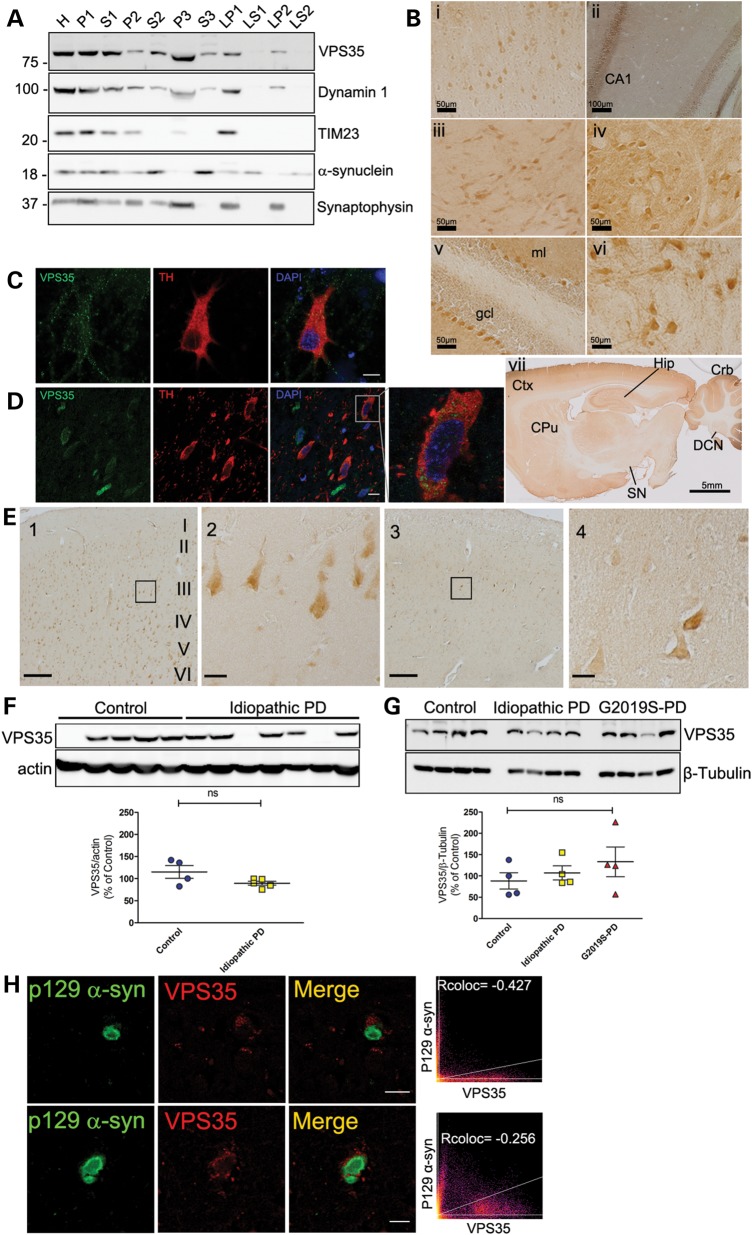
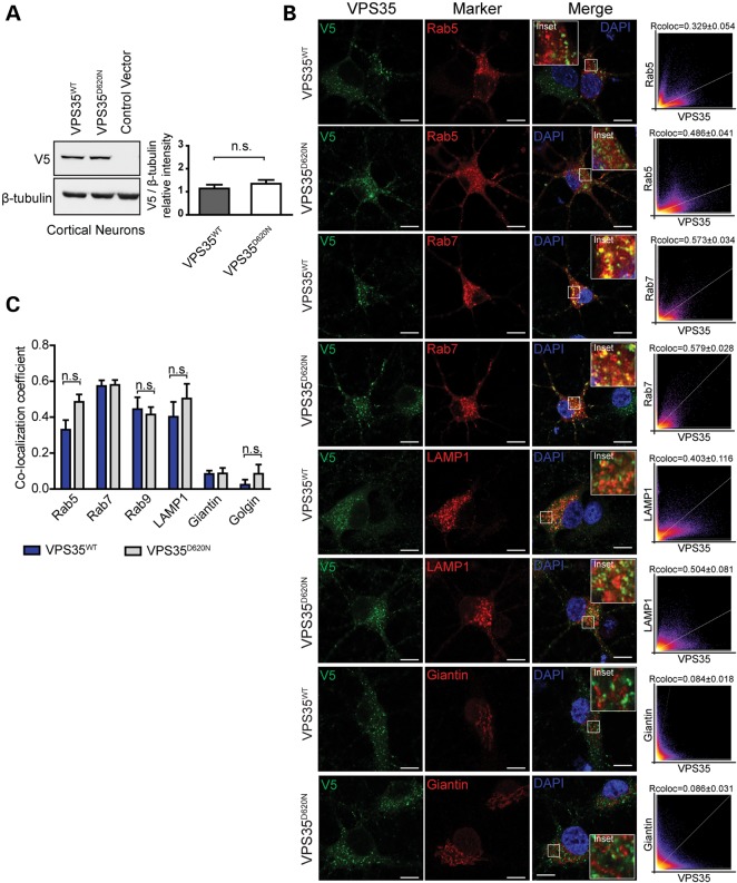
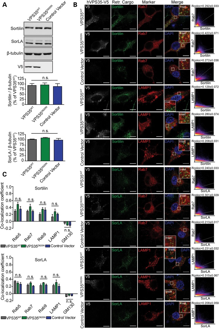
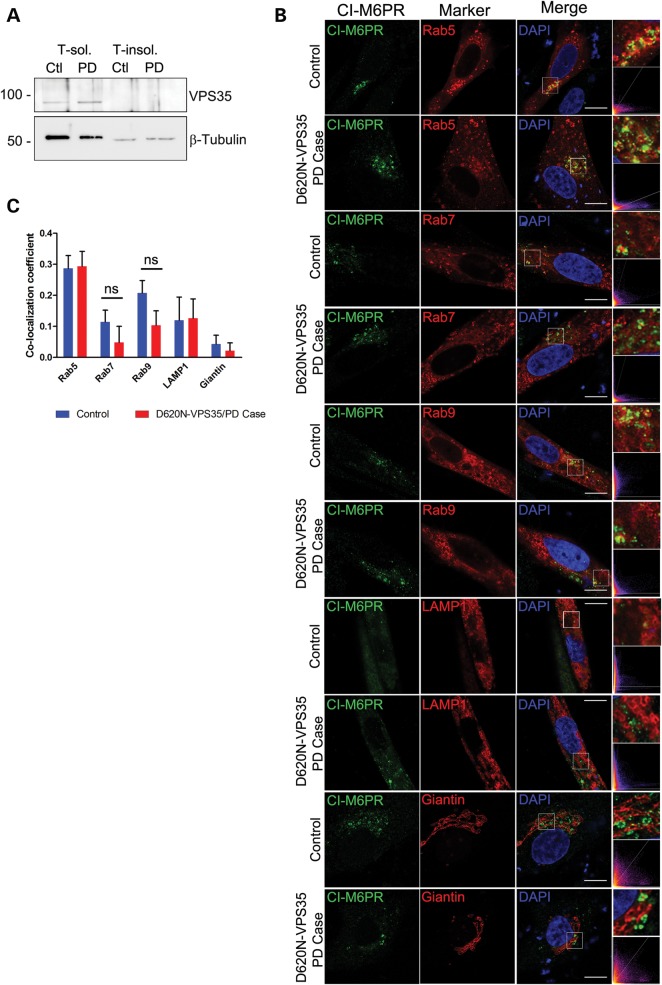
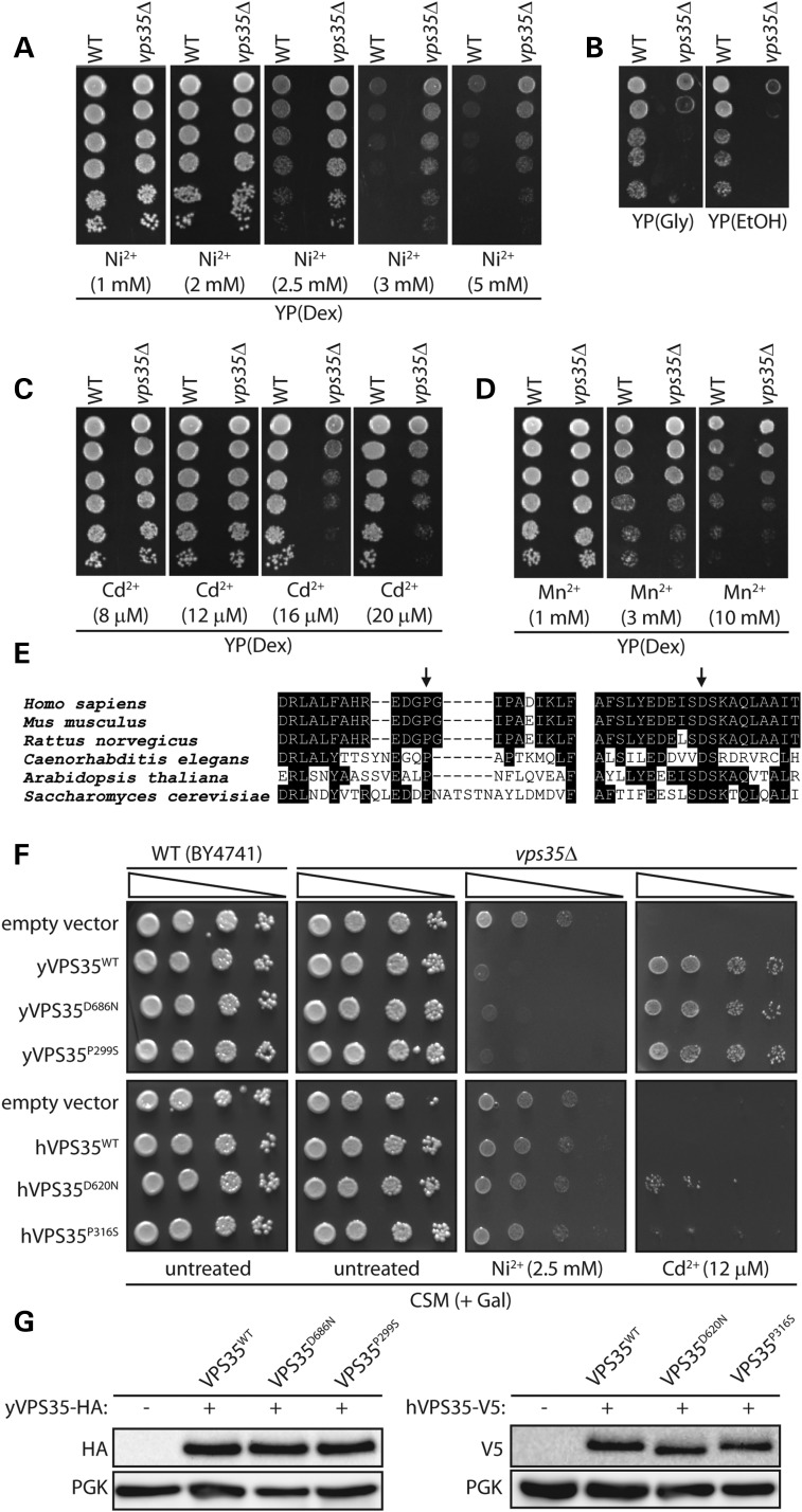
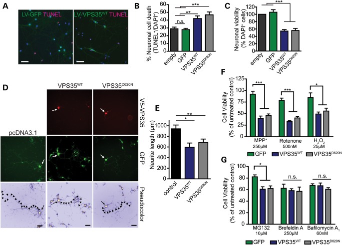
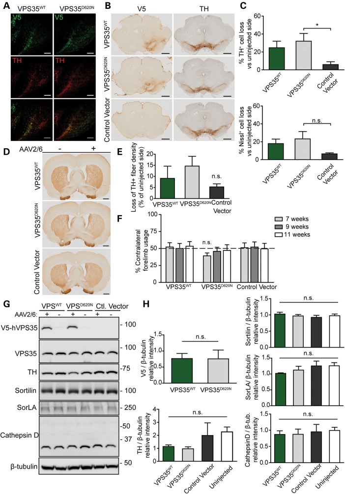
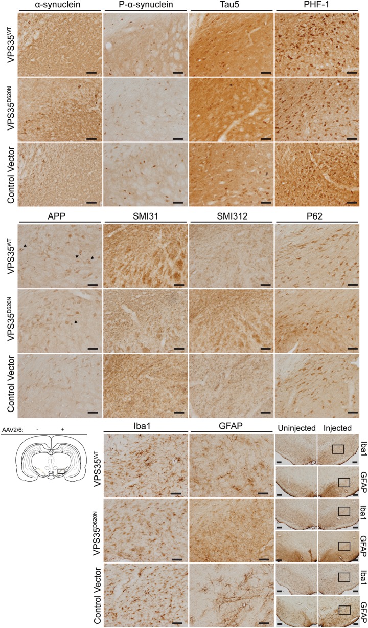
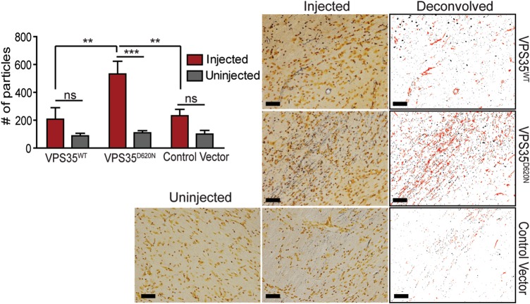
Mutations in the vacuolar protein sorting 35 homolog (VPS35) gene at the PARK17 locus, encoding a key component of the retromer complex, were recently identified as a new cause of late-onset, autosomal dominant Parkinson's disease (PD). Here we explore the pathogenic consequences of PD-associated mutations in VPS35 using a number of model systems. VPS35 exhibits a broad neuronal distribution throughout the rodent brain, including within the nigrostriatal dopaminergic pathway. In the human brain, VPS35 protein levels and distribution are similar in tissues from control and PD subjects, and VPS35 is not associated with Lewy body pathology. The common D620N missense mutation in VPS35 does not compromise its protein stability or localization to endosomal and lysosomal vesicles, or the vesicular sorting of the retromer cargo, sortilin, SorLA and cation-independent mannose 6-phosphate receptor, in rodent primary neurons or patient-derived human fibroblasts. In yeast we show that PD-linked VPS35 mutations are functional and can normally complement VPS35 null phenotypes suggesting that they do not result in a loss-of-function. In rat primary cortical cultures the overexpression of human VPS35 induces neuronal cell death and increases neuronal vulnerability to PD-relevant cellular stress. In a novel viral-mediated gene transfer rat model, the expression of D620N VPS35 induces the marked degeneration of substantia nigra dopaminergic neurons and axonal pathology, a cardinal pathological hallmark of PD. Collectively, these studies establish that dominant VPS35 mutations lead to neurodegeneration in PD consistent with a gain-of-function mechanism, and support a key role for VPS35 in the development of PD.


Figures

Figure 1.
Figure 1.
Cellular distribution and levels of endogenous VPS35 in normal and pathological mammalian brain. (A) Subcellular fractionation of endogenous VPS35 in mouse cerebral cortex. VPS35 is enriched in the microsomal (P3), synaptosomal (LP1) and synaptic vesicle (LP2) membrane fractions. Dynamin 1, TIM23, α-synuclein and synaptophysin serve as markers for microsomes, mitochondria, synaptic vesicle cytosolic and synaptosomal/synaptic vesicle membranes, respectively. Molecular mass is indicated in kDa. (B) Immunolabeling of endogenous VPS35 in the rat brain. VPS35 is detected in (i) pyramidal neurons of cortical layer III, (ii) pyramidal neurons of the hippocampus (CA1 region), (iii) ventral midbrain, (iv) brainstem (superior olivary complex), (v) Purkinje neurons in the cerebellum (granule cell layer, gcl; molecular layer, ml), (vi) deep cerebellar nuclei, and (vii) a sagittal section of rat brain (cerebral cortex, Ctx; hippocampal formation, Hip; cerebellum, Crb; deep cerebellar nuclei, DCN; caudate putamen, CPu; substantia nigra, SN. (C) Confocal microscopy analysis of rat primary midbrain cultures immunolabeled with VPS35 and the dopaminergic marker, tyrosine hydroxylase (TH). Nuclei are labeled with DAPI. VPS35 localizes to punctate intracellular vesicular structures within the soma and neuritic processes of TH-positive dopaminergic neurons. Scale bar: 10 μm. (D) Co-localization of endogenous VPS35 with TH-positive dopaminergic neurons in the substantia nigra pars compacta of adult rats. Scale bar: 10 μm. (E) Immunolabeling of endogenous VPS35 in the human cingulate cortex of control (1) and PD/DLB (3) subjects. Scale bar: 200 μm. High-magnification images of pyramidal neurons from cortical layer III are shown corresponding to the boxed area from control (2) and PD/DLB (4) brains. Scale bar: 50 μm. (F–G) Western blot analysis of soluble extracts from F human caudate putamen of control and idiopathic PD/DLB subjects, and G human frontal cortex of control, idiopathic PD (PD) or G2019S LRRK2-linked PD subjects, with antibodies to VPS35, and actin or β-tubulin as protein loading controls. Densitometric analysis of VPS35 normalized to actin or β-tubulin levels for individual subjects are shown expressed as a percent of the mean of control subjects. Horizontal bars represent mean ± SEM (n = 4–5 subjects/group) for each subject group. Subjects without VPS35 expression in F were excluded from the analysis (control = 4 from 5; PD = 5 from 7). ns, non-significant by one-way analysis of variance (ANOVA) with Dunnett's post hoc test. (H) Confocal microscopic analysis of VPS35 co-localization with Lewy bodies labeled with phospho-Ser129-α-synuclein in cortical layer III neurons from a PD/DLB subject. Correlation coefficients (Rcoloc) and cytofluorograms indicate a lack of co-localization of VPS35 with Lewy bodies. Scale bars: 20 μm (top panels) or 5 μm (bottom panels).
Figure 2.
Figure 2.
Normal steady-state levels and vesicular localization of VPS35D620N in cortical neurons. (A) Western blot analysis of soluble extracts from primary cortical neurons infected with lentiviral vectors expressing V5-tagged human VPS35 (WT or D620N) or control virus with anti-V5 or β-tubulin antibodies. Densitometric analysis of human VPS35 normalized to β-tubulin levels indicates the equivalent expression of WT and D620N variants (mean ± SEM, n = 4 experiments). n.s., non-significant by unpaired, two-tailed Student's t-test. (B) Representative confocal microscopic images of primary cortical neurons co-labeled for WT or D620N human VPS35 (V5) and RFP-Rab5, GFP-Rab7, RFP-LAMP1 or trans-Golgi protein Giantin, and DAPI. Inset indicates enlarged boxed area in merged images. Cytofluorograms and correlation coefficients (Rcoloc, mean ± SEM, n ≥ 5 neurons) indicate the degree of co-localization of fluorescence signals for V5 and each marker. Scale bars: 10 μm. (C) Graph showing co-localization coefficients (mean ± SEM, n ≥ 5 neurons/group) of WT or D620N VPS35 with each vesicular marker in cortical neurons. n.s., non-significant by unpaired, two-tailed Student's t-test as indicated.
Figure 3.
Figure 3.
Protein levels and vesicular sorting of the retromer cargo sortilin and SorLA are not altered by VPS35D620N expression in primary cortical neurons. (A) Western blot analysis of soluble extracts from primary cortical neurons infected with lentiviral vectors expressing V5-tagged human VPS35 (WT or D620N) or a control virus, with antibodies to sortilin, SorLA and V5 or β-tubulin as a protein loading control. Graphs indicate densitometric analysis of sortilin or SorLA normalized to β-tubulin levels and expressed as percent of the VPS35WT condition (mean ± SEM, n = 4 experiments). (B) Representative confocal microscopic images of primary cortical neurons co-labeled for human VPS35 (V5), sortilin or SorLA and each vesicular marker (GFP-Rab7 or RFP-LAMP1), and DAPI. Inset indicates enlarged boxed area in merged images. Cytofluorograms and correlation coefficients (Rcoloc, mean ± SEM, n ≥ 7 neurons) indicate the degree of co-localization of fluorescence signals for sortilin or SorLA and each vesicular marker. Scale bars: 10 μm. (C) Graph showing co-localization coefficients (mean ± SEM, n ≥ 7 neurons/group) of sortilin (upper) or SorLA (lower) with each vesicular marker (RFP-Rab5, GFP-Rab7, GFP-Rab9, RFP-LAMP1 or Golgi protein GM130) in cortical neurons expressing human VPS35 (WT or D620N) or a control vector. n.s., non-significant by one-way ANOVA with Newman–Keuls post hoc analysis.
Figure 4.
Figure 4.
Normal VPS35 Levels and Vesicular Sorting of the Retromer Cargo CI-M6PR in Primary Human Fibroblasts Derived from a D620N Mutant PD Subject. (A) Western blot analysis of 1% Triton-soluble (T-sol.) and Triton-insoluble (T-insol.) fractions of primary fibroblasts derived from a Parkinson's disease subject harboring the D620N VPS35 mutation (PD) and a healthy control (Ctl). Blots are probed with antibodies to VPS35 and β-tubulin as a protein loading control. Molecular mass is indicated in kDa. (B) Representative confocal microscopic images and cytofluorograms of human primary fibroblasts (control or D620N) co-labeled for cation-independent mannose-6-phosphate receptor (CI-M6PR) and vesicular markers (RFP-Rab5, GFP-Rab7, GFP-Rab9, RFP-LAMP1 or trans-Golgi protein Giantin). Enlarged boxed areas of merged images are shown. Scale bar: 10 μm. (C) Graph indicates the co-localization coefficients (mean ± SEM, n ≥ 5 cells) of CI-M6PR with each vesicular marker in primary fibroblasts from control or D620N PD subjects. n.s., non-significant by unpaired, two-tailed Student's t-test, as indicated.
Figure 5.
Figure 5.
Analogous PD-like mutations in yeast VPS35 can functionally complement growth phenotypes in VPS35 null yeast cells. (A–D) Haploid yeast cells (BY4741, MATa), either wild-type (WT) or with a deletion of VPS35 (vps35Δ), were spotted onto YP(Dex) rich media containing different concentrations of (A) nickel (Ni2+), (C) cadmium (Cd2+) or (D) manganese (Mn2+), or YP media containing (B) non-fermentable carbon sources, glycerol (Gly) or ethanol (EtOH), and grown for 2–3 days at 30°C. Shown are 5-fold serial dilutions (from top to bottom) starting with equal numbers of cells. (E) Protein sequence alignment of VPS35 in the region encompassing the human P316 and D620 residues across several model species, indicating the high conservation of these two residues. (F) WT or vps35Δ yeast strains were transformed with galactose-inducible low-copy expression vectors containing HA-tagged yeast VPS35 (WT, D686N or P299S) or V5-tagged human VPS35 (WT, D620N or P316S) variants, or with an empty vector (p416GAL1). Equivalent numbers of cells were spotted as five-fold serial dilutions (from left to right) onto complete synthetic media lacking uracil (CSM) and containing galactose as the sole carbon source with or without additional Ni2+ (2.5 mM) or Cd2+ (12 µM), as indicated. (G) Western blot analysis of soluble extracts from WT yeast cells transformed with yeast or human VPS35 variants following growth on CSM-URA media containing galactose to induce VPS35 expression. VPS35 is detected using anti-HA or anti-V5 antibodies with an antibody to 3-phosphoglycerate kinase (PGK) used as a protein loading control.
Figure 6.
Figure 6.
Overexpression of human VPS35 induces neuronal cell death, impairs neurite outgrowth and increases neuronal vulnerability to cellular stress. (A) Representative immunofluorescent images of primary cortical neurons at DIV 14 infected with GFP or VPS35WT lentivirus (green) and co-labeling of apoptotic (TUNEL-positive, red) or total (DAPI-positive, cyan) nuclei to assess neuronal cell death. Scale bar: 50 μm. (B) Cortical neurons infected with lentiviral vectors expressing GFP or human VPS35 variants were assessed for apoptotic cell death by quantifying the number of TUNEL-positive neurons as a percent of total neurons (DAPI-positive nuclei) for each condition (mean ± SEM, n = 5 independent cultures). (C) Neuronal viability was assessed in cortical cultures infected with lentiviral vectors at DIV 14 by counting the average number of neurons (DAPI-positive nuclei) for each condition, and expressed as a percent of an empty control virus (mean ± SEM, n = 5 independent cultures). (D) Representative fluorescent microscopic images of cortical neurons at DIV 7 co-labeled with V5-tagged human VPS35 (WT or D620N) and GFP. GFP images were pseudocolored with ICA to identify neuronal soma (arrowhead) and axonal processes (arrows) for neurite length measurements. Scale bar: 200 μm. (E) Quantitation of axonal process length from GFP-positive neurons expressing human VPS35 (WT or D620N) compared with control neurons (empty vector). Bars represent neurite length (mean ± SEM, n = 5 independent cultures) from 170–189 neurons. (F) Primary cortical neurons infected with lentiviral vectors expressing human VPS35 (WT or D620N) or GFP were treated at DIV 14 with cellular toxins (MPP+, rotenone or H2O2) for 24 h and assessed by alamarBlue cell viability assay. Bars represent cell viability expressed as a percent of untreated cultures for each condition (mean ± SEM, n ≥ 3 independent cultures). (G) Cortical neurons infected with lentiviral vectors as in F were treated with cellular toxins (MG132, Brefeldin A or Bafilomycin A1) for 24 h prior to cell viability assays. Cell viability relative to untreated cultures is shown (mean ± SEM, n ≥ 3 independent cultures). Data were analyzed by one-way ANOVA with Newman–Keuls post hoc analysis, as indicated (*P<0.05, **P<0.01 or ***P<0.001). n.s., non-significant.
Figure 7.
Figure 7.
Dopaminergic neuronal degeneration induced by AAV2/6-mediated expression of D620N VPS35 in the substantia nigra of adult rats. (A) Photomicrographs showing immunofluorescent co-labeling of substantia nigra with anti-V5 and anti-TH antibodies indicating the equivalent expression of V5-tagged human VPS35WT and VPS35D620N proteins within dopaminergic neurons at 12 weeks following stereotactic injection of AAV2/6 vectors. Scale bar: 200 μm. (B) Representative photomicrographs of anti-V5 and anti-TH immunohistochemical analysis in adjacent sections of rat substantia nigra at 12 weeks following the stereotactic injection of AAV2/6 vectors expressing human VPS35WT and VPS35D620N or a control vector. V5 labeling in the substantia nigra is accompanied by a decrease in TH immunostaining. Scale bar: 1 mm. (C) Stereological analysis of TH-positive dopaminergic or total Nissl-positive neurons in the substantia nigra expressed as percent cell loss relative to the contralateral uninjected nigra. Bars represent mean ± SEM (n = 6 animals/group). *P<0.05 by one-way ANOVA with Dunnett's post hoc analysis. n.s., non-significant. (D) Representative photomicrographs of immunostaining for TH-positive nerve terminal in the striatum at 12 weeks following AAV2/6 delivery. Scale bar: 1 mm. (E) Quantitation of optical density of TH immunostaining in the striatum. Data are expressed as percent loss of TH-positive fibers relative to the contralateral uninjected side. Bars represent the mean ± SEM (n ≥ 4 animals/group). Data were assessed by one-way ANOVA with Dunnett's post hoc analysis. (F) Cylinder asymmetry test performed at 7, 9 and 11 weeks postsurgery assessing forelimb contacts with the cylinder wall over a 5 min period. Forelimb use contralateral to the injected nigra is expressed as a percent of total forelimb contacts (left + right) on the cylinder wall. Dashed line indicates the expected forelimb usage under normal conditions. Bars represent mean ± SEM (n = 8 animals/group). Data were assessed by one-way ANOVA with Newman–Keuls post hoc analysis. (G) Western blot analysis of soluble extracts from ventral midbrain at 12 weeks following stereotactic injection of rats with AAV2/6 vectors, separated into ipsilateral (+) and contralateral (−) hemispheres. Blots were probed with antibodies to V5, VPS35, TH, sortilin, SorLA and cathepsin D, with β-tubulin as a loading control. Molecular masses are indicated in kDa. (H) Densitometric analysis of V5, TH, sortilin, SorLA or cathepsin D levels normalized to β-tubulin levels on blots containing ventral midbrain extracts. Bars represent mean ± SEM (n = 3–4 animals/group). Data were analyzed by one-way ANOVA with Newman–Keuls post hoc analysis for multiple comparisons, or by unpaired, two-tailed Student's t-test for pair-wise comparisons, as indicated. n.s., non-significant.
Figure 8.
Figure 8.
AAV2/6-mediated expression of human VPS35 in the rat substantia nigra induces axonal pathology. Immunohistochemical analysis of substantia nigra at 12 weeks following the stereotactic injection of AAV2/6 vectors expressing human VPS35WT and VPS35D620N or a control vector. Antibodies detecting total α-synuclein, α-synuclein phosphorylated at Ser129 (P-α-synuclein), total tau (Tau5), tau phosphorylated at Ser396 and Ser404 (PHF-1), APP isoforms, phosphorylated neurofilament H (SMI31), phosphorylated neurofilament M/H (SMI312), autophagy substrate p62/sequestosome 1, GFAP and Iba1 were employed. Representative photomicrographs are taken from the injected substantia nigra as indicated in the schematic diagram. Scale bar: 50 μm. Macroscopic images of GFAP and Iba1 in the injected and non-injected substantia nigra are shown for comparison. Boxes indicate the region used for high-power images of the injected substantia nigra. Scale bar: 50 μm.
Figure 9.
Figure 9.
Nigral axonal degeneration induced by AAV2/6-mediated expression of D620N VPS35. Representative photomicrographs of substantia nigra sections processed with Gallyas silver stain, from rats injected with AAV2/6 vectors expressing human VPS35WT and VPS35D620N or a control vector, to reveal degenerating axons and nerve terminals (indicated in black). Scale bar: 50 μm. Images were subjected to deconvolution, color separation and filtering (circularity index ≤0.6 to remove nuclei/soma) using ImageJ to isolate black particles representative of neuritic processes and terminals for quantitation. Examples of particles analyzed are shown pseudocolored in red. Data are expressed as the number of black particles (silver-positive neurites/terminals) and bars represent the mean ± SEM (n = 6 animals/group). **P<0.01 or ***P<0.001 by one-way ANOVA with Tukey's post hoc analysis as indicated. n.s., non-significant.

References

    1. Lang A.E., Lozano A.M. Parkinson’s disease—first of two parts. N. Engl. J. Med. 1998;339:1044–1053. - PubMed
    1. Lang A.E., Lozano A.M. Parkinson’s disease. Second of two parts. N. Engl. J. Med. 1998;339:1130–1143. - PubMed
    1. Spillantini M.G., Schmidt M.L., Lee V.M., Trojanowski J.Q., Jakes R., Goedert M. Alpha-synuclein in Lewy bodies. Nature. 1997;388:839–840. - PubMed
    1. Gasser T. Mendelian forms of Parkinson’s disease. Biochim. Biophys. Acta. 2009;1792:587–596. - PubMed
    1. Moore D.J., West A.B., Dawson V.L., Dawson T.M. Molecular pathophysiology of Parkinson’s disease. Annu. Rev. Neurosci. 2005;28:57–87. - PubMed

Publication types

MeSH terms

Substances