. 2014 Sep 1;23(17):4612-20.
doi: 10.1093/hmg/ddu176. Epub 2014 Apr 15.

Dissecting the mechanisms underlying the accumulation of mitochondrial DNA deletions in human skeletal muscle

Affiliations

Dissecting the mechanisms underlying the accumulation of mitochondrial DNA deletions in human skeletal muscle

Georgia Campbell et al. Hum Mol Genet. .

Abstract

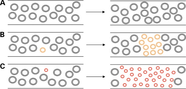
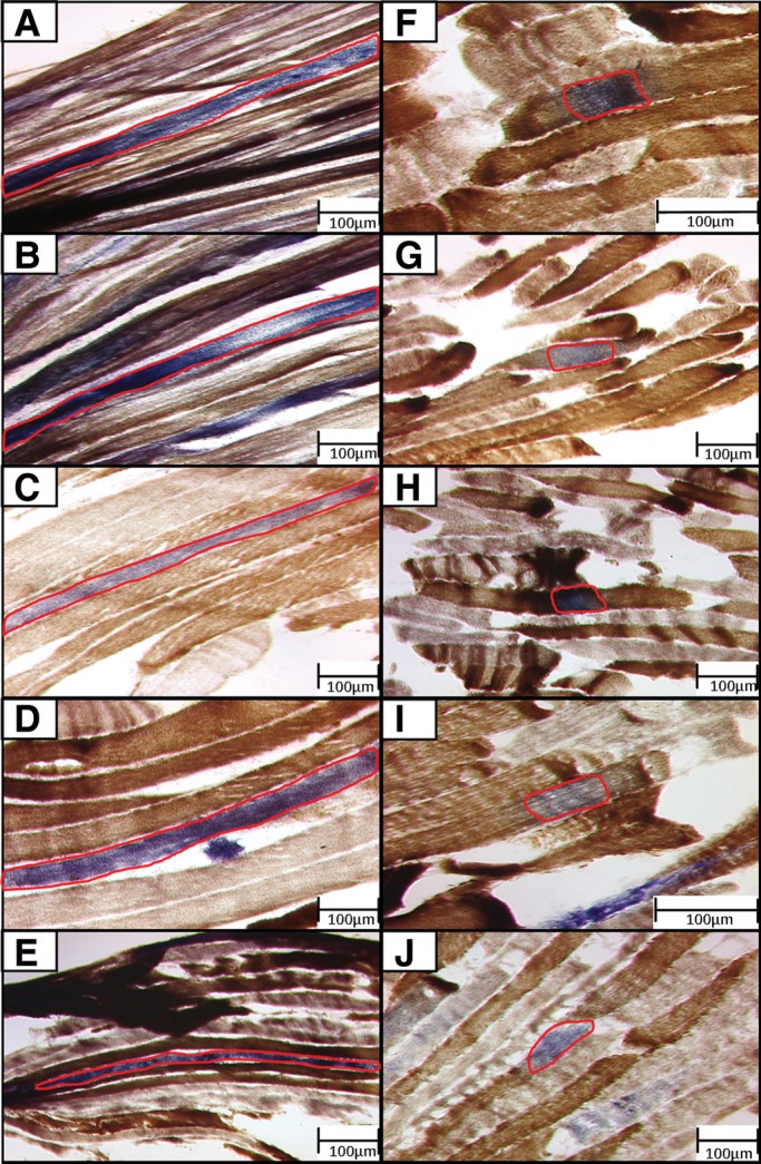
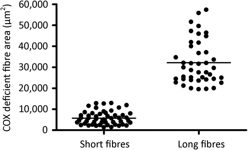
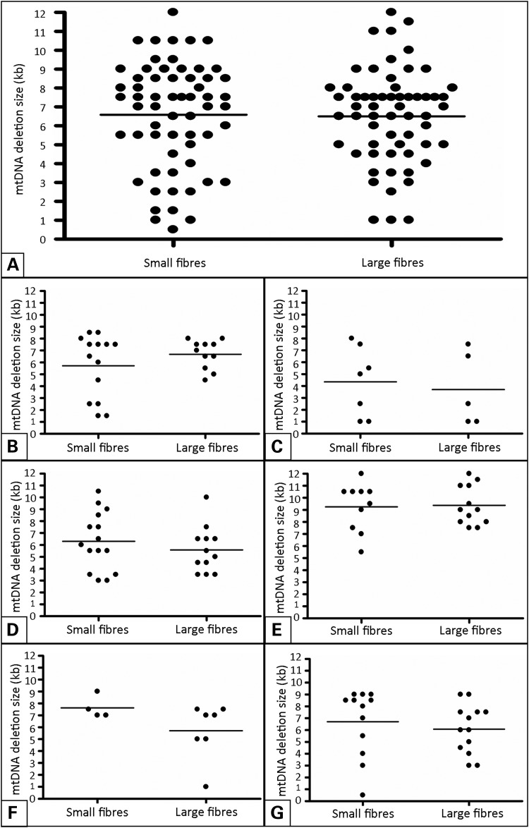
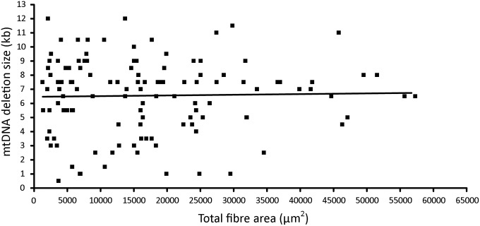
Large-scale mitochondrial DNA (mtDNA) deletions are an important cause of mitochondrial disease, while somatic mtDNA deletions cause focal respiratory chain deficiency associated with ageing and neurodegenerative disorders. As mtDNA deletions only cause cellular pathology at high levels of mtDNA heteroplasmy, an mtDNA deletion must accumulate to levels which can result in biochemical dysfunction-a process known as clonal expansion. A number of hypotheses have been proposed for clonal expansion of mtDNA deletions, including a replicative advantage for deleted mitochondrial genomes inferred by their smaller size--implying that the largest mtDNA deletions would also display a replicative advantage over smaller mtDNA deletions. We proposed that in muscle fibres from patients with mtDNA maintenance disorders, which lead to the accumulation of multiple mtDNA deletions, we would observe the largest mtDNA deletions spreading the furthest longitudinally through individual muscle fibres by means of a greater rate of clonal expansion. We characterized mtDNA deletions in patients with mtDNA maintenance disorders from a range of 'large' and 'small' cytochrome c oxidase (COX)-deficient regions in skeletal muscle fibres. We measured the size of clonally expanded deletions in 62 small and 60 large individual COX-deficient f regions. No significant difference was observed in individual patients or in the total dataset (small fibre regions mean 6.59 kb--large fibre regions mean 6.51 kb). Thus no difference existed in the rate of clonal expansion throughout muscle fibres between mtDNA deletions of different sizes; smaller mitochondrial genomes therefore do not appear to have an inherent replicative advantage in human muscle.


Figures

Figure 1.
Figure 1.
Proposed mechanism for accumulation of smaller mtDNA molecules longitudinally through muscle fibres. Illustration of increased longitudinal accumulation of larger mtDNA deletions through muscle fibres, as predicted by the ‘survival of the smallest’ hypothesis (9). (A) In the presence of no mtDNA deletions, wild-type mtDNA copy number is maintained throughout a muscle fibre over time. (B) Following mtDNA deletion formation a replicative advantage over wild-type mtDNA, due to the smaller size of the deleted molecule, causes an increase in the mtDNA deletion level and a spread of the deleted species through the muscle fibre. (C) The larger the size of the mtDNA deletion, the smaller the remaining genome and the greater the replicative advantage over wild-type mtDNA. A larger mtDNA deletion size therefore leads to a faster accumulation of the deleted species over the same time span and a greater spread through a muscle fibre .
Figure 2.
Figure 2.
Images of ‘large’ and ‘small’ COX-deficient fibre regions selected for extraction by laser microdissection. Panel of 10 images of longitudinal muscle sections examined using COX/SDH histochemistry, captured prior to laser microdissection—these are representative of typical cutting areas. (A–E) Depict five large COX-deficient fibre regions, where the outlined region of COX-deficiency exceeds 500 µm in length. (FJ) Depict five small COX-deficient fibre regions, where the outlined region of COX-deficiency remains under 200 µm in length. All LMD cutting areas for single fibre isolation are outlined in red.
Figure 3.
Figure 3.
Assessment of COX-deficient fibres captured by laser microdissection. Scatter plot of all captured COX-deficient fibre areas (µm2), as measured by the LMD system. Captured fibres were grouped into two types; short (under 200 µm in length) and large (over 500 µm in length). Captured area was assessed to ensure no overlap existed in terms of size between these two groups. Small COX-deficient fibres: mean COX-deficient fibre area = 5725 ± 3194 µm2. Large COX-deficient fibres: mean COX-deficient fibre area = 32 120 ± 3194 µm2. The two groups of COX-deficient muscle fibre areas were found to be significantly different using a Mann–Whitney test (P < 0.0001).
Figure 4.
Figure 4.
mtDNA deletion sizes present in (A) the total dataset, and (B–G) datasets from each patient. Assessment of mtDNA deletion sizes present in large and small COX-deficient fibres, depicted by scatter plot and carried out by Mann–Whitney U test (non-Gaussian data distribution) or unpaired t-test (normally distributed data). (A) Small COX-deficient fibres, mean mtDNA deletion size = 6589 bp (±2788 bp, N = 62); large COX-deficient fibres, mean mtDNA deletion size = 6508 bp (±2478 bp, N = 60). No significant difference was found to exist between the two fibre groups (P = 0.5506), with a similar range of mtDNA deletion sizes displayed in both. Similar results are seen for each of the six patient datasets (B–G), with no significant difference in deletion size between the two fibre groups in any single case (P = 0.7009, 0.7751, 0.4136, 0.8272, 2303 and 0.3406, respectively).
Figure 5.
Figure 5.
Correlation between mtDNA deletion size and COX-deficient fibre area. Spearman's correlation analysis carried out on the total data set of 122 COX-deficient skeletal muscle fibre regions, to determine if any relationship exists between mtDNA deletion size and the size of the resulting area of biochemical deficiency. Relationship between these variables depicted here by scatter plot and linear regression line. No significant correlation was found to exist between these two parameters (P = 0.7792).

References

    1. Schaefer A.M., McFarland R., Blakely E.L., He L., Whittaker R.G., Taylor R.W., Chinnery P.F., Turnbull D.M. Prevalence of mitochondrial DNA disease in adults. Ann. Neurol. 2008;63:35–39. - PubMed
    1. Reeve A.K., Krishnan K.J., Turnbull D. In: Mitochondria and Oxidative Stress in Neurodegenerative Disorders. Gibson G.E., Ratan R.R., Beal M.F., editors. Vol. 1147. Oxford: Blackwell Publishing; 2008. pp. 21–29.
    1. Larsson N. Somatic mitochondrial DNA mutations in mammalian aging. Annu. Rev. Biochem. 2010;79:683–706. - PubMed
    1. Kraytsberg Y., Kudryavtseva E., McKee A.C., Geula C., Kowall N.W., Khrapko K. Mitochondrial DNA deletions are abundant and cause functional impairment in aged human substantia nigra neurons. Nat. Genet. 2006;38:518–520. - PubMed
    1. Bender A., Krishnan K.J., Morris C.M., Taylor G.A., Reeve A.K., Perry R.H., Jaros E., Hersheson J.S., Betts J., Klopstock T., et al. High levels of mitochondrial DNA deletions in substantia nigra neurons in aging and Parkinson disease. Nat. Genet. 2006;38:515–517. - PubMed

Publication types

Substances