. 2014 Jul;52(7):657-70.
doi: 10.1002/dvg.22781. Epub 2014 May 2.

Epidermal growth factor-like domain 7 is a marker of the endothelial lineage and active angiogenesis

Affiliations

Epidermal growth factor-like domain 7 is a marker of the endothelial lineage and active angiogenesis

Kathryn Bambino et al. Genesis. 2014 Jul.

Abstract

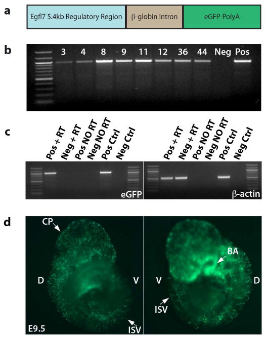
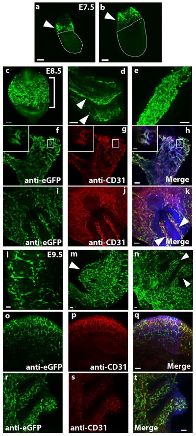
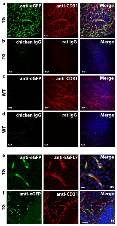
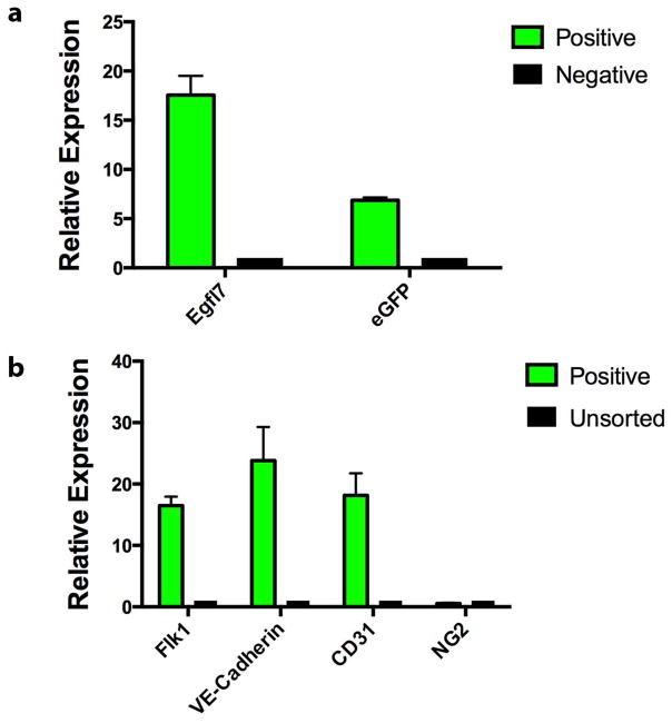
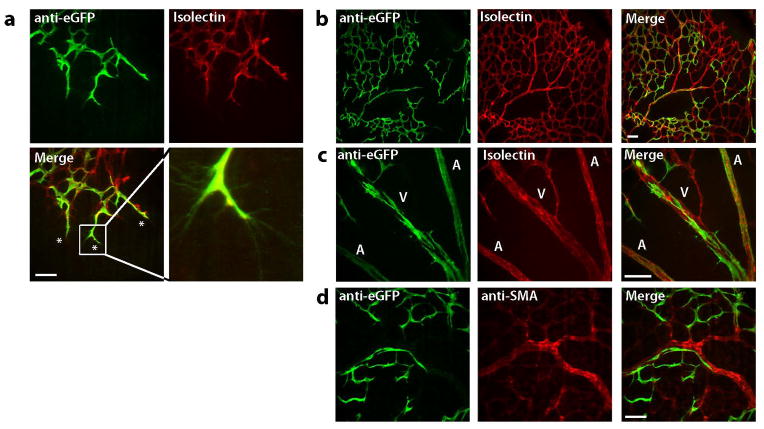
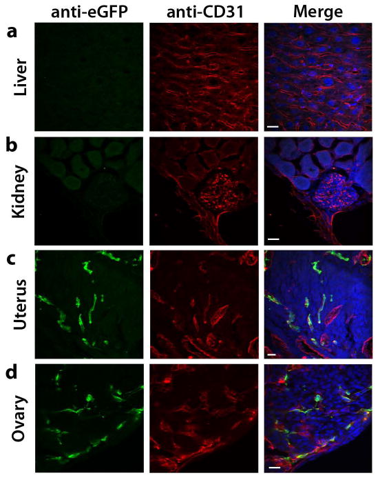
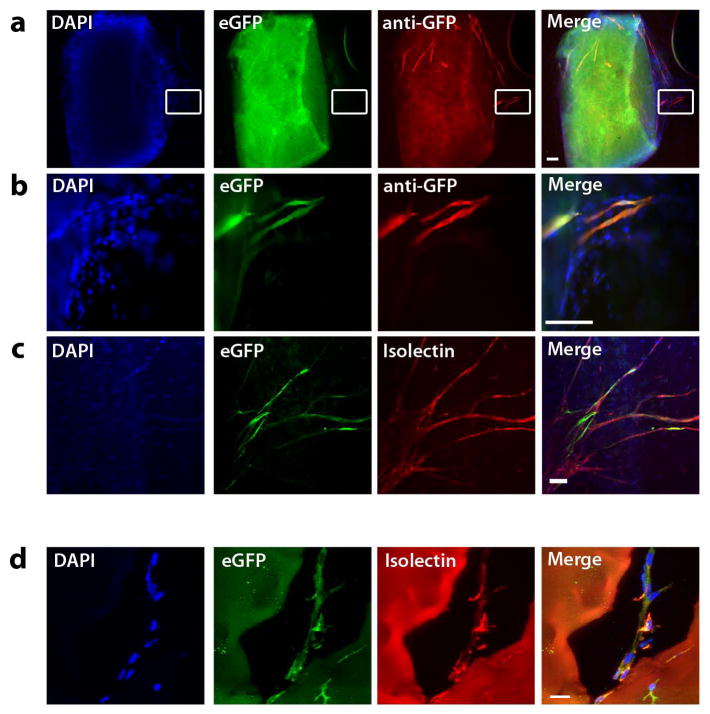
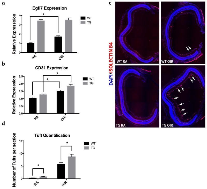
Epidermal growth factor-like domain 7 (Egfl7) expression in the developing embryo is largely restricted to sites of mesodermal progenitors of angioblasts/hemangioblasts and the vascular endothelium. We hypothesize that Egfl7 marks the endothelial lineage during embryonic development, and can be used to define the emergence of endothelial progenitor cells, as well as to visualize newly-forming vasculature in the embryo and during the processes of physiologic and pathologic angiogenesis in the adult. We have generated a transgenic mouse strain that expresses enhanced green fluorescent protein (eGFP) under the control of a minimal Egfl7 regulatory sequence (Egfl7:eGFP). Expression of the transgene recapitulated that of endogenous Egfl7 at sites of vasculogenesis and angiogenesis in the allantois, yolk sac, and in the embryo proper. The transgene was not expressed in the quiescent endothelium of most adult organs. However, the uterus and ovary, which undergo vascular growth and remodeling throughout the estrus cycle, expressed high levels of Egfl7:eGFP. Importantly, expression of the Egfl7:eGFP transgene was induced in adult neovasculature. We also found that increased Egfl7 expression contributed to pathologic revascularization in the mouse retina. To our knowledge, this is the first mouse model that enables monitoring of endothelial cells at sites of active vasculogenesis and angiogenesis. This model also facilitated the isolation and characterization of EGFL7(+) endothelial cell populations by fluorescence activated cell sorting (FACS). Together, our results demonstrate that the Egfl7:eGFP reporter mouse is a valuable tool that can be used to elucidate the mechanisms by which blood vessels form during development and under pathologic circumstances.

Keywords: eGFP; endothelial reporter; transgenic mice; vascular development.


Conflict of interest statement

Disclosure of Conflicts of Interest

The authors declare no competing financial interests.

Figures

Figure 1
Figure 1. Generation of Egfl7:eGFP transgenic mice
a) Schematic of the Egfl7:eGFP transgene. 5.4kb upstream of exon 1b, containing two ETS response elements, was cloned upstream of the β-globin intron, eGFP cDNA, and poly-adenylation sequence. b) Genotyping of potential founders. Genomic DNA was isolated from tail snips of potential founder pups, and genotyped by PCR for the presence of the eGFP sequence. Eight potential founders were identified. c) Transcript expression in E9.5 line 12 embryos by semi-quantitative RT-PCR. Only those embryos that expressed eGFP by live fluorescence expressed the eGFP transcript by semi-quantitative RT-RCR. d) Live expression of eGFP in a representative transgenic E9.5 embryo. D = dorsal, V = ventral, CP = choroid plexus, ISV = intersomitic vessel(s), BA = branchial arches.
Figure 2
Figure 2. Egfl7:eGFP is expressed at sites of mesodermal progenitors of endothelial cells and the developing vasculature
Whole mount staining of Egfl7:eGFP transgenic embryos. Three litters were analyzed at each time point and for each staining condition. All images are compressed z-stacks unless otherwise noted. a–e) Anti-eGFP staining. (a–b) E7.5 embryos. Arrowheads point to extraembryonic mesoderm (EEM), (scale bars = 100 μm). (c–e) E8.5 embryos. Bracket in panel (c) indicates the yolk sac, arrowheads in panel (d) indicate head folds, and an allantois is pictured in panel (e). f–k) Anti-eGFP (green) and anti-CD31 (red) co-staining of E8.5 embryos. Allantois is pictured in (f–h) (scale bar = 50 μm) with high magnification image of a single optical section shown in inset (scale bar = 20 μm). Embryo within yolk sac is pictured in (i–k). Arrowheads point to paired dorsal aortae (scale bar = 50 μm). l–n) Anti-eGFP staining of E9.5 embryos. Intersomitic vessels are shown in panel (l), arrowhead in panel (m) indicates remnants of umbilical and vitelline arteries, and arrowheads in panel (n) point to branchial arches (scale bars = 50 μm). o–t) Anti-eGFP (green) and anti-CD31 (red) co-staining of E9.5 embryos. Trunk vasculature including intersomitic vessels is shown in panels (o–q) (scale bar = 100 μm). Branchial arches are shown in panels (r–t) (scale bar = 50 μm).
Figure 3
Figure 3. Egfl7:eGFP marks the developing vasculature and faithfully recapitulates endogenous EGFL7 expression
a–b) Whole mount staining of E10.5 transgenic (TG) embryos (scale bar = 50 μm), head vasculature shown. a) Anti-eGFP and anti-CD31. b) IgG controls. c–d) Whole mount staining of E10.5 wild type (WT) embryos (scale bar = 50 μm), head vasculature shown. c) Anti-eGFP and anti-CD31. d) IgG controls. e) Anti-EGFL7 (red) and anti-eGFP (green) immunofluorescence staining of sections of an E9.5 transgenic embryo (scale bar = 10 μm). eGFP expression is restricted to EGFL7-expressing cells. as = aortic sac. f) Anti-CD31 (red) and anti-eGFP (green) immunofluorescence staining of sections of and E9.5 transgenic embryo (scale bar = 100 μm). tr = trunk.
Figure 4
Figure 4. Embryonic Egfl7:eGFP+ cells express endothelial genes
Gene expression analysis of eGFP+ cells sorted from E9.5 embryos. eGFP+ and eGFP, as well as unsorted control cells were isolated and analyzed for expression of Egfl7, eGFP (a), Flk1, VE-Cadherin, CD31, and NG2 transcripts (b). Graph contains data from three independent sorting experiments. Error bars represent SEM.
Figure 5
Figure 5. Egfl7:eGFP is expressed in sites of physiologic angiogenesis
Expression of Egfl7:eGFP in the neonatal retina. P6 and P7 retinas were isolated and whole-mount stained for eGFP (green) and Isolectin B4 orα-SMA (red). a) eGFP is expressed in tip cells at the sprouting vascular front (scale bar = 50 μm) and in the endothelium (b–c, scale bar = 100 μm), but not in smooth muscle cells of the remodeling vascular plexus (d, scale bar = 50 μm). A = artery, V = vein.
Figure 6
Figure 6. Egfl7:eGFP is downregulated in the quiescent adult vasculature
Anti-CD31 (red) and anti-eGFP (green) immunofluorescence staining on sections of organs harvested from 10–12 week-old Egfl7:eGFP transgenic mice (scale bar = 20 μm). a) Liver. b) Kidney. c) Uterus. d) Ovary.
Figure 7
Figure 7. Egfl7:eGFP+ cells contribute to neovasculature
a) Aortic Ring assay. Aortae from Egfl7:eGFP transgenic adult mice embedded in collagen I gels for eight days develop eGFP+ endothelial sprouts from the quiescent aorta. Endogenous eGFP (green), anti-eGFP immunofluorescence (red) (scale bar = 100 μm). b) Enlargement of boxed area in panels in A (scale bar = 100 μm). c) Anti-eGFP immunofluorescence (green) and DyLight-594-conjugated isolectin B4 labeling (red) (scale bar = 100 μm). d) Matrigel implants. Day 10 Matrigel implants harvested from Egfl7:eGFP transgenic mice. Anti-eGFP immunofluorescence (green) and DyLight-594-conjugated isolectin B4 labeling (red) (scale bar = 20 μm).
Figure 8
Figure 8. Egfl7:eGFP is induced during retinal revascularization
a) Retinas were isolated from P17 pups and stained using anti-eGFP antibody (green) and DyLight-594-labeled isolectin B4 (red) (n = 11) (scale bar = 100 μm). b) After exposure to 75% oxygen, mice were returned to room air and retinas were isolated at P17, and stained and imaged as in panel A (n = 9). c) Quantification of eGFP+/isolectin+ pixels. Values are shown as mean +/− SD. d) Gene expression analysis of P17 retinas. Egfl7 and CD31 transcripts are slightly induced in OIR retinas compared to room air controls. Values are represented as mean +/− SEM (RA n = 11, OIR n = 9).
Figure 9
Figure 9. Increased Egfl7 expression contributes to pathological neovascularization in mouse model of ROP
a) Expression of Egfl7 transcript in WT and Tie2-Egfl7 TG room air and OIR pups at P17. b) Expression of CD31 transcript in WT and Tie2-Egfl7 TG room air and OIR pups at P17. c) Retinas were isolated from P17 pups, sectioned, and stained using DyLight-594-labeled isolectin B4 (red). Representative figures contain composites of 20x tiled images of the entire retina. Arrows indicate pathological vascular tufts. d) Quantification of pathological vascular tufts in P17 retinas (five sections per retina were quantified). Values are represented as mean +/− SEM; *p<0.05. n = 6 for each genotype and each condition.

Similar articles

Cited by

References

    1. Arora R, Papaioannou VE. The murine allantois: a model system for the study of blood vessel formation. Blood. 2012;120:2562–2572. - PMC - PubMed
    1. Baker M, Robinson SD, Lechertier T, Barber PR, Tavora B, D’Amico G, Jones DT, Vojnovic B, Hodivala-Dilke K. Use of the mouse aortic ring assay to study angiogenesis. Nat Protoc. 2012;7:89–104. - PubMed
    1. Campagnolo L, Leahy A, Chitnis S, Koschnick S, Fitch MJ, Fallon JT, Loskutoff D, Taubman MB, Stuhlmann H. EGFL7 is a chemoattractant for endothelial cells and is up-regulated in angiogenesis and arterial injury. Am J Pathol. 2005;167:275–284. - PMC - PubMed
    1. Carmeliet P. Mechanisms of angiogenesis and arteriogenesis. Nat Med. 2000;6:389–395. - PubMed
    1. Carmeliet P. Angiogenesis in health and disease. Nat Med. 2003;9:653–660. - PubMed

Publication types

MeSH terms