. 2014 Apr 16;34(16):5399-405.
doi: 10.1523/JNEUROSCI.5202-13.2014.

Seeing scenes: topographic visual hallucinations evoked by direct electrical stimulation of the parahippocampal place area

Affiliations

Seeing scenes: topographic visual hallucinations evoked by direct electrical stimulation of the parahippocampal place area

Pierre Mégevand et al. J Neurosci. .

Abstract

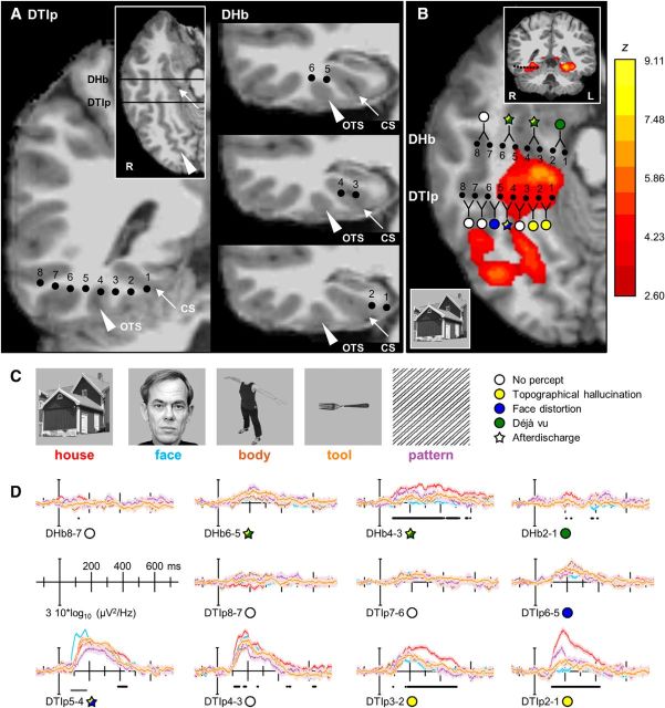
In recent years, functional neuroimaging has disclosed a network of cortical areas in the basal temporal lobe that selectively respond to visual scenes, including the parahippocampal place area (PPA). Beyond the observation that lesions involving the PPA cause topographic disorientation, there is little causal evidence linking neural activity in that area to the perception of places. Here, we combined functional magnetic resonance imaging (fMRI) and intracranial EEG (iEEG) recordings to delineate place-selective cortex in a patient implanted with stereo-EEG electrodes for presurgical evaluation of drug-resistant epilepsy. Bipolar direct electrical stimulation of a cortical area in the collateral sulcus and medial fusiform gyrus, which was place-selective according to both fMRI and iEEG, induced a topographic visual hallucination: the patient described seeing indoor and outdoor scenes that included views of the neighborhood he lives in. By contrast, stimulating the more lateral aspect of the basal temporal lobe caused distortion of the patient's perception of faces, as recently reported (Parvizi et al., 2012). Our results support the causal role of the PPA in the perception of visual scenes, demonstrate that electrical stimulation of higher order visual areas can induce complex hallucinations, and also reaffirm direct electrical brain stimulation as a tool to assess the function of the human cerebral cortex.

Keywords: direct electrical stimulation; functional magnetic resonance imaging; intracranial electroencephalography; parahippocampal place area; scene perception; visual hallucination.


Figures

Figure 1.
Figure 1.
Location of the intracranial electrodes, house-specific visual responses identified by fMRI, iEEG, and results of electrical brain stimulation. A, The location of the intracranial electrodes of shafts DTIp and DHb is displayed on the patient's high-resolution T1 MRI. Inset, Illustration of the coronal slices along the rostral-caudal axis. All DTIp electrodes were in the same coronal plane. For DHb, contacts 4 and 3 were on the coronal slice located in the inset; contacts 6 and 5 were on the coronal slice immediately rostral; and contacts 2 and 1 on the slice immediately caudal. The white arrow highlights the collateral sulcus; white arrowhead, the occipitotemporal sulcus. B, Regions that displayed greater BOLD responses to houses than other visual stimuli are overlaid on the patient's isometric axial T1 MRI. The basal temporal electrodes DHb and DTIp are also displayed. The inset shows a coronal slice in the plane of DTIp. The effect of direct electrical stimulation of pairs of neighboring electrodes is depicted as colored circles, or stars if there were afterdischarges as a result of the stimulation. The color codes for the symbols are detailed in C. The axial slice shown was in the plane of contacts DTIp5 through 2 and of contacts DHb4 and 3. DTIp7 and 1 and DHb6 and 5 were on the slice immediately (1 mm) dorsal and were projected down; DTIp8 was 2 mm dorsal and was also projected down; DHb2 and 1 were 1 mm ventral and were projected up. C, Left, Exemplars of stimuli used in iEEG paradigm to detect visual category-selective cortex. The fMRI paradigm included houses, faces, tools and patterns but not body parts. Right, Color codes for the symbols used to summarize the results of electrical stimulation in B and D (see Results; Table 1). D, Event-related changes in iEEG broadband gamma/high gamma power (40–160 Hz) following presentation of five categories of images. Different categories are represented by different colors (C). Shading indicates SE. Black dotted lines below each plot indicate significantly (padj < 0.01) higher responses to places than other visual stimuli. Gray lines indicate significantly (padj < 0.01) higher responses to faces than other visual stimuli. The symbols next to the electrode names summarize the effects of direct electrical stimulation at each site.

Comment in

References

    1. Aguirre GK, Zarahn E, D'Esposito M. An area within human ventral cortex sensitive to “building” stimuli: evidence and implications. Neuron. 1998;21:373–383. doi: 10.1016/S0896-6273(00)80546-2. - DOI - PubMed
    1. Bancaud J, Brunet-Bourgin F, Chauvel P, Halgren E. Anatomical origin of deja vu and vivid “memories” in human temporal lobe epilepsy. Brain. 1994;117:71–90. doi: 10.1093/brain/117.1.71. - DOI - PubMed
    1. Bar M, Aminoff E. Cortical analysis of visual context. Neuron. 2003;38:347–358. doi: 10.1016/S0896-6273(03)00167-3. - DOI - PubMed
    1. Bartolomei F, Barbeau E, Gavaret M, Guye M, McGonigal A, Régis J, Chauvel P. Cortical stimulation study of the role of rhinal cortex in deja vu and reminiscence of memories. Neurology. 2004;63:858–864. doi: 10.1212/01.WNL.0000137037.56916.3F. - DOI - PubMed
    1. Bastin J, Committeri G, Kahane P, Galati G, Minotti L, Lachaux JP, Berthoz A. Timing of posterior parahippocampal gyrus activity reveals multiple scene processing stages. Hum Brain Mapp. 2013a;34:1357–1370. doi: 10.1002/hbm.21515. - DOI - PMC - PubMed

Publication types

LinkOut - more resources