. 2014 Apr 16;34(16):5486-96.
doi: 10.1523/JNEUROSCI.4861-12.2014.

Leptin modulates the intrinsic excitability of AgRP/NPY neurons in the arcuate nucleus of the hypothalamus

Affiliations

Leptin modulates the intrinsic excitability of AgRP/NPY neurons in the arcuate nucleus of the hypothalamus

Scott B Baver et al. J Neurosci. .

Abstract

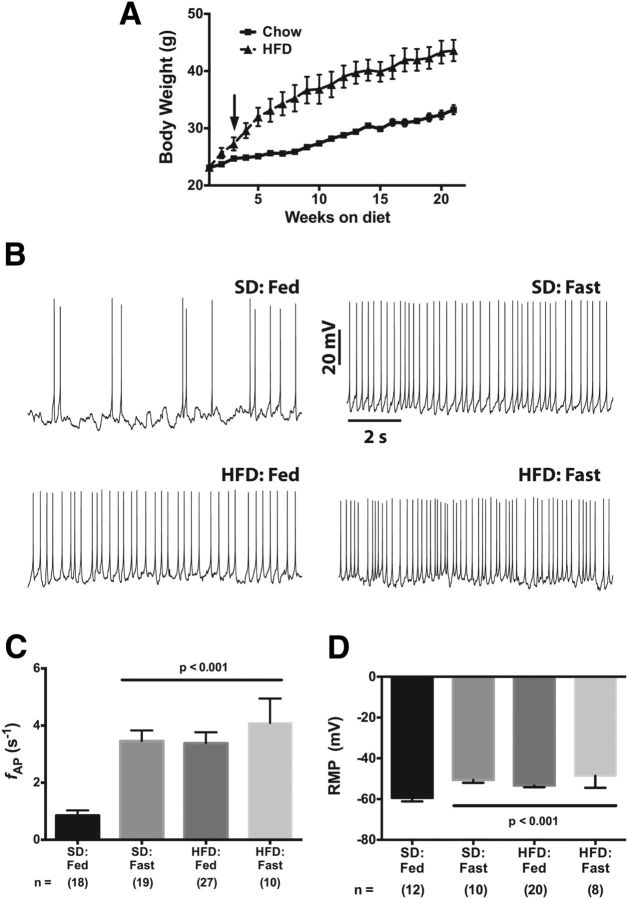
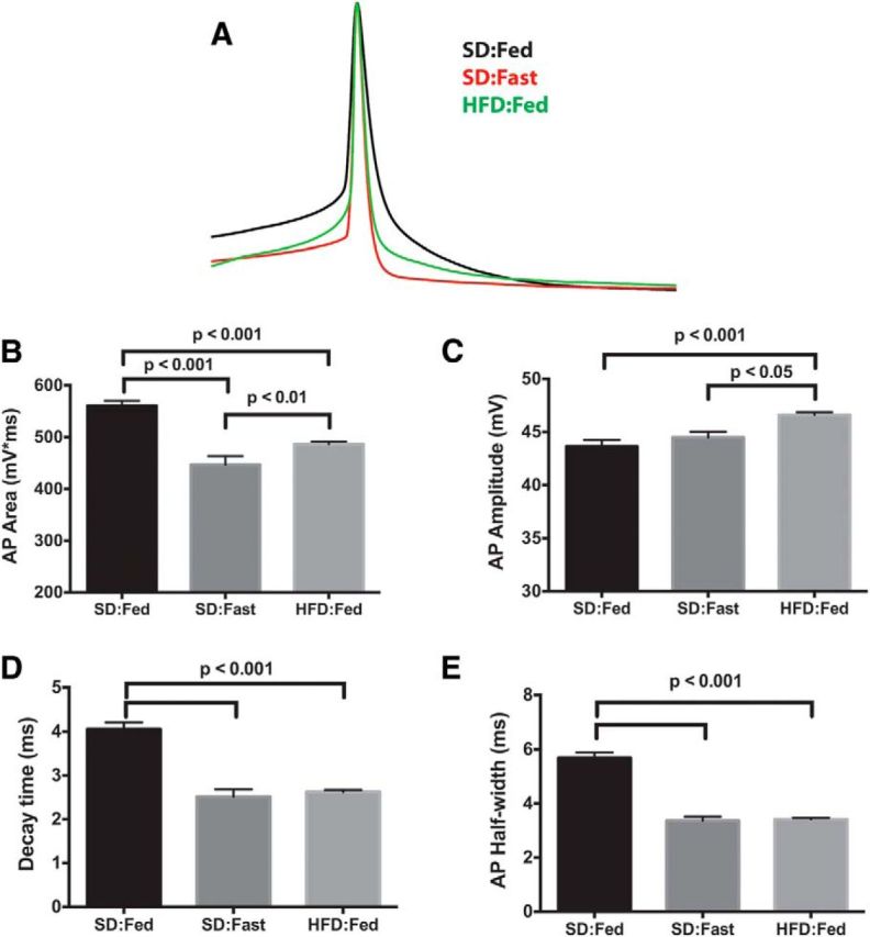
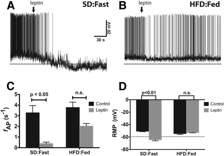
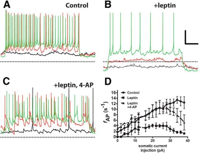
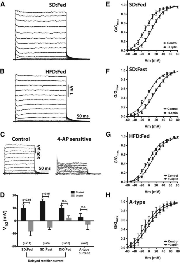
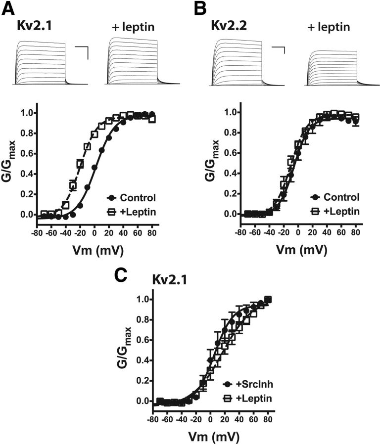
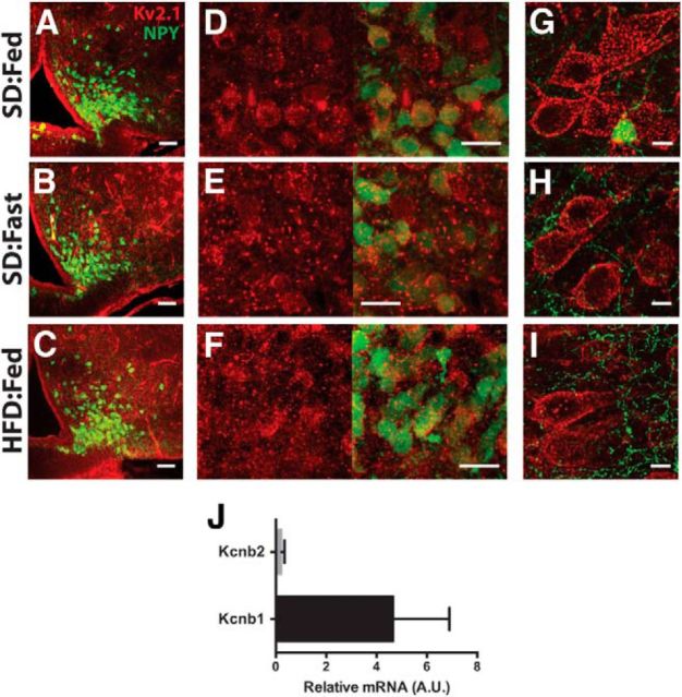
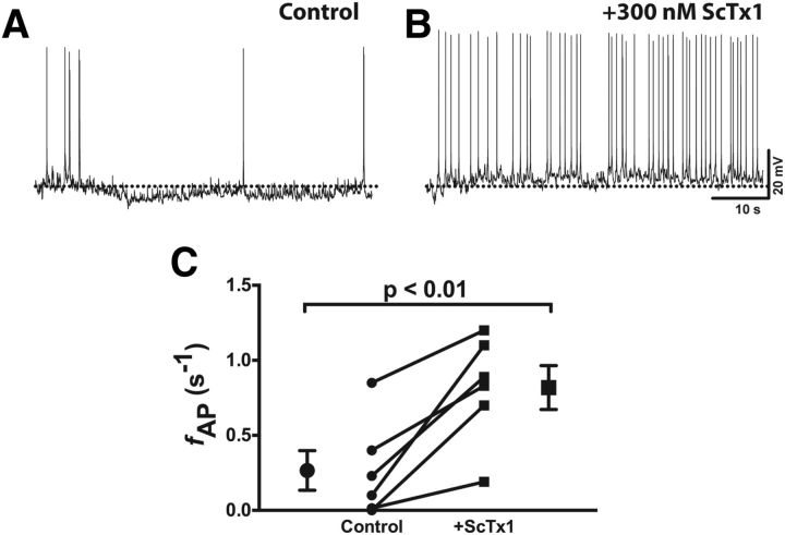
The hypothalamic arcuate nucleus (ARH) is a brain region critical for regulation of food intake and a primary area for the action of leptin in the CNS. In lean mice, the adipokine leptin inhibits neuropeptide Y (NPY) and agouti-related peptide (AgRP) neuronal activity, resulting in decreased food intake. Here we show that diet-induced obesity in mice is associated with persistent activation of NPY neurons and a failure of leptin to reduce the firing rate or hyperpolarize the resting membrane potential. However, the molecular mechanism whereby diet uncouples leptin's effect on neuronal excitability remains to be fully elucidated. In NPY neurons from lean mice, the Kv channel blocker 4-aminopyridine inhibited leptin-induced changes in input resistance and spike rate. Consistent with this, we found that ARH NPY neurons have a large, leptin-sensitive delayed rectifier K(+) current and that leptin sensitivity of this current is blunted in neurons from diet-induced obese mice. This current is primarily carried by Kv2-containing channels, as the Kv2 channel inhibitor stromatoxin-1 significantly increased the spontaneous firing rate in NPY neurons from lean mice. In HEK cells, leptin induced a significant hyperpolarizing shift in the voltage dependence of Kv2.1 but had no effect on the function of the closely related channel Kv2.2 when these channels were coexpressed with the long isoform of the leptin receptor LepRb. Our results suggest that dynamic modulation of somatic Kv2.1 channels regulates the intrinsic excitability of NPY neurons to modulate the spontaneous activity and the integration of synaptic input onto these neurons in the ARH.

Keywords: Kv channels; Kv2.1; arcuate nucleus; hypothalamus; leptin; neuropeptide Y.


Figures

Figure 1.
Figure 1.
Diet-induced obesity increases the firing rate and resting membrane potential of arcuate NPY neurons. A, Body-weight curve for male hrGFP-NPY mice fed either a standard chow or a high-fat diet. The arrow indicates the point at which the mean body weight of the HFD:Fed cohort was statistically greater than the SD:Fed cohort. B, Effect of calorie restriction and diet on spontaneous action potential frequency of NPY neurons in acute brain slices from lean satiated (SD:Fed), fasted (SD:Fast), and DIO (HFD:Fed and HFD:Fast) mice. C, D, Summary plots of firing rate (C) and resting membrane potential (D). All data are presented as mean ± SEM. Statistical significance for the firing rate data was determined using a nonparametric Kruskal–Wallis ANOVA with a post hoc Dunn's multiple comparisons test. A standard parametric ANOVA was used to determine significance of the RMP data. The number of GFP-positive NPY neurons in each group is given in parentheses below each column.
Figure 2.
Figure 2.
Action potential repolarization is altered in NPY neurons from food-deprived and DIO mice. A, Mean AP waveforms were generated by averaging 15 individual APs from each group. The resulting waveforms were normalized to the peak amplitude and overlaid using the peak for alignment. B, Mean AP area from the point of threshold crossing to return to baseline (positive area only). C, Mean AP amplitude calculated from threshold to peak. D, Mean time for AP to decay e-fold from peak. E, Mean AP width at 50% repolarization.
Figure 3.
Figure 3.
Leptin fails to decrease the spontaneous firing rate or induce hyperpolarization of the RMP in ARH NPY neurons from DIO mice. A, B, Spontaneous APs from NPY neurons in acute brain slices from a lean, fasted mouse (A) or a DIO mouse (B). Leptin (100 nm) was added to the bath solution at the indicated time (arrow). The dotted line is at −65 mV. C, D, Summary bar graph of firing rate (C) and RMP (D). Statistical significance within dietary groups was determined using a paired Student's t test. All data presented as mean ± SEM.
Figure 4.
Figure 4.
Blockade of Kv channels prevents leptin-induced modulation of the intrinsic membrane properties of NPY neurons. A–C, Current-clamp recordings from an ARH NPY neuron in an acute brain slice from a fasted mouse in normal aCSF (A), with 100 nm leptin (B), and with 100 nm leptin and 20 mm 4-AP (C). Black trace, 0 pA; red trace, +10 pA stimulus; green trace, +30 pA stimulus. The dotted line indicates −60 mV. D, Stimulus–response curve for all three conditions. Data are presented as mean ± SEM.
Figure 5.
Figure 5.
Diet-induced obesity mitigates the leptin sensitivity of the delayed rectifier current in NPY neurons. A, B, Representative K+ currents from NPY neurons in brain slices from a lean, SD:Fed mouse (A) and an HFD:Fed mouse (B). C, The relative contribution of delayed rectifier and A-type currents to the total K+ current was ascertained by measurement of voltage-dependent K+ currents before and after bath application of 5 mm 4-AP, followed by off-line subtraction of the 4-AP-sensitive current from the control currents. D, Mean activation midpoints with and without leptin derived from the G–V curves in E–H. The A-type K+ current was measured as the peak current within 10 ms of the onset of depolarization, and the delayed rectifier current was measured as the peak current at the end of the test pulse. Statistical significance within groups was determined by the paired Student's t test, and data are presented as mean ± SEM. The number of GFP-positive NPY neurons in each group is given in parentheses.
Figure 6.
Figure 6.
Leptin sensitivity of Kv2-family delayed rectifier channels. A, Top, Representative K+ currents from HEK cells expressing GFP-Kv2.1 with LepRb before and after the addition of 100 nm leptin. Calibration: 5 nA, 50 ms. Bottom, Mean G–V curves for the Kv2.1 current with and without leptin. B, Top, Representative K+ currents from HEK cells expressing GFP-Kv2.2 with LepRb before and after the addition of 100 nm leptin. Calibration: 5 nA, 50 ms. Bottom, Mean G--V curves for the Kv2.2 current with and without leptin. C, Mean G–V curve for Kv2.1 current after Src inhibition by the coaddition of 1 μm SrcI1 and 100 nm PP1 with and without leptin.
Figure 7.
Figure 7.
Kv2.1 immunoreactivity in ARH neurons from hrGFP-NPY mice. A–C, Low-magnification view of Kv2.1 (red) in ARH for anatomical reference. Green indicates GFP expression in NPY neurons. Scale bars, 50 μm. D–F, High-magnification image of Kv2.1 alone (red, left) and Kv2.1 with GFP (right). Scale bars, 20 μm. G–I, High-magnification image of Kv2.1 in cortical layer 2/3 pyramidal neurons. Scale bars, 10 μm. J, Relative transcript levels of Kcnb1 (Kv2.1) and Kcnb2 (Kv2.2) in ARH normalized to S19 expression.
Figure 8.
Figure 8.
Stromatoxin-1 increases the spontaneous firing rate of NPY neurons in brain slices from lean, fed mice. A, B, Representative spontaneous APs recorded in normal aCSF (A) and after bath application of 300 nm ScTx1 (B). C, Summary firing rate data before and after ScTx1 application. Statistical significance was determined using a paired Student's t test. Data are presented as mean ± SEM.

References

    1. Aponte Y, Atasoy D, Sternson SM. AGRP neurons are sufficient to orchestrate feeding behavior rapidly and without training. Nature. 2010;14:351–356. doi: 10.1038/nn.2739. - DOI - PMC - PubMed
    1. Atasoy D, Betley JN, Su HH, Sternson SM. Deconstruction of a neural circuit for hunger. Nature. 2012;488:172–177. doi: 10.1038/nature11270. - DOI - PMC - PubMed
    1. Baver SB, O'Connell KM. The C-terminus of neuronal Kv2.1 channels is required for channel localization and targeting but not for NMDA-receptor-mediated regulation of channel function. Neuroscience. 2012;217:56–66. doi: 10.1016/j.neuroscience.2012.04.054. - DOI - PMC - PubMed
    1. Bouret SG, Bates SH, Chen S, Myers MG, Jr, Simerly RB. Distinct roles for specific leptin receptor signals in the development of hypothalamic feeding circuits. J Neurosci. 2012;32:1244–1252. doi: 10.1523/JNEUROSCI.2277-11.2012. - DOI - PMC - PubMed
    1. Cerda O, Trimmer JS. Analysis and functional implications of phosphorylation of neuronal voltage-gated potassium channels. Neurosci Lett. 2010;486:60–67. doi: 10.1016/j.neulet.2010.06.064. - DOI - PMC - PubMed

MeSH terms