. 2014 Jul;88(13):7464-73.
doi: 10.1128/JVI.00794-14. Epub 2014 Apr 16.

Characterization of the nucleocytoplasmic shuttle of the matrix protein of influenza B virus

Affiliations

Characterization of the nucleocytoplasmic shuttle of the matrix protein of influenza B virus

Shuai Cao et al. J Virol. 2014 Jul.

Abstract

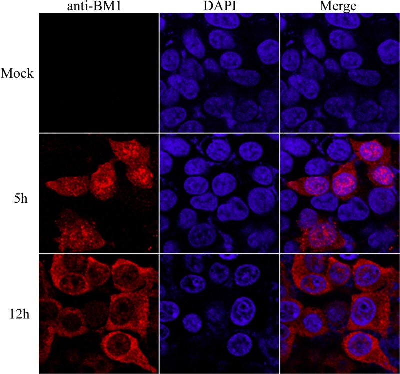
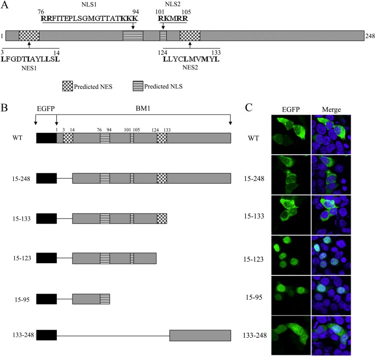
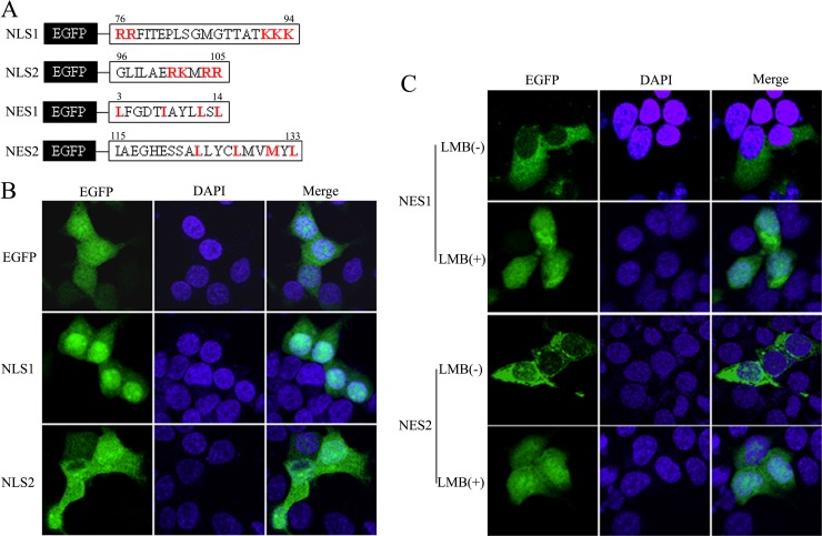
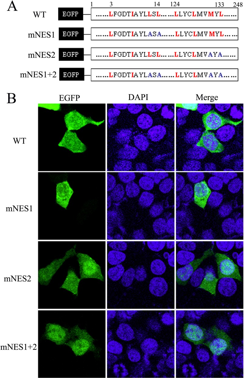
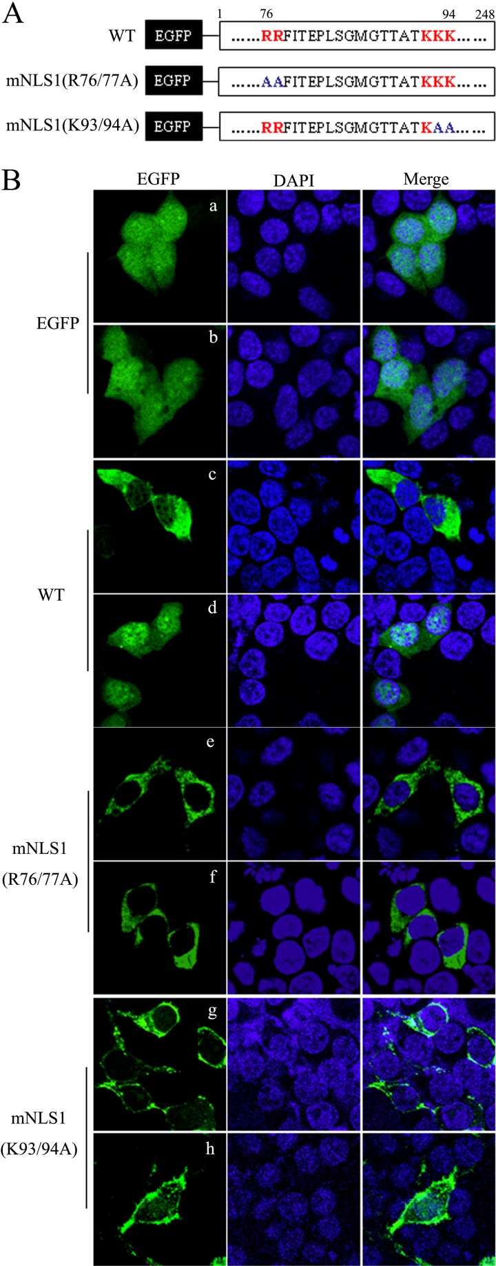
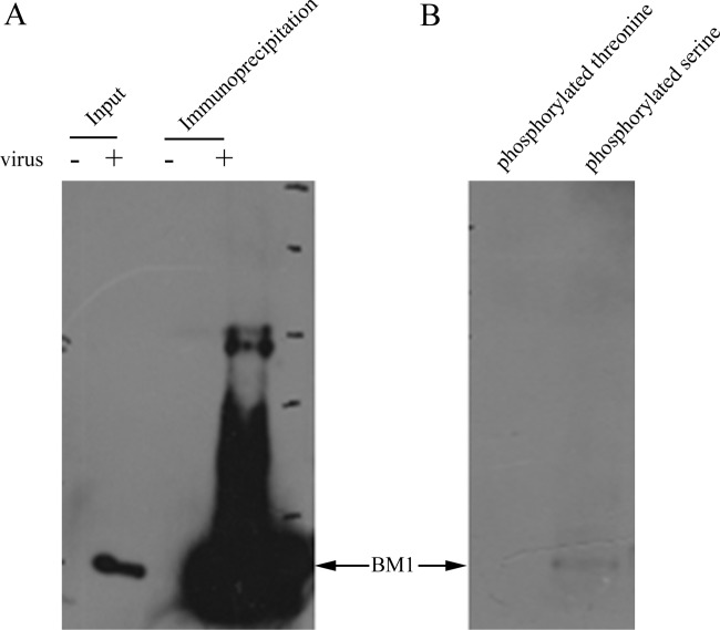
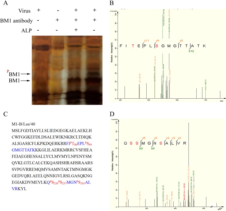
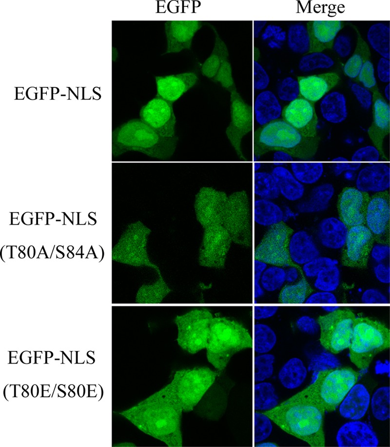
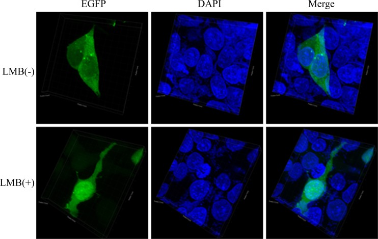
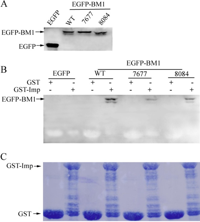
Influenza B virus is an enveloped negative-strand RNA virus that contributes considerably to annual influenza illnesses in human. The matrix protein of influenza B virus (BM1) acts as a cytoplasmic-nuclear shuttling protein during the early and late stages of infection. The mechanism of this intracellular transport of BM1 was revealed through the identification of two leucine-rich CRM1-dependent nuclear export signals (NESs) (3 to 14 amino acids [aa] and 124 to 133 aa), one bipartite nuclear localization signal (NLS) (76 to 94 aa), and two phosphorylation sites (80T and 84S) in BM1. The biological function of the NLS and NES regions were determined through the observation of the intracellular distribution of enhanced green fluorescent protein (EGFP)-tagged signal peptides, and wild-type, NES-mutant, and NLS-mutant EGFP-BM1. Furthermore, the NLS phosphorylation sites 80T and 84S, were found to be required for the nuclear accumulation of EGFP-NLS and for the efficient binding of EGFP-BM1 to human importin-α1. Moreover, all of these regions/sites were required for the generation of viable influenza B virus in a 12-plasmid virus rescue system.

Importance: This study expands our understanding of the life cycle of influenza B virus by defining the dynamic mechanism of the nucleocytoplasmic shuttle of BM1 and could provide a scientific basis for the development of antiviral medication.


Figures

FIG 1
FIG 1
BM1 shuttled between the cytoplasm and nucleus of 293T cells during infection. 293T cells were incubated with or without influenza B virus diluted in DMEM (1 μg of TPCK-trypsin/ml) at an MOI of ∼1. After a 1-h absorption at 37°C, the culture medium was replaced with DMEM (2% FBS), and the cells were further incubated at 37°C with 5% CO2. The cells were then fixed with PBS (4% paraformaldehyde) at the indicated time points postinfection. Intracellular localization of BM1 and staining with anti-BM1 polyclonal antibody were performed, followed by imaging with a confocal laser scanning fluorescence microscope (Olympus LSCMFV500).
FIG 2
FIG 2
Schematic diagram and intracellular distribution of WT and truncated BM1 constructs. (A) Diagram of predicted NESs and NLSs in the context of BM1. (B) Schematic representation of the constructs encoding EGFP-tagged WT or truncated BM1. (C) 293T cells were transfected with constructs encoding the EGFP tagged WT or truncated BM1, respectively. At 20 h posttransfection, cycloheximide (1 μg/ml) was added to the culture medium. After a 1-h incubation, the cells were fixed PBS (4% paraformaldehyde) and stained with DAPI. The intracellular localization of the indicated proteins was imaged by confocal laser scanning fluorescence microscopy (Olympus LSCMFV500).
FIG 3
FIG 3
Verification of the biological activities of the predicted NESs and NLSs using EGFP fusion proteins. (A) The predicted NLSs and NESs peptides in BM1 were fused to the C terminus EGFP. 293T cells were transfected with constructs coding the predicted NLS1 and NLS2 (B) and NES1 and NES2 (C), fused with EGFP, respectively. At 20 h posttransfection, LMB was added to the culture medium of the indicated cells to the concentration of 11 nM. After a 3-h incubation, the cells were fixed PBS (4% paraformaldehyde) and stained with DAPI. The intracellular localization of the indicated proteins was imaged by confocal laser scanning fluorescence microscopy (Olympus LSCMFV500).
FIG 4
FIG 4
The nuclear export of BM1 depended on NES1 and NES2. (A) The NES1 and/or NES2 were mutated in the context of full-length EGFP tagged BM1. (B) The constructs encoding the WT or mutant EGFP-BM1 were transfected into 293T cells, respectively. At 20 h posttransfection, the cells were fixed PBS (4% paraformaldehyde) and stained with DAPI. The intracellular localization of the indicated proteins was imaged by confocal laser scanning fluorescence microscopy (Olympus LSCMFV500).
FIG 5
FIG 5
NLS1 mediated the nuclear import of BM1. (A) The critical residues in NLS1 were substituted by alanine in the context of full-length EGFP tagged BM1. (B) The constructs encoding EGFP, the WT and mutant EGFP-BM1 were transfected into 293T cells, respectively. At 20 h posttransfection, the cells were treated with (b, d, f, and h) or without (a, c, e, and g) LMB (11 nM). After a 3-h incubation, the cells were fixed PBS (4% paraformaldehyde) and stained with DAPI. The intracellular localization of the indicated proteins was imaged by confocal laser scanning fluorescence microscopy (Olympus LSCMFV500).
FIG 6
FIG 6
Serine-phosphorylated BM1 was detected by specific antibodies against phosphorylated serine. 293T cells were infected with (+) or without (−) influenza B virus at an MOI of 1 for 36 h. The cell lysates were incubated with anti-BM1 polyclonal antibody at 4°C overnight and then precipitated with protein G-beads. (A) The cell lysates and the precipitated proteins were analyzed by Western blotting with anti-BM1 monoclonal antibody. (B) The precipitated proteins were also detected with antibodies specific against phosphorylated serine and phosphorylated threonine, respectively.
FIG 7
FIG 7
Identification of the phosphorylated residues in BM1 by MS. (A) Cell lysates of mock- or influenza B virus-infected MDCK cells were treated with or without ALP, immune precipitated with or without monoclonal anti-BM1 antibody, and subjected to gel electrophoresis. The band of candidate phosphorylated BM1 was collected from the gel and analyzed by LC-MS/MS. The candidate band was identified as BM1, and two peptides detected using MS (B and D) revealed the phosphorylated residues. (C) The peptides detected using MS (blue) and phosphorylated residues (red) are indicated in the context of BM1.
FIG 8
FIG 8
Role of phosphorylated S80 and T84 in the nuclear import of BM1. 293T cells were transfected with constructs encoding EGFP-tagged WT or mutant NLS1. At 20 h posttransfection, the cells were fixed PBS (4% paraformaldehyde) and stained with DAPI. The intracellular localization of the indicated proteins was imaged by confocal laser scanning fluorescence microscopy (Leica SP8).
FIG 9
FIG 9
Role of phosphorylated S80 and T84 in the nuclear import of BM1. 293T cells were transfected with constructs encoding EGFP-tagged full-length BM1 with alanine substitution of S80 and T84. At 20 h posttransfection, LMB was added to the culture medium of the indicated cells to the concentration of 11 nM. After a 3-h incubation, the cells were fixed PBS (4% paraformaldehyde) and stained with DAPI. The intracellular localization of the EGFP-BM1–S80/T84A was imaged by confocal laser scanning fluorescence microscopy (LSCM Leica SP8). The three-dimensional images were created with the corresponding two-dimensional images taken along the Z-axis (software Imaris).
FIG 10
FIG 10
Phosphorylated S80 and T84 and the critical arginines in NLS1 were crucial for the binding of BM1 to importin-α1. (A) 293T cells were transfected with EGFP-tagged WT, R76/77A mutant, and S80/T84A mutant BM1, respectively. The cells were collected at 36 h posttransfection, and the indicated proteins in cell lysates were detected by Western blotting with monoclonal anti-GFP antibody. (B) The cell lysates containing the EGFP-tagged WT or mutant BM1 were incubated with purified GST or GST-tagged human importin-α1 at 4°C for 2 h and then precipitated with immobilized glutathione. The WT or mutant EGFP-tagged BM1 precipitated together with GST–importin-α1 were detected by Western blotting with monoclonal anti-GFP antibody. (C) The nitrocellulose blotting membrane was stained with Coomassie brilliant blue.

Similar articles

Cited by

References

    1. Horvath CM, Williams MA, Lamb RA. 1990. Eukaryotic coupled translation of tandem cistrons: identification of the influenza B virus BM2 polypeptide. EMBO J. 9:2639–2647 - PMC - PubMed
    1. Almond JW, Haymerle HA, Felsenreich VD, Reeve P. 1979. The structural and infected cell polypeptides of influenza B virus. J. Gen. Virol. 45:611–621. 10.1099/0022-1317-45-3-611 - DOI - PubMed
    1. Fischer W, Pitkeathly M, Wallace B, Forrest L, Smith G, Sansom M. 2000. Transmembrane peptide NB of influenza B: a simulation, structure, and conductance study. Biochemistry 39:12708–12716. 10.1021/bi001000e - DOI - PubMed
    1. Nakamura K, Kitame F, Homma M. 1981. A comparison of proteins among various influenza B virus strains by one-dimensional peptide mapping. J. Gen. Virol. 56:315. 10.1099/0022-1317-56-2-315 - DOI - PubMed
    1. Ng AKL, Lam MKH, Zhang H, Liu J, Au SWN, Chan PKS, Wang J, Shaw PC. 2012. Structural basis for RNA binding and homo-oligomer formation by influenza B virus nucleoprotein. J. Virol. 86:6758–6767. 10.1128/JVI.00073-12 - DOI - PMC - PubMed

Publication types

MeSH terms

Substances