Review
. 2014 Mar;48(2):136-44.
doi: 10.4103/0019-5413.128747.

Tubercular spondylitis in children

Affiliations
Review

Tubercular spondylitis in children

Anil K Jain et al. Indian J Orthop. 2014 Mar.

Abstract

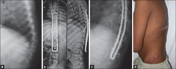
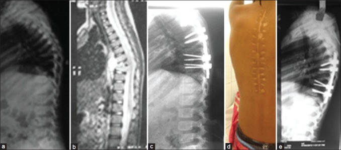
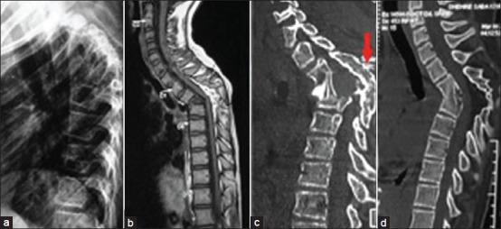
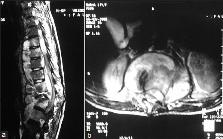
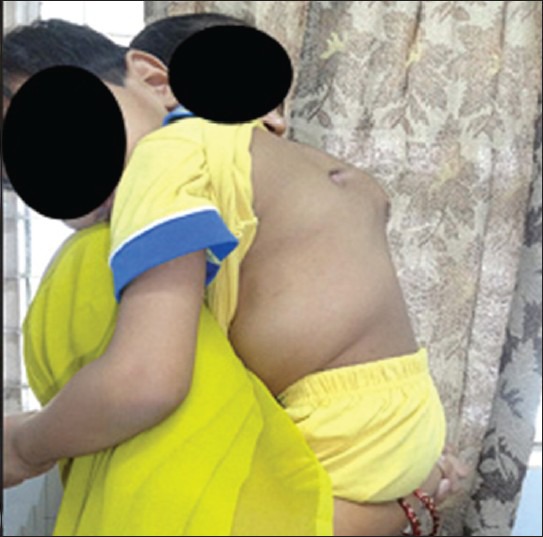
Spine of the child has unique anatomy and growth potential to grow to adult size. Tuberculosis (TB) spine results in bone loss as well as disturbed growth potential, hence spinal deformities may progress as the child grows. The growth potential is also disturbed when the disease focus is surgically intervened. Surgery is indicated for complications such as deformity, neurological deficit, instability, huge abscess, diagnostic dilemma and in suspected drug resistance to mycobacterium tuberculosis. The child on antitubercular treatment needs to be periodically evaluated for weight gain and drug dosages need to be adjusted accordingly. The severe progressive kyphotic deformity should be surgically corrected. Mild to moderate cases should be followed up until maturity to observe progression/improvement of spinal deformity. The surgical correction of kyphotic deformity in active disease is less hazardous than in a healed kyphosis. The internal kyphectomy by extra pleural approach allows adequate removal of internal salient in paraplegic patients with healed kyphotic deformity.

Keywords: Kyphus correction; osteoarticular tuberculosis; pediatric tuberculosis; tuberculosis of spine.


Conflict of interest statement

Conflict of Interest: None

Figures

Figure 1
Figure 1
Lateral x-rays of spine in 3 children shows (a, b) moderate kyphotic deformity (c) severe kyphotic deformity following TB of spine. (d, e) Lateral x-rays and line diagramme showing the proximal vertebrae resting on anterior surface on to distal vertebrae
Figure 2
Figure 2
(a) Computed tomography (CT) scan (sagittal section) of upper dorsal spine in a 3 year old child shows 5 vertebral body disease. The vertebral body almost disappeared (b) on 3D reconstruction and (c) axial CT shows no trace of bone in VB
Figure 3
Figure 3
(a) Clinical photograph of a 6 year old child reported with severe kyphosis following long segment tuberculosis of spine. (b) lateral x-rays shows severe kyphosis with destruction of D5-9 vertebral bodies (c) the lateral x-rays of the same patient taken 3 months before (which patient was carrying) also shows widening of prevertebral soft tissue shadow seen as deviation of tracheal shadow infront of upper dorsal spine (white arrows). The diagnosis was missed at this stage
Figure 4
Figure 4
A 5 year old child with TB of spine on anti tubercular treatment for 6 months presented with a radiological signs of progressive kyphotic deformity (a) Standard extra pleural anterolateral decompression was done with stabilization using a closed Hartshill (b and c) was done (d) clinical photograph showing the surgical scar over iliac crest and healed posterior T-incision
Figure 5
Figure 5
A 5 year female with (a) kyphus deformity (b) preoperative X-ray and (c) magnetic resonance imaging after 6 months of anti tubercular treatment and (d) postoperative X-ray showing kyphus correction using pedicle screws (e) healed posterior midline incision (e) 2 years followup x-rays of the same patient, the anterior vertebral body height has increased on followup
Figure 6
Figure 6
Preoperative (a) X-ray lateral view (b) sagittal T1WI MRI show kyphus deformity internal gibbus at D3 in an 11-year-old girl following old childhood diseases. The lesion has no activity. (c) immediate postoperative sagittal reconstruction image shows internal kyphectomy and anterior transposition of the spinal cord (d) same image at one year followup shows incorporated bone graft
Figure 7
Figure 7
(a) T2W1 magnetic resonance imaging sagittal section shows a 9 vertebral body affection with kyphosis correction was done by pedicle screw insertion, each hole for pedicle screw showed pus, hence hartshill fixation was performed where sublaminar wires took purchase against healthy lamina (b) Intraosseous abscess communicating with anterior prevertebral collection seen in axial section
Figure 8
Figure 8
Clinical photograph of a child taken on shoulder by mother. The patient was operated for kyphus correction with Hartshill fixation. The kyphus has progressed as Hartshill has given way

References

    1. Sarwark J, Aubin CE. Growth considerations of the immature spine. J Bone Joint Surg Am. 2007;89:8–13. - PubMed
    1. Soames RW. Skeletal system. In: Williams LH, Berry MM, Collins P, Dyson M, Dussek J, Ferguson MW, editors. Gray's Anatomy: The Anatomical Basis of Medicine and Surgery. 38th ed. New York: Churchill Livingstone; 1995. pp. 510–36.
    1. Dimeglio A, Ferran JL. Three-dimensional analysis of the hip during growth. Acta Orthop Belg. 1990;56:111–4. - PubMed
    1. Roaf R. Vertebral growth and its mechanical control. J Bone Joint Surg Br. 1960;42-B:40–59. - PubMed
    1. Taylor JR. Growth of human intervertebral discs and vertebral bodies. J Anat. 1975;120(Pt 1):49–68. - PMC - PubMed

LinkOut - more resources