Review
. 2014 Mar 27:9:1537-57.
doi: 10.2147/IJN.S53593. eCollection 2014.

Receptor binding peptides for target-selective delivery of nanoparticles encapsulated drugs

Affiliations
Review

Receptor binding peptides for target-selective delivery of nanoparticles encapsulated drugs

Antonella Accardo et al. Int J Nanomedicine. .

Abstract

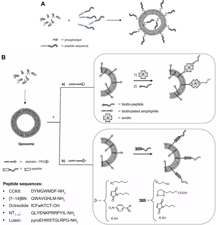
Active targeting by means of drug encapsulated nanoparticles decorated with targeting bioactive moieties represents the next frontier in drug delivery; it reduces drug side effects and increases the therapeutic index. Peptides, based on their chemical and biological properties, could have a prevalent role to direct drug encapsulated nanoparticles, such as liposomes, micelles, or hard nanoparticles, toward the tumor tissues. A considerable number of molecular targets for peptides are either exclusively expressed or overexpressed on both cancer vasculature and cancer cells. They can be classified into three wide categories: integrins; growth factor receptors (GFRs); and G-protein coupled receptors (GPCRs). Therapeutic agents based on nanovectors decorated with peptides targeting membrane receptors belonging to the GPCR family overexpressed by cancer cells are reviewed in this article. The most studied targeting membrane receptors are considered: somatostatin receptors; cholecystokinin receptors; receptors associated with the Bombesin like peptides family; luteinizing hormone-releasing hormone receptors; and neurotensin receptors. Nanovectors of different sizes and shapes (micelles, liposomes, or hard nanoparticles) loaded with doxorubicin or other cytotoxic drugs and externally functionalized with natural or synthetic peptides are able to target the overexpressed receptors and are described based on their formulation and in vitro and in vivo behaviors.

Keywords: active targeting; drug delivery; nanoparticles; receptors binding peptides; supramolecular aggregates.


Figures

Figure 1
Figure 1
Introduction of bioactive peptides on aggregate surfaces. Notes: (A) The bioactive peptide may be introduced on the aggregate surface directly during nanostructure preparation by coupling the peptide to an amphiphilic moiety according to a pre-functionalization strategy; with this approach, however, the bioactive peptide is displayed on the external liposome surface as well as in the inner compartment. (B) Alternatively, peptide introduction can be performed after nanostructures have been obtained, according to a post-functionalization strategy. For the second approach, peptide coupling after liposome or nanoparticle preparation involves the introduction of suitable activated functional groups onto the external side of liposomes or nanoparticles for covalent or non-covalent peptide binding. To guarantee correct orientation of the targeting ligand, biorthogonal and site-specific surface reactions are necessary. Functional groups commonly used are: 1) amine for the amine-N-hydroxysuccinamide coupling method, 2) maleimide for Michael addition, 3) azide for Cu(I)-catalized Huisgen cycloaddition (CuAAC), 4) biotin for non-covalent interaction with avidin or triphosphines for Staudinger ligation, and hydroxylamine for oxime bond. In the inset are reported the peptide sequences. Abbreviations: BN, bombesin; CCK8, cholecystokinin-8; NT, neurotensin; PEG, polyethylene glycol.
Figure 2
Figure 2
In vivo imaging of tumor-bearing mice after administration of Cy-7 loaded DAHC micelles (A) and Cy-7 loaded OPD(20%)-DAHC micelles (B) at 1, 6, 12, and 24 hours. Note: Reprinted from Biomaterials, 33(27), Huo M, Zou A, Yao C, et al, Somatostatin receptor-mediated tumor-targeting drug delivery using octreotide-PEG-deoxycholic acid conjugate-modified N-deoxycholic acid-O, N-hydroxyethylation chitosan micelles, 6393–6407, Copyright (2012) with permission from Elsevier. Abbreviations: DAHC, N-deoxycholic acid-O,N-hydroxyethylation chitosan; OPD, OCT(Phe)-PEG-DOCA; h, hours.
Figure 3
Figure 3
Antitumor efficiency of different treatments in MCF-7-bearing subcutaneous tumor models in nude mice. (A) Tumor volumes versus time. Data represent mean ± standard deviation (n=6). (B) Tumors excised at the end of the tests. Note: Springer and Pharm Res, 29, 2012, 2902–2911, Spatiotemporally controlled co-delivery of anti-vasculature agent and cytotoxic drug by octreotide-modified stealth liposomes, Dai W, Jin W, Zhang J, et al, Figure 10. With kind permission from Springer Science and Business Media. Abbreviations: PBS, phosphate buffer solution; OCT, octreotide.
Figure 4
Figure 4
Scheme of Naposomes formulation and their in vitro and in vivo behavior. Flow cytometric analysis of association of liposomal DOX and free DOX with human cells. Notes: A431 cells (A) and HUVEC cells (B) at a density of 1.3 · 106 cells/mL were incubated with CCK8/DOTA-DOX, DOTA-DOX, and free DOX at a final concentration of 1 μ±g DOX/mL for 1 hour at 48°C. Untreated cells served as negative control while free doxorubicin solution was used as positive control. The untreated cells (negative controls) and cells incubated with non-specific DOTA-DOX give identical behavior with overlapping curves. (C and D) Cytotoxicity of liposomal DOX against human cells on 431 cells and HUVEC, respectively. Cells were incubated with CCK8/DOTA-DOX and DOTA-DOX at different concentration ranging between 0 and 1,000 ng/mL at 37°C. After 8 hours, the medium was removed and after an additional 72 hours, an MTT assay was performed. Data are expressed as percent of negative control. (E) γ-camera image (dorsal view) obtained prior to dissection of one of the animals 18 h after injection of radiolabeled aggregates clearly shows higher concentration of the radiolabel in the receptor positive xenograft (+, left flank) compared with the control tumor (−, right flank). (A–D) Reproduced with permission from John Wiley and Sons. Morisco A, Accardo A, Tesauro D, Palumbo R, Benedetti E, Morelli G. Peptide-labeled supramolecular aggregates as selective doxorubicin carriers for delivery to tumor cells. Biopolymers. 2011;96:88–96. Copyright © 2011 Wiley Periodicals, Inc. (E) Reproduced with permission from John Wiley and Sons. Accardo A, Tesauro D, Aloj L, et al. Peptide-containing aggregates as selective nanocarriers for therapeutics. ChemMedChem. 2008;3(4):594–602. Copyright © 2008 WILEY-VCH Verlag GmbH & Co. KGaA, Weinheim. Abbreviations: CCK, cholecystokinin; DOTA, 1,4,7,10-tetraazacyclododecane-N,N’,N’’,N’’’-tetraacetic acid; DOX, doxorubicin; HUVEC, human umbilical vein endothelial cell; MTT, 3-(4,5-dimethyl-2-thiazolyl)-2,5-diphenyl-2H-tetrazolium bromide.
Figure 5
Figure 5
Liposomes encapsulating mitoxantrone uptake in various cell lines followed by confocal laser scanning fluorescence microscopy. Cell lines were treated with either LHRH-MTX-SL or MTX-SL for 4 hours at 37°C. (A) LHRH-MTX-SL in LHRH receptor high-expressing MCF-7 cells; (B) MTX-SL in LHRH receptor high-expressing MCF-7 cells; (C) MCF-7 cells treated with drug-free medium used as a control; (D) LHRH-MTX-SL in LHRH receptor low-expressing SK-OV-3 cells; (E) MTX-SL in LHRH receptor low-expressing SK-OV-3 cells; (F) SKOV-3 cells treated with drug-free medium used as a control. Note: Copyright © 2010. Reproduced with permission of Dove Medical Press. He Y, Zhang L, Song C. Luteinizing hormone-releasing hormone receptor-mediated delivery of mitoxantrone using LHRH analogs modified with PEGylated liposomes. Int J Nanomedicine. 2010;5:697–705. Abbreviations: LHRH, luteinizing hormone releasing hormone; MTX, methotrexate; SL, loaded liposomes.
Figure 6
Figure 6
Confocal microscopy of (A) HT29 and (B) TE671 cells incubated with DOPC-(C18)2Lys(NT8-13)4-DOX liposomes (200 nm, right) and with DOPC-DOX liposomes (200 nm, left) for 2 hours at 37°C. Plasma membranes were stained with lectin-FITC (green). Note: Reproduced with permission from Falciani C, Accardo A, Brunetti J, et al. Target-selective drug delivery through liposomes labeled with oligobranched neurotensin pep tides. ChemMedChem. 2011;6(4):678–685. Copyright © 2011 WILEY-VCH Verlag GmbH & Co. KGaA, Weinheim. Abbreviations: DOPC, 1,2-dioleoyl-sn-glycero-3-phosphocholine; DOX, doxorubicin; FITC, fluorescein isothiocyanate.

Similar articles

Cited by

References

    1. Richardson PF. Nanotechnology therapeutics in oncology-recent Developments and future outlook. Annu Rep Med Chem. 2012;47:239–252.
    1. Levchenko TS, Hartner WC, Torchilin VP. Liposomes for cardiovascular targeting. Ther Deliv. 2012;3(4):501–514. - PubMed
    1. Maradana MR, Thomas R, O’Sullivan BJ. Targeted delivery of curcumin for treating type 2 diabetes. Mol Nutr Food Res. 2013;57(9):1550–1556. - PubMed
    1. Levesque JP, Winkler IG. It takes nerves to recover from chemotherapy. Nat Med. 2013;19(6):669–671. - PubMed
    1. Matsumura Y, Maeda H. A new concept for macromolecular therapeutics in cancer chemotherapy: mechanism of tumoritropic accumulation of proteins and the antitumor agent smancs. Cancer Res. 1986;46(12 Pt 1):6387–6392. - PubMed

Publication types

MeSH terms