. 2014 Apr 1;1(4):258-271.
doi: 10.1002/acn3.54.

(-)-Epicatechin protects hemorrhagic brain via synergistic Nrf2 pathways

Affiliations

(-)-Epicatechin protects hemorrhagic brain via synergistic Nrf2 pathways

Che-Feng Chang et al. Ann Clin Transl Neurol. .

Abstract

Objective: In the wake of intracerebral hemorrhage (ICH), a devastating stroke with no effective treatment, hemoglobin/iron-induced oxidative injury leads to neuronal loss and poor neurologic outcomes. (-)-Epicatechin (EC), a brain-permeable flavanol that modulates redox/oxidative stress via the NF-E2-related factor (Nrf) 2 pathway, has been shown to be beneficial for vascular and cognitive function in humans. Here, we examined whether EC can reduce early brain injury in ICH mouse models and investigated the underlying mechanisms.

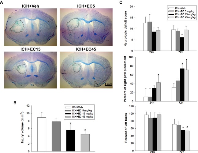
Methods: ICH was induced by injecting collagenase, autologous blood, or thrombin into mouse striatum. EC was administered orally at 3 h after ICH and then every 24 h. Lesion volume, neurologic deficits, brain edema, reactive oxygen species, and protein expression and activity were evaluated.

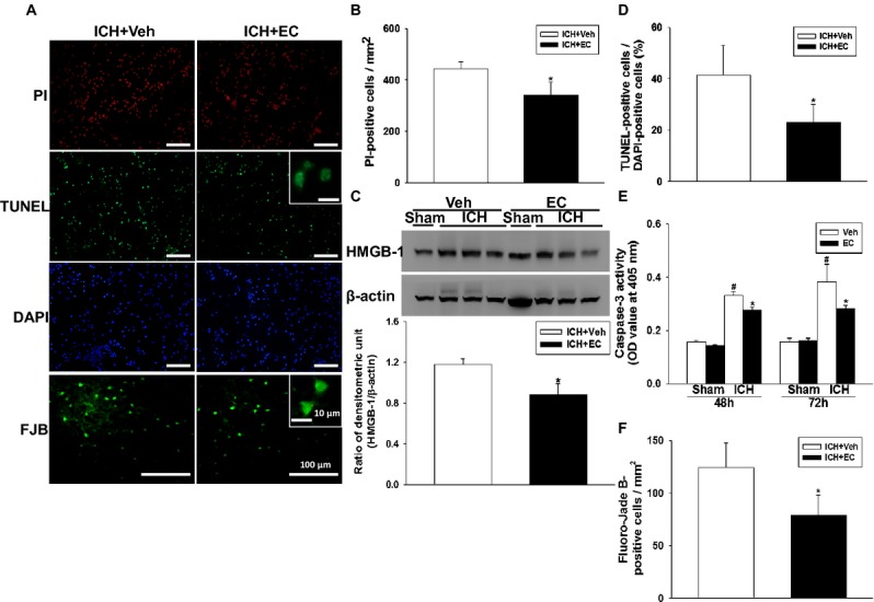
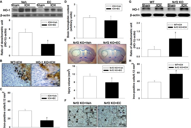
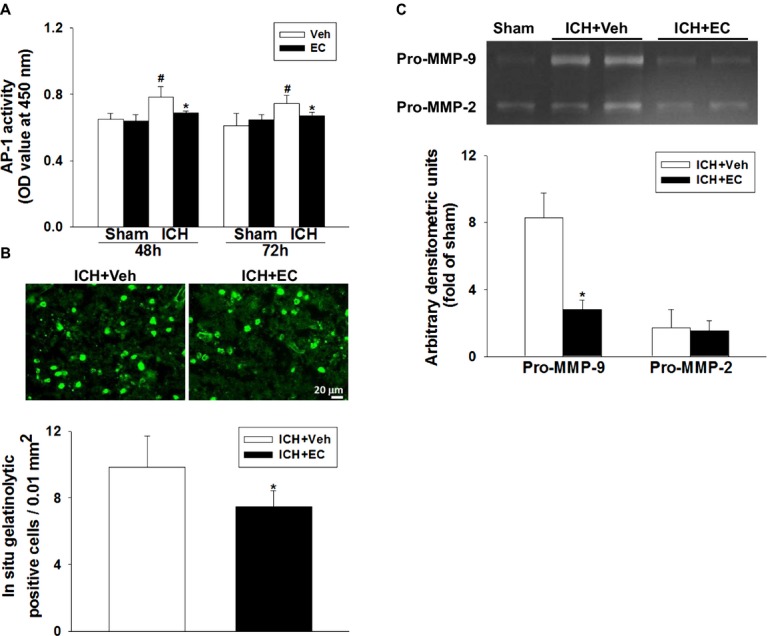
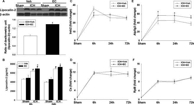
Results: EC significantly reduced lesion volume and ameliorated neurologic deficits in both male and female ICH mice. Cell death and neuronal degeneration were decreased in the perihematomal area and were associated with reductions in caspase-3 activity and HMGB-1 level. These changes were accompanied by attenuation of oxidative insults, increased phase II enzyme expression, and increased Nrf2 nuclear accumulation. Interestingly, in addition to providing neuroprotection via Nrf2 signaling, EC diminished heme oxygenase-1 induction and brain iron deposition via an Nrf2-independent pathway that downregulated ICH-induced activating protein-1 activation and decreased matrix metalloproteinase 9 activity, lipocalin-2 levels, iron-dependent cell death, and ferroptosis-related gene expression.

Interpretation: Collectively, our data show that EC protects against ICH by activation of Nrf2-dependent and -independent pathways and may serve as a potential intervention for patients with ICH.

Keywords: (−)-Epicatechin; NF-E2-related factor 2; ferroptosis; heme oxygenase-1; iron.


Figures

Figure 1
Figure 1
Effects of EC on brain injury volume and neurologic function in mice subjected to ICH. (A) Representative Luxol fast blue/Cresyl violet-stained brain sections at 72 h post-ICH; injured areas lack staining and are circled in white. Scale bar: 1 mm. (B) Quantification analysis shows significantly smaller brain injury volumes in EC-treated groups than in the vehicle-treated group at 72 h post-ICH. (C) EC posttreatment improved neurologic deficit score (top), forelimb placing (middle), and corner turn test performance (bottom) of mice subjected to ICH. Values are mean ± SD; n = 10 mice per group. *P < 0.05 versus vehicle.
Figure 2
Figure 2
Identification of cell death, apoptosis, and neuronal degeneration in mice subjected to ICH. (A) Representative propidium iodide (PI)-, TUNEL-, DAPI-, and Fluoro-Jade B (FJB)-stained brain sections. The insets are representative TUNEL- and FJB-positive cells at higher magnification. Scale bars: 100 μm; 10 μm (inset). (B) Quantification analysis indicates that 15 mg/kg EC (EC 15) significantly reduced PI-positive cells at 72 h post-ICH. (C) Top: Representative immunoblots of the HMGB-1 protein in ipsilateral hemispheres from sham-operated and ICH mice treated with or without EC 15. Bottom: Densitometric analysis shows that HMGB-1 expression in the ipsilateral hemispheres was significantly lower in EC 15 mice than in vehicle-treated mice 72 h after ICH. (D) Quantification analysis shows that the percentage of TUNEL-positive cells was significantly lower in the EC 15 mice than in the vehicle-treated mice 72 h after ICH. (E) Bar graphs showing caspase-3 enzyme activity in EC and vehicle groups at 48 and 72 h after sham surgery or ICH induction. ICH significantly increased caspase-3 enzyme activity in both EC- and vehicle-treated mice. The hemorrhagic hemispheres in the EC-treated group exhibited significantly less caspase-3 activity than did those in the vehicle-treated group. (F) Quantification analysis of FJB staining indicates that EC-treated mice had significantly fewer degenerating neurons than did vehicle-treated mice 72 h after ICH. Values are mean ± SD; n = 6–9 mice per group. *P < 0.05 versus vehicle; #P < 0.05 versus sham.
Figure 3
Figure 3
EC alleviates ICH-induced reactive oxygen species (ROS) production. (A) Top: Ethidium fluorescent signal was evident in brain sections from mice 24 h after ICH. Scale bar: 50 μm. Bottom: Quantification analysis of fluorescence intensity indicated that EC significantly reduced ROS production at 1 day post-ICH. (B) Bar graphs showing malondialdehyde (MDA) level at 48 and 72 h after sham surgery or ICH induction. ICH significantly increased MDA level in hemorrhagic brain tissue of both EC- and vehicle-treated mice. EC-treated mice had less MDA than did vehicle-treated mice. (C) Representative immunoblot of hemorrhagic brain tissue shows that EC-treated mice had less protein carbonylation than did vehicle-treated mice. β-actin served as a loading control. The arrows show the carbonylated proteins at different molecular weights. (D) Top: Representative immunoblot of ipsilateral hemispheres from mice treated with or without 15 mg/kg EC; brains were collected 6 h after sham surgery or ICH. Bottom: Bar graph of densitometric analysis shows Nrf2 protein level in nuclear extracts from ipsilateral hemispheres of EC- and vehicle-treated mice. (E and F) Line graphs show SOD-1 and NQO-1 mRNA expression in EC- and vehicle-treated mice at 6, 24, and 72 h after ICH. Compared with vehicle treatment, EC significantly increased SOD-1 mRNA expression at 24 and 72 h and NQO-1 mRNA expression at 6 and 72 h in the ipsilateral hemisphere. Values are mean ± SD; n = 5–6 mice per group. *P < 0.05 versus vehicle; #P < 0.05 versus sham.
Figure 4
Figure 4
Effects of EC on HO-1 protein expression, iron deposition, and hemorrhagic injury volume in WT, Nrf2 KO, and HO-1 KO mice subjected to ICH. (A) Top: Representative immunoblot of HO-1 protein from sham-operated and ICH mice treated with or without EC. Bottom: Densitometric analysis shows that HO-1 protein level in the ipsilateral hemisphere was significantly less in EC-treated mice than in vehicle-treated mice at 72 h after ICH. (B) Representative Perls-stained brain sections from WT and HO-1 KO mice 72 h post-ICH. In the perihematomal region, many iron-positive cells can be seen in the WT mice; based on cell morphology, most of them are likely activated microglia/macrophages with cytoplasmic iron deposits (arrows). The iron-positive cells with similar morphology are rarely detectable in the HO-1 KO mice. (C) Ferric iron accumulation in the perihematomal region of mice at 72 h post-ICH was significantly less in EC-treated mice than in vehicle-treated mice. (D) Hemoglobin levels were not significantly different in brains of vehicle- and EC-treated mice at 24 h post-ICH. (E) Representative brain sections from Nrf2 KO mice 72 h post-ICH were stained with Luxol fast blue/Cresyl violet. Areas of injury lack staining and are circled with a black line. Bottom: Quantification analysis shows significantly smaller brain injury volumes in EC-treated than in vehicle-treated mice. (F) Representative Perls-stained brain sections from Nrf2 KO mice with or without EC administration 72 h post-ICH. Fewer iron-positive cells are present in the EC-treated mice. The inset images show neuron-like and microglia/macrophage-like morphology (Left) and iron-positive (P) and -negative (N) cells (Right). (G) Top: Representative immunoblot of HO-1 protein from WT and Nrf2 KO mice subjected to sham operation or ICH induction. Bottom: Bar graph of densitometric analysis shows a significant increase in HO-1 expression in the ipsilateral hemispheres of Nrf2 KO mice at 72 h after ICH compared with that in WT mice. (H) Nrf2 KO mice had significantly more Perls-positive cells than did WT mice in the perihematomal region 72 h post-ICH. Scale bars: 50 μm (B); 1 mm (E); 20 μm (F). Values are mean ± SD; n = 5–10 mice per group. *P < 0.05 versus vehicle or WT.
Figure 5
Figure 5
Effects of EC on AP-1 and MMP activity in mice subjected to ICH. (A) AP-1 activity was significantly greater in the ipsilateral hemisphere of ICH mice than in that of sham-operated mice. EC administration significantly reduced AP-1 activity at 48 and 72 h post-ICH. (B) Top: Representative gelatin in situ zymography fluorescent images from ICH mice treated with vehicle or EC. Bottom: Quantification shows that gelatinolytic activity was lower in EC-treated mice than in vehicle-treated mice at 72 h post-ICH. (C) Top: Representative gel zymography of MMP-9 and MMP-2 activity from vehicle- and EC-treated mice at 72 h post-ICH. The gelatinase activity of pro-MMP-9 was increased in brains from both vehicle- and EC-treated mice. Bottom: Quantification analysis indicated that pro-MMP-9 activity was decreased by EC administration. Pro-MMP-2 gelatinase activity was weak in all groups. Values are mean ± SD; n = 5–6 mice per group. *P < 0.05 versus vehicle; #P < 0.05 versus sham.
Figure 6
Figure 6
Effects of EC on lipocalin-2 (LCN2) and ferroptosis-related genes in mice subjected to ICH. (A) Top: Representative immunoblot of LCN2 protein expression in hemorrhagic tissue from sham-operated and ICH mice treated with or without EC. Bottom: Densitometric analysis shows a significant increase in LCN2 protein expression in the ipsilateral hemisphere of EC-treated mice 72 h after ICH compared with that in vehicle-treated mice. (B) Bar graph shows LCN2 protein concentration in the ipsilateral hemispheres of vehicle- and EC-treated mice at 6 and 24 h post-ICH. ICH significantly increased LCN2 protein levels in both groups, but LCN2 protein level was significantly higher in the EC-treated group than in the vehicle-treated group. (C–F) Line graphs show mRNA expression of Ireb2, Cs, Atp5g3, and Rpl8 in the hemorrhagic hemispheres of vehicle- and EC-treated mice at 6, 24, and 72 h post-ICH. EC treatment significantly decreased Ireb2 mRNA expression at 6 h and increased Atp5g3 mRNA expression at 6, 24, and 72 h after ICH compared with vehicle treatment. Cs and Rpl8 mRNA expression did not differ significantly between the two groups. Values are mean ± SD; n = 5–6 mice per group. *P < 0.05 versus vehicle; #P < 0.05 versus sham.

References

    1. Donnan GA, Hankey GJ, Davis SM. Intracerebral haemorrhage: a need for more data and new research directions. Lancet Neurol. 2010;9:133–134. - PubMed
    1. Xi G, Keep RF, Hoff JT. Mechanisms of brain injury after intracerebral haemorrhage. Lancet Neurol. 2006;5:53–63. - PubMed
    1. Wang J. Preclinical and clinical research on inflammation after intracerebral hemorrhage. Prog Neurobiol. 2010;92:463–477. - PMC - PubMed
    1. Zecca L, Youdim MB, Riederer P, et al. Iron, brain ageing and neurodegenerative disorders. Nat Rev Neurosci. 2004;5:863–873. - PubMed
    1. Wang X, Mori T, Sumii T, et al. Hemoglobin-induced cytotoxicity in rat cerebral cortical neurons: caspase activation and oxidative stress. Stroke. 2002;33:1882–1888. - PubMed

LinkOut - more resources