. 2014 Apr 17:12:28.
doi: 10.1186/1741-7007-12-28.

Reprogramming of the chick retinal pigmented epithelium after retinal injury

Affiliations

Reprogramming of the chick retinal pigmented epithelium after retinal injury

Agustin Luz-Madrigal et al. BMC Biol. .

Abstract

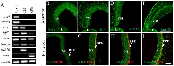
Background: One of the promises in regenerative medicine is to regenerate or replace damaged tissues. The embryonic chick can regenerate its retina by transdifferentiation of the retinal pigmented epithelium (RPE) and by activation of stem/progenitor cells present in the ciliary margin. These two ways of regeneration occur concomitantly when an external source of fibroblast growth factor 2 (FGF2) is present after injury (retinectomy). During the process of transdifferentiation, the RPE loses its pigmentation and is reprogrammed to become neuroepithelium, which differentiates to reconstitute the different cell types of the neural retina. Somatic mammalian cells can be reprogrammed to become induced pluripotent stem cells by ectopic expression of pluripotency-inducing factors such as Oct4, Sox2, Klf4, c-Myc and in some cases Nanog and Lin-28. However, there is limited information concerning the expression of these factors during natural regenerative processes. Organisms that are able to regenerate their organs could share similar mechanisms and factors with the reprogramming process of somatic cells. Herein, we investigate the expression of pluripotency-inducing factors in the RPE after retinectomy (injury) and during transdifferentiation in the presence of FGF2.

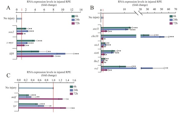
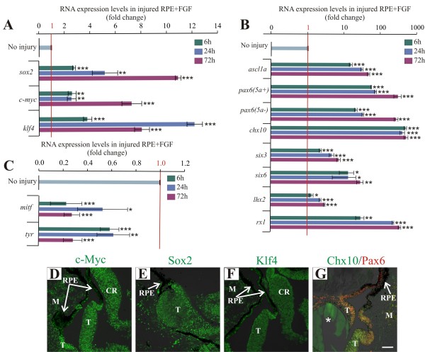
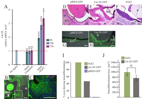
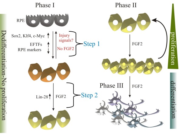
Results: We present evidence that upon injury, the quiescent (p27(Kip1)+/BrdU-) RPE cells transiently dedifferentiate and express sox2, c-myc and klf4 along with eye field transcriptional factors and display a differential up-regulation of alternative splice variants of pax6. However, this transient process of dedifferentiation is not sustained unless FGF2 is present. We have identified lin-28 as a downstream target of FGF2 during the process of retina regeneration. Moreover, we show that overexpression of lin-28 after retinectomy was sufficient to induce transdifferentiation of the RPE in the absence of FGF2.

Conclusion: These findings delineate in detail the molecular changes that take place in the RPE during the process of transdifferentiation in the embryonic chick, and specifically identify Lin-28 as an important factor in this process. We propose a novel model in which injury signals initiate RPE dedifferentiation, while FGF2 up-regulates Lin-28, allowing for RPE transdifferentiation to proceed.


Figures

Figure 1
Figure 1
Expression of pluripotency-inducing factors in the developing chick eye. (A) RT-PCR analysis of pluripotency-inducing factors at Stage 8 to 9 and ciliary margin (CM) and retinal pigmented epithelium (RPE) at Stage 24 (E4). Expression of the housekeeping gene gapdh was used as an internal control. rdh10 and mitf are specifically expressed in the CM and RPE respectively and were used as controls for the specificity of the tissues. Immunohistochemical staining using antibodies against (B) Sox2, (C) Klf4, (D) c-Myc and (E) Lin-28 in the anterior region of E4 eyes (in green). In the posterior region, (F) Sox2, (G) Klf4, (H) c-Myc and (I) Lin-28 were tested along with Mitf (microphthalmia-associated transcriptional factor; in red). The scale bar in panel E represents 100 μm and also applies to panels B, C and D. Scale bar in panel I represents 50 μm and applies to panels F, G and H. CM, ciliary margin; L: lens; NE: neuroepithelium; RPE, retinal pigmented epithelium.
Figure 2
Figure 2
Pluripotency inducing factors sox2, c-myc and klf4, and eye field transcriptional factor expression is increased in the retinal pigmented epithelium after retina removal. Quantitative RT-PCR analysis at 6, 24 and 72 hours post-retinectomy (injury) of (A)sox2, c-myc and klf4; (B) eye field transcriptional factors six3, six6, lhx2 and rx1 and the progenitor markers ascl1 and chx10; and (C) Retinal pigmented epithelium (RPE)-specific factors mitf and tyr. The expression levels were normalized with intact RPE (no injury). The analysis was performed using three independent biological samples (n = 3) in triplicate and the comparative cycle threshold (2-ΔΔCt) method was used to determine relative changes in transcripts compared with gapdh mRNA levels. Significance was determined with unpaired Student’s t-test by comparing each time point with the intact RPE (no injury). Error bars represent standard error of the mean. *P < 0.05; **P < 0.01; ***P < 0.001, compared with intact RPE. NS, non-significant; RPE, retinal pigmented epithelium.
Figure 3
Figure 3
The alternative splice variants of pax-6 are differentially regulated in the injured retinal pigmented epithelium. Quantitative RT-PCR analysis of the splice variants of pax6 (5a- and 5a+) at 6, 24 and 72 h post-retinectomy (injury). The expression levels were normalized with intact retinal pigmented epithelium (no injury). The analysis was performed using three independent biological samples (n = 3) in triplicate and the comparative cycle threshold (2-ΔΔCt) method was used to determine relative changes in transcripts compared with gapdh mRNA levels. Significance was determined with unpaired Student’s t-test by comparing each time point with the non-injured retinal pigmented epithelium. Error bars represent standard error of the mean. *P < 0.05; ***P < 0.001, compared with intact RPE. NS, non-significant; RPE, retinal pigmented epithelium.
Figure 4
Figure 4
Pluripotency-inducing factors sox2, c-myc and klf4 and eye field transcriptional factor expression is sustained in the injured retinal pigmented epithelium in the presence of FGF2. (A-C) Quantitative RT-PCR analysis of sox2, c-myc and klf4(A); eye field transcriptional factors pax6 (5a+), pax6 (5a-), six3, six6 and lhx2, rx1 and the progenitor markers ascl1and chx10(B); and retinal pigmented epithelium (RPE)-specific markers mitf and tyr(C) at 6, 24 and 72 h PR in the presence of FGF2. The expression levels were normalized with intact RPE (no injury). The analysis was performed using three independent biological samples (n = 3) in triplicate and the comparative cycle threshold (2-ΔΔCt) method was used to determine relative changes in transcripts compared with gapdh mRNA levels. Significance was determined with unpaired Student’s t-test by comparing each time point with the intact RPE (no injury). Error bars represent standard error of the mean. *P < 0.05; **P < 0.01; ***P < 0.001, compared with intact RPE. (D-G) Immunofluorescence analysis of c-Myc (D), Sox2 (E), Klf4 (F) and progenitor markers Pax6 and Chx10 (G) in transdifferentiated RPE. The asterisk represents the FGF2-soaked heparin bead. The scale bar in panel G represents 50 μm and applies to panels D-G. CR, ciliary regeneration; M, mesenchyme; RPE: retinal pigmented epithelium; T, transdifferentiated RPE.
Figure 5
Figure 5
Lin-28 is sufficient to induce retinal pigmented epithelium transdifferentiation. (A) Quantitative RT-PCR analysis at 6, 24 and 72 h post-retinectomy (PR) shows the relative levels of lin-28 expression in the injured retinal pigmented epithelium (RPE) in the absence or presence of FGF2. The expression levels were normalized with intact RPE (no injury). The analysis was performed using three independent biological samples (n = 3) in triplicate and the comparative cycle threshold (2-ΔΔCt) method was used to determine relative changes in transcripts compared with gapdh mRNA levels. The Student’s t-test was used to determine significance. Error bars represent standard error of the mean. **P < 0.01; ***P < 0.001, compared with intact RPE. (B) Lin-28 immunofluorescence in the transdifferentiated RPE 72 h PR in presence of FGF2. (C) Magnification of the boxed area in B stained for Lin-28 (green) and DAPI (blue). (D-F) Hematoxylin-and-eosin-stained sections at 72 h PR of electroporated eyes with pcDNA3.1 + pIRES-GFP (D), pCLIN-28 + pIRES-GFP (Lin-28 + GFP) (E) or treated with FGF2 (F). (G,H) GFP immunofluorescence analysis of electroporated eyes with pIRES-GFP (G) or pCLIN-28 + pIRES-GFP (Lin-28 + GFP) (H). (I) Percentage of eyes showing transdifferentiation at 72 h PR in the presence of FGF2 (n = 12, 100%); electroporated with pCLIN-28 + pIRES-GFP (Lin-28a + GFP) (n = 33, 48%, including the thickened depigmented RPE to full RPE transdifferentiation); or pIRES-GFP (n = 17, 0%). (J) Quantitative analysis of transdifferentiated areas observed in histological sections from eyes treated with FGF2 or electroporated with pCLIN-28 + pIRES-GFP. Error bars represent standard error of the mean. The asterisk represents the FGF2-soaked heparin bead. The scale bar in panels B, E and F represents 50 μm. The scale bars in panels C, D, and G and H represent 300 μm, 100 μm, and 20 μm respectively. L: lens; M: mesenchyme; NS: non-significant; RPE: retinal pigmented epithelium; T: transdifferentiated RPE.
Figure 6
Figure 6
Model representing the process of chick retinal pigmented epithelium transdifferentiation. Phase I includes dedifferentiation. During this phase, there is no proliferation. During step 1, injury signals are produced in response to retinectomy; pluripotency-induced factors Sox2, c-Myc and Klf4 that are also present in retina progenitors are up-regulated as well as eye field transcriptional factors (EFTFs), along with a down-regulation of RPE specific markers. During step 2, after the addition of exogenous FGF2, Lin-28, is up-regulated; however, during this stage there is no cell proliferation. In Phase II, in the presence of FGF2, proliferation is initiated. Lastly, on Phase III, differentiation of retinal cells takes place.

References

    1. Barbosa-Sabanero K, Hoffmann A, Judge C, Lightcap N, Tsonis PA, Del Rio-Tsonis K. Lens and retina regeneration: new perspectives from model organisms. Biochem J. 2012;12:321–334. doi: 10.1042/BJ20120813. - DOI - PubMed
    1. Fuhrmann S, Zou C, Levine EM. Retinal pigment epithelium development, plasticity, and tissue homeostasis. Exp Eye Res. 2013. [Epub ahead of print] - PMC - PubMed
    1. Coulombre JL, Coulombre AJ. Regeneration of neural retina from the pigmented epithelium in the chick embryo. Dev Biol. 1965;12:79–92. doi: 10.1016/0012-1606(65)90022-9. - DOI - PubMed
    1. Hamburger VHH. A series of normal stages in the development of the chick embryo. J Morphol. 1951;12:49–92. doi: 10.1002/jmor.1050880104. - DOI - PubMed
    1. Spence JR, Madhavan M, Ewing JD, Jones DK, Lehman BM, Del Rio-Tsonis K. The hedgehog pathway is a modulator of retina regeneration. Development. 2004;12:4607–4621. doi: 10.1242/dev.01298. - DOI - PubMed

Publication types

MeSH terms