Review
. 2014 Apr 16;82(2):261-78.
doi: 10.1016/j.neuron.2014.03.029.

Representing sex in the brain, one module at a time

Affiliations
Review

Representing sex in the brain, one module at a time

Cindy F Yang et al. Neuron. .

Abstract

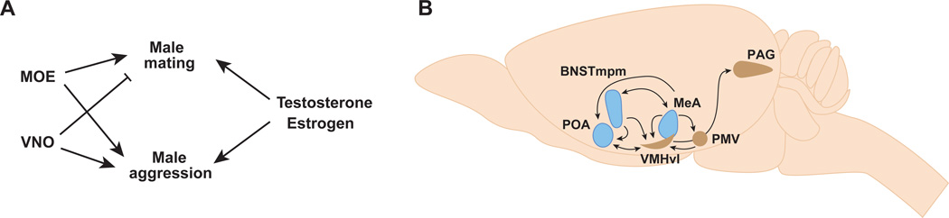
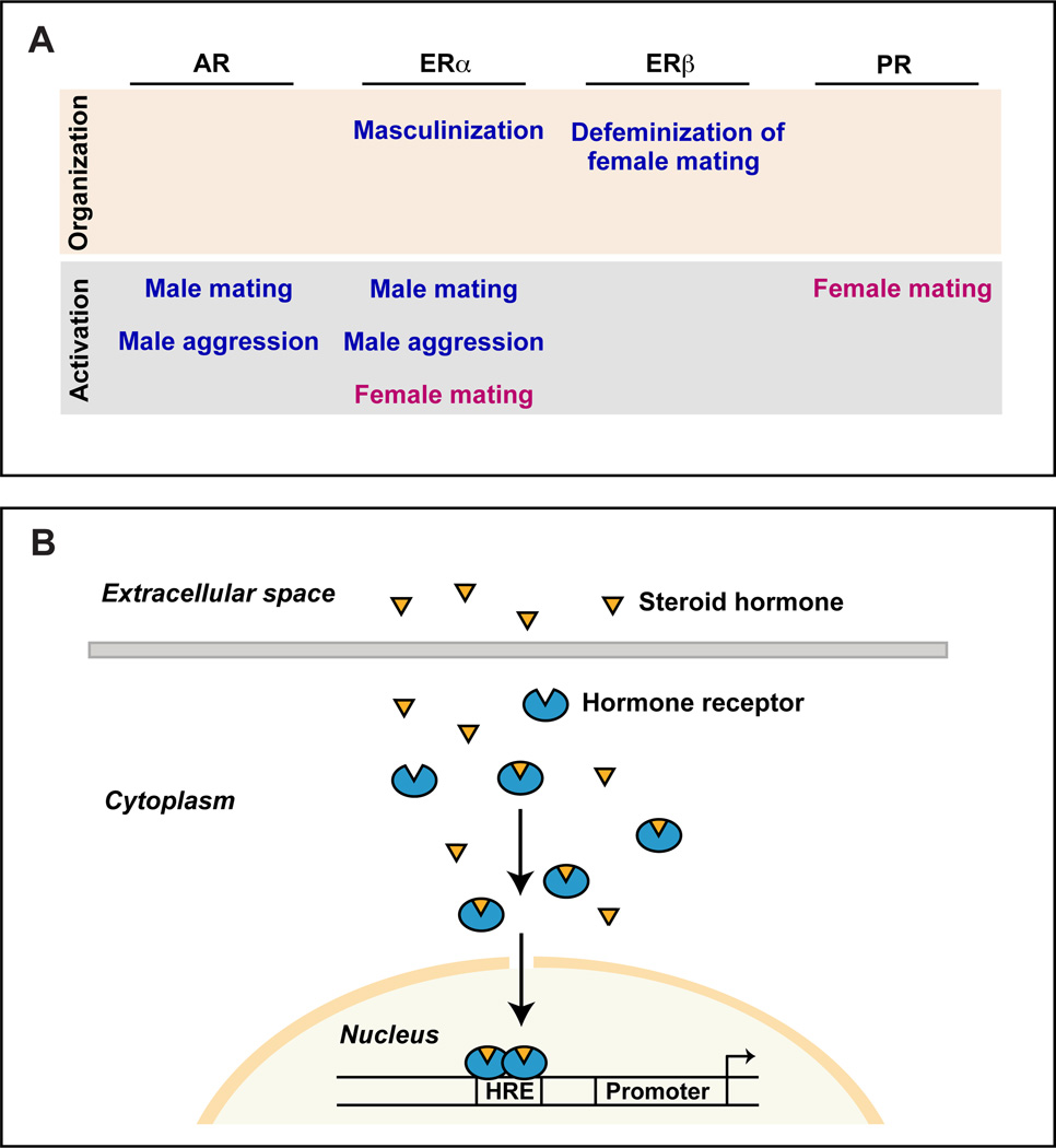
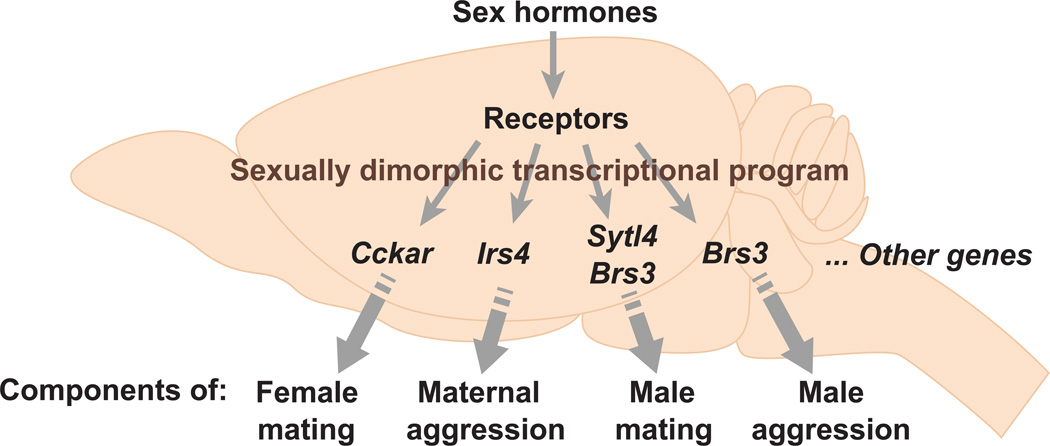
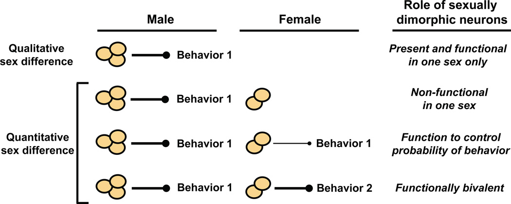
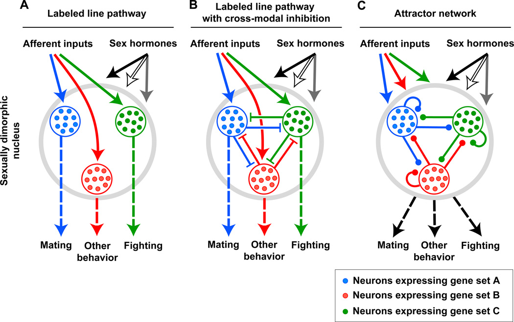
Sexually dimorphic behaviors, qualitative or quantitative differences in behaviors between the sexes, result from the activity of a sexually differentiated nervous system. Sensory cues and sex hormones control the entire repertoire of sexually dimorphic behaviors, including those commonly thought to be charged with emotion such as courtship and aggression. Such overarching control mechanisms regulate distinct genes and neurons that in turn specify the display of these behaviors in a modular manner. How such modular control is transformed into cohesive internal states that correspond to sexually dimorphic behavior is poorly understood. We summarize current understanding of the neural circuit control of sexually dimorphic behaviors from several perspectives, including how neural circuits in general, and sexually dimorphic neurons in particular, can generate sexually dimorphic behaviors, and how molecular mechanisms and evolutionary constraints shape these behaviors. We propose that emergent themes such as the modular genetic and neural control of dimorphic behavior are broadly applicable to the neural control of other behaviors.


Figures

Figure 1
Figure 1. Neural circuits that can generate sexually dimorphic behaviors
Simplified wiring diagram of some neural circuit configurations that can generate sexually dimorphic behaviors. Although only circuits driving male-specific behaviors are shown for clarity, similar circuits will exist for female-specific behaviors. The axon termini of all neurons except those of motor neurons end in small solid circles to show that they may transmit effectively excitatory, inhibitory, or neuromodulatory output. Termini of male motor neurons are shown as arrows to illustrate stimulation of the muscle groups required for the behavioral display. By contrast, female motor neurons are not shown to have termini to depict lack of activation of the male-specific behavioral program. (A) The entire neural circuit for generating a male-specific behavior is only present in males. (B) Sensory neurons unique to males feed into a shared neural circuit to activate a male-typical behavior. (C) Motor neurons unique to males are regulated by a shared neural circuit to activate a male-typical behavioral response. (D) Sensory and motor neurons are shared between the sexes but there are sex differences in intermediary neuronal populations. Most sex differences in intermediary neurons appear to be quantitative rather than qualitative in mice; in other words, the comparable neuronal population is shared between the sexes, but it displays cellular or molecular sexual dimorphisms that permit activation of the behavior only in males.
Figure 2
Figure 2. Sex determination and sexual differentiation of behavior
(A) In mice the presence or absence of Sry drives the differentiation of the bipotential gonad into testes or ovaries, respectively. Sex hormones released into the circulation by the gonads act on their cognate receptors to organize the brain during development and to control the activation of sex-typical behaviors in the adult. In males, estrogen organizes the neural substrates for behavior neonatally, and both estrogen and testosterone activate these pathways for male-typical behavior in adults. In the absence of the neonatal organizational effect of estrogen, the default differentiation program of the brain is female although this hormone may be important in adolescence for maturation of the neural substrates underlying female sexual receptivity (not shown) (Bakker et al., 2002; Brock et al., 2011). Both estrogen and progesterone activate the neural circuit underlying this behavior in adult females. (B) The sex of a fruit fly is determined in a cell autonomous manner, with the expression of sex lethal (Sxl) specifying a female differentiation program. Sex-specific splice forms of doublesex (Dsx) and fruitless direct the cell autonomous differentiation of neurons that control sex-typical behaviors. Ix, intersex, and Tra, transformer.
Figure 3
Figure 3. Sex hormone control of sexually dimorphic behaviors
Sex hormones produced in the gonads cross the blood-brain barrier and bind to hormone receptors in neurons to regulate sex-typical behaviors. In males, testosterone directs behavior by binding to its receptor AR or it is converted via aromatase into estrogen, which binds to its cognate receptors ERα and ERβ. In females, estrogen and progesterone direct behavior via their cognate receptors ERα and ERβ and PR, respectively.
Figure 4
Figure 4. Pheromone sensing pathways
Schematic representing that pheromone sensing neurons in the main olfactory epithelium and vomeronasal organ activate distinct neural pathways. Dashed oval represents the AOB. Thin arrows to the MeA and BNSTmpm depict relatively minor projections to these areas from the MOB and AOB, respectively.
Figure 5
Figure 5. Sensory and hormonal control of sexually dimorphic behaviors
(A) Control of male pattern mating and aggression by chemosensory input and sex hormones. (B) Schematic representing extensive interconnections between hypothalamic and amygdalar nuclei that regulate sexually dimorphic behaviors. These areas process pheromonal information, and subsets of adult neurons within each of these regions express sex hormone receptors; neurons within some of these regions (blue) also express aromatase. PAG, peri-aqueductal gray; PMV, ventral pre-mamillary nucleus; POA, preoptic hypothalamus; VMHvl, ventrolateral component of the ventromedial hypothalamus.
Figure 6
Figure 6. Mechanism and function of sex hormone action
(A) Function of sex hormone receptors in the nervous system during development (organization) and adult life (activation). Not shown is the requirement of estrogen to feminize sexual behavior, which occurs likely via ERα subsequent to the neonatal organizational phase but prior to adulthood (Bakker et al., 2002; Brock et al., 2011). (B) Schematic illustrating sex hormone action via nuclear hormone receptors. Sex hormones are steroids that can cross the blood-brain barrier and cell membranes to bind their cognate receptors. Hormone-bound receptor translocates to the nucleus, where it can regulate transcription of target genes by directly binding to specific DNA sequences. HRE, hormone response element.
Figure 7
Figure 7. Modular genetic control of sexually dimorphic behaviors by sex hormones
This model proposes that sex hormones control a sexually dimorphic transcriptional program in the nervous system such that individual dimorphically expressed genes control one or a few components of a sex-typical behavior. This model is supported by work showing that genes downstream of sex hormone signaling (Brs3, Cckar, Irs4, Sytl4) are required for the normal display of sexual or aggressive displays (Xu et al., 2012). Many genes downstream of sex hormone signaling still remain to be identified.
Figure 8
Figure 8. Function of sexually dimorphic neuronal populations
Neurons present only in one sex (qualitative sex difference) may either activate or inhibit a sexually dimorphic behavior in that sex. More commonly in mice and other vertebrates, a neuronal population is present in both sexes but presents sex differences (quantitative sex difference) in gene expression, cell number, or other cytological feature. In such cases, the neurons may be non-functional in one sex, regulate the probability of displaying a sexually dimorphic behavior, or control the display of different sexually dimorphic behaviors in the two sexes (functionally bivalent).
Figure 9
Figure 9. The relation between molecular heterogeneity in and functional pleiotropy of sexually dimorphic brain regions
Some models that can be used to relate molecular heterogeneity to functional diversity are shown. (A) Afferent and sex hormone inputs drive labeled line pathways to control different behaviors. Such a labeled line pathway is a simplified version of a multi-layered feed forward network. (B) Afferent and sex hormone inputs drive labeled line pathways with cross-modal inhibition to control different behaviors. (C) The heterogeneous neurons constitute an attractor type network with local and recurrent connections. Afferent and sex hormone inputs in conjunction with local circuits lead to a stable state of the network that elicits behavior. Molecular heterogeneity may afford a single neural circuit to utilize labeled line, labeled line with cross-modal inhibition, and attractor network pathways at distinct synaptic stations. Alternately, a single neural circuit may consist entirely of one or the other of these pathways.

References

    1. Allen E, Francis BF, Robertson LL, Colgate CE, Johnston CG, Doisy EA, Kountz WB, Gibson HV. The hormone of the ovarian follicle; its localization and action in test animals, and additional points bearing upon the internal secretion of the ovary. Am. J. Anat. 1924;34:133–181.
    1. Amateau SK, Alt JJ, Stamps CL, McCarthy MM. Brain estradiol content in newborn rats: sex differences, regional heterogeneity, and possible de novo synthesis by the female telencephalon. Endocrinology. 2004;145:2906–2917. - PubMed
    1. Antliff HR, Young WC. Behavioral and tissue responses of male guinea pigs to estrogens and the problem of hormone specificity. Endocrinology. 1956;59:74–82. - PubMed
    1. Arnold AP. The organizational-activational hypothesis as the foundation for a unified theory of sexual differentiation of all mammalian tissues. Horm. Behav. 2009;55:570–578. - PMC - PubMed
    1. Asahina K, Watanabe K, Duistermars BJ, Hoopfer E, González CR, Eyjólfsdóttir EA, Perona P, Anderson DJ. Tachykinin-Expressing Neurons Control Male-Specific Aggressive Arousal in Drosophila. Cell. 2014;156:221–235. - PMC - PubMed

Publication types

LinkOut - more resources